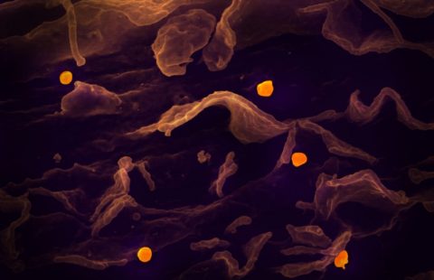
Associated with an aging population and the development of metabolic syndrome, atherosclerosis is a leading cause of death worldwide. Researchers from Inserm Unit 970, the “Paris Cardiovascular Research Center” (Inserm/Université Paris Descartes), have succeeded in revealing the mechanisms underlying the formation of atherosclerotic plaques. In particular, they have discovered the protective role of autophagy, a mechanism for cleaning and recycling cell components, in the cells that line the inner artery wall. These results, published in PNAS on September 25, 2017, provide us with an improved understanding of the initial stages of plaque development, and open up the possibility of developing a preventive treatment.
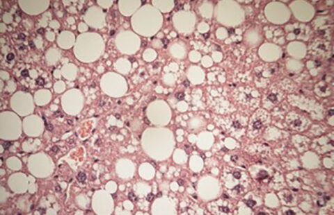
Atherosclerosis is a cardiovascular disease characterized by the build-up of plaque primarily made up of lipids (atheroma) on the inner artery wall. While some of these plaques remain stable, others erode or break apart with dramatic consequences for the patient, including heart attack or stroke.
The risk factors for cardiovascular disease are multiple and include diabetes, obesity, smoking, and hypertension. Although these factors are general, atherosclerotic plaques primarily develop in very specific areas of the circulatory system: the arterial bifurcations and curvatures. These areas are subject to low frictional forces from the flowing blood. In contrast, areas of the arteries exposed to higher frictional forces are protected from atherosclerosis. The mechanisms involved in this protective role of the frictional forces on the development of atherosclerosis remain poorly understood.
A recent study led by Chantal Boulanger and Pierre-Emmanuel Rautou from Inserm Unit 970, the “Paris Cardiovascular Research Center”, has filled this gap in our knowledge of atherosclerosis. Their research has shown the key role played by endothelial autophagy, i.e. the ability of the cells lining the inner wall of the arteries to break down and recycle their own components.
Autophagy is an intracellular process through which the cell breaks down part of its cytoplasm (the contents of the cell between the membrane and the nucleus) in response to stress or a lack of nutrients. The research team observed that the friction exerted by the blood strongly stimulates autophagy on the surface of the arterial wall. This phenomenon enables the endothelial cells to maintain a healthy physiological state, and prevents the development of atherosclerosis by protecting them from cell death, senescence, and inflammation.
The researchers then blocked the mechanism of autophagy on the surface of the arterial wall and observed increased plaque formation in the arterial areas usually protected from the development of atherosclerosis. Endothelial autophagy thus represents the previously missing link between atherosclerosis and the forces exerted by the flowing blood.
“These results prove that inhibiting autophagy is not beneficial in the case of atherosclerosis. Specific stimulation of autophagy, on the other hand, could enable us to prevent the formation of atheroma and thus reduce the risk of heart attack and stroke, which are major public health challenges, ” concludes Chantal Boulanger.
These contents could be interesting :
Medias
Sources
Autophagy is required for endothelial cell alignment and atheroprotection under physiological blood flow A.-C. Viona,b,1, M. Kheloufia,b,c,1, A. Hammoutenea,b, J. Poissona,b, J. Lasselina,b, Cecile Devuea,b, I. Pica,b, N. Dupontb,d,e, J. Bussef, Konstantin Starkf, J. Lafaurie-Janvoreg, A. I. Barakatg, X. Loyera,b, M. Souyrih, B. Violletb,i,j, P. Juliab,k, A. Tedguia,b, P. Codognob,d,e, C. M. Boulangera,b,2, and P.-E. Rautoua,b,c,l,2 a. INSERM, U970, Paris Cardiovascular Research Center, 75015 Paris, France;
b. Université Paris Descartes, Sorbonne Paris Cité, 75006 Paris, France;
c. Université Paris Diderot, Sorbonne Paris Cité, 75013 Paris, France;
d. INSERM U1151, Institut Necker-Enfants Malades-INEM, 75014 Paris, France;
e. CNRS UMR 8253, 75014 Paris, France;
f. Medizinische Klinik I, Klinikum der Universität München, 81377 Munich, Germany;
g. Mechanics & Living Systems, Cardiovascular Cellular Engineering, Laboratoire d’Hydrodynamique, Ecole Polytechnique, UMR 7646, 91128 Palaiseau, France;
h. INSERMUMR_S1131/IHU/Université Paris Diderot, 75013 Paris, France;
i. INSERM U1016, Institut Cochin, 75014 Paris, France;
j. CNRS, UMR 8104, 75014 Paris, France;
k. Service de Chirurgie Cardiaque et Vasculaire, Hôpital Européen Georges Pompidou, AP-HP, 75015 Paris, France; and
l. Département Hospitalo-Universitaire Unity, Pôle des Maladies de l’Appareil Digestif, Service d’Hépatologie, Centre de Référence des Maladies Vasculaires du Foie, Hôpital Beaujon, Assistance Publique-Hopitaux de Paris, 92110 Clichy, France 1. -C.V. and M.K. contributed equally to this work.
2. M.B. and P.-E.R. contributed equally to this work.