Menu

New molecules discovered to tackle cancer resistance

A French-Italian team headed by researchers from CNRS and Inserm (1) has recently discovered a new family of compounds that could make it possible to treat numerous cancers, particularly brain tumours and skin cancers. These substances, patented by CNRS, block a chain of molecular reactions known as the Hedgehog signalling pathway, the disruption of which could be involved in several cancers. These compounds could become new medicines in the long term. In the short term, they should prove to be precious tools in elucidating the role of the Hedgehog pathway in the development of these tumours and their resistance to treatment. This work, carried out in collaboration with the Laboratoire d’Innovations Thérapeutiques (CNRS / Université de Strasbourg), is published in the Journal of Medicinal Chemistry.

The Hedgehog signalling pathway is a cascade of complex biochemical reactions. Very active during embryogenesis, it participates in cell proliferation and differentiation, as well as in the development of numerous tissues. In adults, it plays a key role, particularly in maintaining stem cells in the brain. The disruption of this pathway could participate in the development of many cancers, especially very aggressive brain tumours in children.

Malfunctions affecting the Hedgehog pathway can be caused by mutations of a membrane receptor known as Smoothened, an essential link permitting the activation of the Hedgehog pathway. Several pharmaceutical laboratories have developed compounds capable of blocking Smoothened. Thanks to these receptor antagonistic compounds (2), they have succeeded in stopping the development of certain tumours. However, experiments conducted on animal models and in humans demonstrate the onset of resistance to these treatments. New mutations of Smoothened in the tumoral cells annihilate the effects of the antagonists responsible for inactivating it. For this reason, it is important to find new antagonists and to better understand the mechanisms underlying this type of resistance.

In order to discover new Smoothened antagonists, the team of researchers coordinated by Martial Ruat adopted a novel strategy, namely the virtual screening of computerized molecular databases.

Among the 500,000 or so molecules included in these databases, they searched for those whose structure could produce the same effect as molecules already known to inhibit Smoothened. The scientists selected one molecule out of about 20 candidates. By slightly modifying its structure in order to optimize it, they discovered a family of compounds known as MRT and tested their biological activity on cultured mouse cells.The team discovered that MRT compounds, and more particularly acylguanidine MRT83, block the proliferation of cells suspected of causing brain tumours. In addition, these new compounds inhibit Smoothened with an activity similar to or exceeding that of already known compounds.

Several years of tests will be necessary before promising new substances such as MRT compounds can be marketed as medicines. Nevertheless, their properties could shed new light on the functioning, the three dimensional structure and the location of Smoothened receptors. These MRT compounds could contribute to understanding the cause of tumour resistance. This study could lead to the discovery of new targets and therapeutic strategies for combating certain cancers.

Footnotes

(1) In the ‘Neurobiology & development’ research unit (CNRS), in collaboration with the University of Sienna (Italy)

(2) When bound to the same cell receptors as one other substance, an antagonist substance blocks all or part of the effects normally produced by the cell. Here the receptor antagonist has an ‘inverse’ effect compared with that of the ‘muted’ receptor.

Post-traumatic stress disorder (PTSD) defined by an inversion of brain activity induced by stress hormones

We remember, right down to the tiniest detail, a car accident that happened over two years ago, when we feared for our lives; and yet, who among us can still remember a very good meal enjoyed just last year, even if it was really very, very good? Today, there is ever-increasing understanding of the biological bases behind this “adaptive” capacity to remember a stressful. However, until now, we knew very little about the state of post-traumatic stress disorder (PTSD). This pathological state is triggered in some individuals after exposure to a highly stressful event. In this state, patients are gripped by fear even when faced with elements that have no objective danger. Is PTSD specific to the human race, influenced by its history and culture? Or is it a state that is found in various species, resulting in common biological changes? These are the questions that Pier-Vincenzo Piazza, Director of the Neurocentre Magendie in Bordeaux (Inserm/ Université Victor Segalen), and his collaborators, have attempted to answer. Details of their results are provided in the Science review (advance publication on-line on Science Express website on 23 February).

It is easier to memorize a stressful event than an agreeable one. Almost all species capable of behaviour are able to recall negative events, proof that this is likely to be a capacity selected during evolution: it ensures survival in a hostile environment.

However, for some individuals, exposure to very stressful events may trigger a pathological state: post-traumatic stress disorder (PTSD) being the most emblematic. In the United States, estimates suggest that this syndrome afflicts 6.8% of the general population and that 30% of veterans from the Vietnam war and 12% of the Gulf war suffer from it (source National Centre for PTSD).

In this state of stress, the memory of the patient is impaired: it is no longer capable of adapting its reaction of fear to the “right” context and to the “right” predictive elements. Sufferers experience fear in situations where there are no threats. These fears become all-consuming, eventually preventing a normal life. “If you are attacked by a lion in the savannah as a flock of birds flew overhead, it would be normal to feel fear if you were to make a return trip to the savannah” explains Pier-Vincenzo Piazza. “However, you should not be gripped by fear if, when strolling around another natural open space, for example a golf green, you hear a bird cry or catch sight of birds on the horizon,” thethe Inserm research director goes on. If this is the case, you may have developed post-traumatic stress disorder as a consequence to the lion attack.

The teams, led by Pier-Vincenzo Piazza and Aline Desmedt, have demonstrated that such memory impairment associated with PTSD is not specific to humans and is also found in mice. To this end, the researchers conditioned mice to a) anticipate a threat (an electric shock of varying intensity) by using the same specific context (an indicating environment) and b) to distinguish this specific context from stimuli, which although present during the conditioning process, did not predict the threat (a sound).

In normal conditions, the mice showed a reaction of fear when exposed to the specific context (the indicator environment) of the threat, but did not react to the sound (not predictive of threat).

Following this conditioning session, the researchers then administered increasing concentrations of glucocorticoid hormones, the main biological response to stress in mammals. When administration of the glucocorticoids followed an intense threat, as is the case with individuals suffering from post-traumatic stress, the mice were no longer able to restrict their fear reaction to the “right” context and to the correct indicators pronouncing a possible threat. The animals began to show fear: they froze in response to indicators, which although present during the stressful situation, were not predictive of the threat in any way. These results show that PTSD is probably due to a simultaneous overproduction of glucocorticoids in some subjects as the traumatic event occurs.

Memory impairment induced by glucocorticoids is accompanied by activity reorganisation in the brain, and, more specifically, the hippocampus-amygdale circuit, which is essential for coding memories associated with fear. In normal conditions, when an individual associates a threat with a context, strong activity is observed in the hippocampus, the structure in the brain required for all knowledge acquisition associating a specific context, area, etc. with an event. However, activity in the amygdale is low. The amygdale is an area of the brain that is also involved in emotional memory, but it memorizes specific indicators, such as sounds, which predict the threat.

When the mice were subject to an increase in glucocorticoids and memory impairment characterising PTSD was observed, activity in the hippocampus was reduced, whereas activity in the amygdale increased. In states of post traumatic stress, the researchers noted an inversion of normal activity in the brain. Normal activity in the amygdale may explain the fact that the subject begins to “over-respond” to presumed indicators, present during the traumatising event, but which are not, themselves, predictive of any danger. Low activity in the hippocampus may explain why subjects no longer recognize the right context: they are therefore incapable of containing their fear to appropriate situations.

PTSD is not only an overbearing memory of the traumatizing situation, but also a memory impairment that prevents patients from containing their reaction of fear to the context that predicts the threat”, explain the researchers. In the case of post-traumatic stress disorder, a vivid memory of the traumatising event is associated with amnesia in terms of the surrounding context of the event. Some contextual elements present during the traumatising event are wrongly considered to be predictive of the event.

To conclude, the authors explain that PTSD-related memory problems seem to be caused by a biological response to abnormal stress suffered by some patients: for these patients, excessive glucocorticoid production at the same time as exposure to intense stress triggers an inversion of normal activity in brain structures that code fear-related memories.

“We have demonstrated that PTSD can occur in species other than humans and that there are shared biological origins. The mice model of this pathology now paves the way for better understanding of the molecular bases of this pathology, which could lead to the development of a treatment,” conclude the Inserm researchers.

Is there a general motivation centre in the depths of the brain?

A team coordinated by Mathias Pessiglione, Inserm researcher at the “Centre de recherche en neurosciences de la Pitié Salpêtrière” (Inserm/UPMC-Université Pierre and Marie Curie/CNRS) have identified the part of the brain driving motivation during actions that combine physical and mental effort: the ventral striatum. The results of their study were published in PLoS Biology on 21 February 2012.

The results of an activity (physical or mental) partly depend on the efforts devoted to it, which may be incentive-motivated. For example, a sportsperson is likely to train with “increased intensity” if the result will bring social prestige or financial gain. The same can be said for students who study for their exams with the objective of succeeding in their professional career. What happens when physical and mental efforts are required to reach an objective?

Mathias Pessiglione and his team from Inserm unit 975 “Centre de recherche en neurosciences de la Pitié-Salpêtrière” examined whether mental and physical efforts are driven by a motivation ‘centre’ or whether they are conducted by different parts of the brain. The researchers studied the neural mechanisms resulting from activities that combine both action and cognition.

To this end, a series of 360 tests, combining mental and physical effort, were performed whilst being monitored by a scanner. The 20 voluntary participants were placed in the supine position, with their heads in a functional MRI scanner. They then had to complete a series of tasks through which they could accumulate winnings. However, in each series the winnings were limited to the first incorrect response. The tasks combined cognitive and motor actions. The participants had to find the highest number from among different-sized numbers and then select it by squeezing a handle located by their left or right hand (depending on the number’s location). At the end of the test, a winnings summary was displayed to motivate the participant.

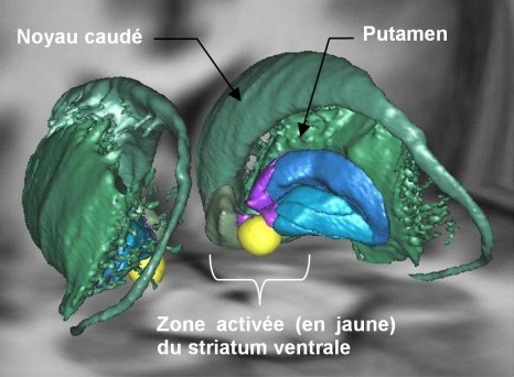
© M. Pessiglione, Inserm

3D representation of motivational system (striatum ventral) activated during physical or mental effort.

Using images obtained from the MRI scans taken during the test, Mathias Pessiglione and his team identified a general motivational system in the depths of the brain, i.e. a structure capable of activating any effort type, both mental (concentrating on the task in hand) or physical (lifting a load). The researchers observed that the ventral striatum was activated in proportion to the amount of money involved: the higher the degree of motivation, the higher the activation level. Furthermore, the ventral striatum is connected to the median part of the striatum (the caudate nucleus) when the task to be performed is cognitively difficult (when the physical size and the numerical value of the numbers did not correspond). This ventral region solicits the lateral part of the striatum (the putamen) when the difficulty is motor-related (when the handle had to be squeezed very tightly).

The researchers suggest that the expectation of a reward is encoded in the ventral striatum, which can then drive either the motor or cognitive part of the striatum, depending on the task, in order to boost performance. “The ventral striatum may commute connections in accordance with the request, i.e. enhance the neuronal activity in the caudate nucleus for a cognitive operation and in the putamen for a physical action” explains Mathias Pessiglione.

When your left hand mimics what your right hand does: it’s in the genes

Research scientists from Inserm, CNRS, UPMC and AP-HP working for the Centre de Recherche de l’Institut du Cerveau et de la Moelle (CRICM) of la Pitié-Salpêtrière, have just discovered mutations that could be the cause of congenital mirror movement disorders. Persons suffering from this disorder are unable to perform different movements with different hands. Genome sequencing in several members of a French family showed the culprit to be the RAD51 gene. Further work carried out on mice suggests that this gene plays a part in motor network cross-over. Cross-over is a key factor in the transmission of brain signals, because it allows the right side of the brain to control the left side of the body and vice versa. This research has been published in The American Journal of Human Genetics.

Congenital mirror movement disorders by Emmanuel Flamand-Roze

Congenital mirror movement is a rare disease transmitted from one generation to another by dominant inheritance. The affected persons lose the ability to carry out different movements with separate hands: when one hand moves in a certain way, the other hand is “forced” to copy the same movement, even if the person does not wish to do so. So people suffering from this disease are totally incapable of bimanual motor activities, such as piano playing for example. This phenomenon has been observed in children, but generally cleared up spontaneously before the age of 10, no doubt due to maturing of the motoneuron networks. However, in people who are affected by the disorder, the illness starts in early childhood and remains unchanged throughout life.

In 2010, research scientists from Quebec analyzed the genome from the members of a large Canadian family and discovered a gene responsible for the disease. Mutations had been detected in the DCC (Deleted in Colorectal Carcinoma) gene. Following this discovery, the team of researchers and doctors coordinated by Emmanuel Flamand-Roze began to search for mutations in this gene in several members of a French family who were also suffering from congenital mirror movements disease, but without success. “The DCC gene was intact”, explained Emmanuel Flamand-Roze. “We thought we were nearly there and instead we had to start searching for mutation in a different gene”, he adds.

Using an approach that combines conventional genetic analysis and “whole exome” analysis (a new-generation genetic analysis technique that involves entirely sequencing the important part of the genome), scientists demonstrated that the RAD51 gene was responsible for congenital mirror movement disease in a large French family and went on to corroborate this result using the same techniques on a German family suffering from the same disorder.

“The RAD51 gene was already known to the scientific community as a potential catalyst for certain types of cancer and in problems of resistance to chemotherapy”, explains Emmanuel Flamand-Roze. “So we wondered whether it had yet another function that could explain the motor symptoms of CMM disease”.

In humans, the motor system is a cross-control system, which means that the left side of the brain controls the motor functions of the right side of the body and vice versa, with the cross-over taking place at the brainstem. While studying the expression of the RAD51 protein during development of the motor system in mice, the research scientists discovered that this gene could be implanted into the cross-over of the motor network that links the brain to the spinal fluid at the brainstem.

This discovery opens up a whole new field of investigation into the development of the motor system and to achieving better understanding of the cerebral mechanisms that control bimanual motricity. It could also shed light on other motricity disorders related to fine movement organization, such as dystonia or certain genetic neurodevelopmental diseases.

Vitamin B and omega-3 supplementation and cancer: new data

Researchers from the Nutritional Epidemiology Joint Research Unit (Inserm-Inra-Cnam-Université Paris 13) have just published a study showing that, in men with a previous history of cardiovascular pathologies, supplementation with B vitamins and omega-3 polyunsaturated fatty acids (the fatty acids present particularly in oily fish and dried fruit) did not significantly increase the occurrence of cancer. However, women with a previous history of cardiovascular pathologies seem to have a higher cancer risk after five years of supplementation. The research is published in detail in the Archives of Internal Medicine.

Although some studies have suggested that supplementation with B vitamins has some benefits for protecting against cancer, the few randomised clinical trials conducted internationally in recent years remain equivocal.

The results of studies of the influence of supplementation with polyunsaturated fatty acids have been mixed.

That is why Valentina Andreeva and Pilar Galan and their staff wanted to study the effects of B vitamins and omega-3 polyunsaturated fatty acids (PUFAs) on cancer by monitoring, for five years from 2003 to 2009, more than 2500 people aged between 45 and 80 who had survived a heart attack, stroke or angina in the last 12 months.

The aim of the trial was to check the hypothesis that supplementation with en5-methyl-THF (and vitamins B6 and B12) and/or omega-3 was of value in preventing the recurrence of ischaemic pathologies in subjects who had suffered a heart attack or stroke.

The results, published this week in the Archives of Internal Medicine, are the outcome of a specific analysis performed as part of the intervention study SU.FOL.OM3, the results of which were published in 2010 in the British Medical Journal (1).

SU.FOL.OM3 is a randomised double-blind placebo-controlled secondary prevention trial in which participants aged from 45 to 80 years with a previous history of cardiovascular pathologies were given daily supplements for five years of B vitamins (3 mg of vitamin B6, 560 μg of folates and 20 μg of vitamin B12) and/or omega-3 PUFAs (600 mg of eicosapentaenoic acid, EPA, and docosapentaenoic acid, DHA, in a ratio of 2:1), according to a factorial design.

It tested the effect of supplementation on cancer development over the five-year monitoring period in 145 men and 29 women.

All types of cancer were monitored. “Despite the low numbers, the results are significant,” the authors believe, because the “methodology used – a randomised double-blind trial – is stringent.

The researchers conclude that “Neither B vitamin supplementation nor omega-3 PUFA supplementation has a significant effect on the occurrence of cancer in men. However, women receiving omega-3 fatty acids tended to present a higher risk of cancer compared with the placebo group (adjusted HR =3.02; CI of 95 % = [1.33; 6.89]). Although the underlying mechanisms are not clear, a potential mediating effect on the metabolism of oestrogens is suggested,” they add.

To confirm or contradict these results, it is necessary to await new studies: randomised trials, cohort studies and mechanistic studies.

Footnote

(1) Supplementation with nutritional doses of B vitamins and omega-3 fatty acids Galan P, Kesse-Guyot E, Czernichow S, Briancon S, Blacher J, Hercberg. Effects of B vitamins and omega 3 fatty acids on cardiovascular diseases:a randomised placebo controlled trial.

BMJ. 2010 341:c6273

Childhood cancer – Factors of genetic susceptibility in Ewing sarcoma discovered

Ewing sarcoma is a rare paediatric bone cancer. However, this condition occurs more frequently in populations of European ancestry. Olivier Delattre (1) and his team, David Cox (2) and Gilles Thomas (3) have sought to understand why. The response may be found in two small regions of the genome: two genetic variants observed most frequently in European populations. Children having one of these two variations have twice the risk of developing a Ewing sarcoma tumour. This discovery was published online in the Nature Genetics issue of 12 February 2012 (4).

Ewing sarcoma is a rare bone tumour occurring in children, teenagers and young adults. For several years now, Dr Olivier Delattre, a specialist in this type of cancer and director of Inserm research, and his team from the Institut Curie, have been researching differences in incidence of this tumour according to geographic origin. To answer this question, they collaborated with Gilles Thomas of the Lyon Cancer Synergie platform and David Cox, both researchers at the Léon Bérard Centre in Lyon.

The majority of Ewing tumours occur in children of European ancestry, with cases very rarely occurring in African or Asian populations. Furthermore, the number of cases in these latter populations remain few even when the populations have immigrated to the United States (0.017 for 105 African-American individuals). “As a result, environmental factors could not be blamed and it was necessary to search for the reasons for this difference in the genome,” explains Olivier Delattre. Such a genetic genetic study, on a rare tumour, was made possible by the development of new tools, the GWAS (Genome-Wide Association Study) in particular, providing for the map representation of individual genetic variations. The analysis was performed using 401 samples of the Ewing tumour, 684 controls from the French population and 3,668 controls from the American population with European ancestry. Of the more than 700,000 genetic variations observed, two of them (rs9430161 and rs224278) are associated with the development of the Ewing tumour. Children with these genetic variants have a twice the risk of developing a Ewing tumour as compared to others,” explains David Cox. This increase in relative risk is important for a better understanding of the disease, but remains insignificant for the carriers of these variants, as the absolute risk remains very slight, with 3 cases per million carriers of the variant. Moreover, these two genetic variants are much more rare in populations of African or Asian ancestry, which partly explains the low incidence of the condition in these populations.

Improving understanding of tumour development

Beyond the identification of the two susceptibility variants, this discovery contributes to the understanding of the cellular mechanism leading to the development of Ewing tumours. The two regions revealed are found near the TARDBP and EGR2 genes. The first has similarities to the gene of which the alteration causes Ewing sarcoma; the second is part of the group of genes regulated by the fusion gene EWS-FLI1, responsible for this tumour. “We can now try to find out how these two genes reinforce the EWS-FLI1 chromosome anomaly responsible for Ewing sarcoma,” adds Olivier Delattre.

The point of view of Olivier Delattre

Olivier Delattre

© Noak/Le Bar Floreal/institut Curie

Olivier Delattre

Genetics of populations to better understand cancer development

“The genetic material contained in the nucleus of each of our cells is a sort of book with 3 trillion characters written with an alphabet of only 4 letters: A, C, G, T. Even if the sequence of these characters is nearly identical from one individual to the next, there is still an average of a 0.1% (several million) difference between two individuals. The diversity of the human population thus lies in the slight variability of our DNA sequence. A frequent variation in a population is called a variant. Certain variants are associated with a condition: they alone do not represent a risk, but they establish a favourable foundation for its development. There are several methods for researching such susceptibilities. In our study on Ewing sarcoma, due to the high variability between one population and the next, the genetics of the populations were the most suitable. The variants have the ability to “modulate” the activity of certain genes contributing to the cancer’s development. Knowledge regarding them thus improves the understanding of tumour mechanisms.”

David Cox’s comments

David Cox

© Photo Centre Léon Bérard

David Cox

Application of genomics in cancer research

“Since the complete sequencing of the human genome in the beginning of the 2000s, our ability to explore our genetic code has exploded. Today, we can research more than 5 million variants, of the ‘polymorphisms’, which differ from one person to the next. These variants are a sort of record of the history of the DNA surrounding them. If a mutation increasing the risk of a disease is present in a population, the variants surrounding it will be transmitted with the mutation for generations. Since we don’t know where these mutations are found, we use the variants to find them, comparing the incidence of the variants of the genome in patients suffering from a disease to individuals of the same population who are healthy.”

Ewing sarcoma tumours

  • With nearly 100 new cases in France each year, the Ewing tumour is the number two primitive malignant bone cancer, in terms of incidence.
  • It occurs in children, teenagers and young adults (up to 30 years old), with the highest incidence at puberty. Also referred to as Ewing sarcoma, it develops mainly in the bones of the pelvis, the ribs, the femur, the fibula and the tibia.
  • Over the past 30 years, the treatment for Ewing sarcoma, relying mainly on radiation therapy, has changed immensely. Today, local forms are mainly treated with an initial combination of chemotherapy and surgery. Post-operation chemotherapy, and sometimes radiation therapy, complete the treatment. The prognosis of Ewing sarcoma has benefited from the contribution of new chemotherapies.
  • It was at the Institut Curie, in the unit of Olivier Delattre, that the chromosome anomaly responsible for this tumourwas discovered in 1984 and characterised in 1992. It consists in a translocation produced, in 90% of cases, between chromosomes 11 and 22, leading to the synthesis of abnormal protein EWS-FLI1, and in 10% of cases between chromosomes 22 and 21, leading to the synthesis of abnormal protein EWS-ERG. There are other alterations, but they are rare. The discovery of these genetic alterations provided for the development of a diagnostic test for the Ewing tumourat the Institut Curie in 1994.

This research was conducted within the framework of a broad European collaboration. They were financed by, in addition to Inserm and the Institut Curie, the French National League against Cancer within the framework of the “2009 Epidemiological Research Project” and by the INC within the framework of the 2008 and 2009 open calls for projects.

Dr Olivier Delattre’s team also receives financial support from the Association of Parents and Friends of Children Treated at the Institut Curie (APAESIC), associations Les Bagouz à Manon, Pas du Géant, Olivier Chape, Les Amis de Claire and Courir pour Mathieu, as well as the Health and Children Federation.

About the same subject

The Institut Curie is a government-recognised public interest foundation bringing together the largest French cancer research centre and two state-of-the-art hospital institutions. A pioneer of several treatments, it is an authority on for breast cancer, paediatric tumours and eye tumours. It provides for the dissemination of medical and scientific innovations on the national and international levels.

Founded in 1909 on a model devised by Marie Curie and still at the cutting edge: “from fundamental research to innovative treatments”, the Institut Curie has 3,000 researchers, physicians, clinicians, technicians and administrative staff. For more information: www.curie.fr

The Léon Bérard Centre is one of the twenty French Centres for the fight against cancer. Based in Lyon and recognised as a reference centre for cancer research in the Rhône-Alpes region, it has three missions: treatment, research and teaching. More than 23,000 patients from all over France are cared for each year in its technical facilities and departments proposing innovative treatments. A total of 1,400 doctors, researchers, caregivers, technicians and administrative staff work at the Léon Bérard Centre. For more information: www.centreleonberard.fr

The Lyon Cancer Research Centre (CRCL)

The Cancer Research Centre of Lyon is a new research establishment, created in January 2011 and approved by Inserm, CNRS, the Université Claude Bernard Lyon 1, the Léon Bérard Centre and working in partnership with the Civil Hospices of Lyon. This body brings together 17 research teams, for 370 members, including 110 researchers and research professors. The work of the CRCL focuses on fundamental cancer research, whiling aiming to support the development of strong translational research to the benefit of the sick.

The French National Institute of Health and Medical Research (Inserm) is a public science and technology institute, jointly supervised by the French Ministry of Health and Ministry of Research. In 2008, Inserm, the only French public research organisation dedicated entirely to human health, became responsible for the strategic, scientific and operational coordination of biomedical research. This central role of coordinator was naturally assigned to the centre due to the scientific quality of its teams and also its capacity to support translational research, also known as bench-to-bed research, describing an approach of applying lessons from the laboratory at the patient’s bedside. For mmore information: http://www.inserm.fr/

Footnotes:

(1) Olivier Delattre, director of the Genetics and Biology of Cancers Unit – Institut Curie/Inserm U830

(2) David Cox, Research Associate at Inserm, researcher in the Breast Cancer Genetics team within the Cancer Research Centre of Lyon UMR Inserm 1052 CNRS 5286 / Léon Bérard Centre / Université Lyon 1

(3) Gilles Thomas, Professor of Universités Lyon 1 – Hospital Practitioner Civil Hospices of Lyon, Director of the Synergie biocomputing platform of Lyon Cancer Léon Bérard Centre / Université Lyon 1 and researcher in the Breast Cancer Genetics team of the Cancer Research Centre of Lyon UMR Inserm 1052 CNRS 5286 / Léon Bérard Centre / Université Lyon 1

(4) “Variants at TARDBP and EGR2/ADO loci associated with Ewing sarcoma susceptibility” Nature Genetics, 12 February 2012, online

How can head injury prevention be improved for cyclists?

Researchers from a team coordinated by Emmanuel Lagarde at Inserm unit 897 “Inserm Epidemiology & Biostatistics research centre”, in collaboration with the Université Victor Segalen Bordeaux 2, have endeavoured to ascertain the factors preventing helmet use and the approaches that could be used to promote it. Although they represent a small proportion of urban travel modes, cyclists make up 5% of deaths and 6% of serious road injuries in towns, with most suffering from head injuries (1). These figures inspired the researchers to study the impact of two promotion strategies (documentation and free helmets) on adults, using a randomised comparative methodology. The results of this study on 1,800 bike users in Bordeaux and its urban area were published in the PLoS ONE review on 15 February.

In 2010 in metropolitan France, 59 cyclists lost their lives and 963 were seriously injured1. Two thirds of those with a life-threatening condition had suffered head injuries (2). How to protect users of a green and healthy mode of transport is today still an open question.

To study significant cycling accident prevention factors, Emmanuel Lagarde, Director of Research at Inserm unit 897 “Inserm Epidemiology & Biostatistics research centre”, and his team, set up a study into the behaviour of some 1,800 cyclists in Bordeaux and its urban area. The study ran from May 2009 to September 2010. The participants, aged between 18 and 75, responded to a questionnaire on their habits and opinions related to bikes and helmets. They were then organised into four groups: the first group received a brochure promoting helmet use; the second received a free helmet; the third a brochure and a helmet; and the fourth group did not receive anything.

Afterwards, all groups were sent a coloured sticker to be placed on their mud-guard, so they could be easily identified in the streets of Bordeaux by a network of seven automated cameras, specially developed by the researchers. The system was used to detect the cyclists anomalously, to record their speeds and to detect whether or not they were wearing helmets.

Figure 1: Azimuth view and automated detected algorithm

Figure 2: mud-guard with participant colour authentication code

Figure 2: mud-guard with participant colour authentication code

Although 90% of all individuals asked to participate in the study thought that helmets do provide head protection, only 13% said they had already worn one. In the first months of the study, participants who had received a free helmet were more than eight times more likely to wear one. “But after four months, there were no longer any discernible differences between the groups: the majority of cyclists had abandoned the helmets” explains Emmanuel Lagarde. He also specifies that “distributing the information brochure had no effect on the use of helmets”.

Using the cameras installed by the researchers, and further to analysis of the questionnaires, the scientists identified two factors that encourage cyclists to wear a helmet:

– Belief that helmets protect the face;

– Encouragement from family to wear one.

This study concludes that promoting the use of helmets among cyclists will remain difficult. According the researchers, “pressure from loved ones is an important factor in terms of helmet use among cyclists”. If prevention is to have a hope of being effective, it must integrate easy access to helmets, parental education and communication on the preventive benefits of wearing a cycle helmet. The safety of cyclists is a complex subject, which goes beyond the issue of helmets. “The behaviour of cyclists and other road users can contribute to the safety of cyclists; road layouts also have a key role” underlines Emmanuel Lagarde.

This study is confunded by the ANR, INPES and the Aquitaine regional council.

Footnotes:

(1) Data from the French national interministerial road safety observatory (ONISR) 2010

(2) Amoros et al. BMC Public Health 2011, 11:653

(French) Dengue : un anticorps prometteur caractérisé

Sorry, this press release is only available in French.

(French) De nouveaux résultats soulignent l’importance des gènes synaptiques dans l’autisme

Sorry, this press release is only available in French.

Serotonin and bones

Serotonin, a well-known brain neurotransmitter, is produced locally in an unexpected place: the bone tissue. This has just been demonstrated by research scientists from the Combined “Bone and joints” research unit 606 (Inserm/Paris Diderot) working jointly with the biochemistry laboratory from the Lariboisière Hospital and the “Cytokines, hematopoiesis and immune response” laboratory (CNRS/University of Paris Descartes) at the Necker Hospital in Paris. Apparently, this locally produced serotonin breaks down the bone tissue. These results, published in the PNAS, suggest that medications that modulate the effects of serotonin, such as anti-depressants or migraine drugs, could in one way or the other modify the delicate balance between the formation and the destruction of bone in the organism.

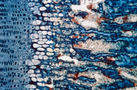
© Inserm, J.-P. Roux

Area showing bone growth and resorption. The osteoclasts, the cells that resorb bone, are shown in red.

Serotonin regulates a wide variety of functions such as mood, behavior, sleep, blood pressure and temperature regulation. It also ensures important functions in several peripheral tissues and regulates vascular and heart functions and gastro-intestinal motility. However only minute levels of serotonin circulate in the body. It is mainly stored in the platelets and is only available for the peripheral organs if it is degorged when the platelets are activated.

Certain researchers decided to look into the effect of serotonin on bone tissue that had been the subject of a recent debate. While some researchers described the negative action of the serotonin circulating in the bone tissue (it apparently prevents bone regeneration by acting on the osteoclasts and reducing their proliferation), others failed to observe any modification to the bones in serotonin-deficient mice.

Osteoclasts or osteoblasts?

Bone remodeling is a highly integrated process. It depends on a fine balance between the growth of bone controlled by osteoblasts and the destruction of bone controlled by osteoclasts. The permanent renewal of bone tissue ensures harmonious growth and maintains and repair bones throughout life.

If this balance is disturbed, excessive activity of the osteoclasts might lead to a marked increase in bone density. On the other hand, increased bone resorption is associated with bone loss and triggers off disorders such as osteoporosis, arthritis and metastatic bone lesions.

So, correct molecular communication between osteoblasts and osteoclasts is necessary in order to regulate the engagement, the proliferation and the differentiation of precursor bone cells.

In view of these contradictory results, Marie Christine De Vernejoul and her colleagues decided to look deeper into this. Thanks to their work with mice, they discovered that this effect on bone tissue was not caused by the circulating serotonin, but to a new serotonin production. “Our work shows that serotonin is produced locally in an unexpected place: bone tissue. It is synthesized by the osteoclasts, the bone cells that resorb bone,” explained the Inserm researcher Marie-Christine De Vernejoul.

Once synthesized, serotonin acts directly on the osteoclasts, the cells that produce it, by increasing their differentiation. This local serotonin production is part of a normal process that also helps to maintain the balance between bone destruction and formation.

“This local serotonin produced by the osteoclasts is much more important for the bone tissue that the circulating serotonin, which seems to explain the different conclusions observed up until now by scientists who had studied to restrictive models” add the writers.

From a functional point of view, the research scientists have discovered that osteoclasts express the serotonin transporter and certain serotonin receptors at their surface. Therefore it seems that drugs that affect the serotonin transporter, such as antidepressants, and serotonin receptors such as migraine drugs, could modify the breakdown of bone tissue and affect this precious balance between bone destruction and bone formation.

At this stage in the research, many perspectives are open to the researchers workers. They are now going to study whether the production of serotonin by the osteoclasts is increased by a lack of estrogen. If this is the case, this could indicate that serotonin plays a part in osteoporosis in post-menopausal women.

fermer