
Press Contact
rf.eiruc@esserp.ecivres01 56 24 55 2
Treating hard and fast seems to be a good way to limit the side effects of radiotherapy. This is the discovery made by researchers at the Curie Institute, Inserm and the Vaud University Hospital, published in Science Translational Medicine on 16 July.
Radiotherapy remains one of the benchmark local treatments for cancer patients: increasingly accurate, it consists of irradiating cancer cells to destroy them while preserving neighbouring healthy tissues and organs as much as possible. By increasing the intensity of the irradiation 1,000 times over a very short time, the researchers have shown that the efficacy remains the same, but healthy tissues are better protected.
“Eradicating the tumour, while limiting side effects, has always been the aim of radiotherapists”, emphasises Vincent Favaudon, researcher at the Curie Institute. Radiotherapy is still one of the most effective approaches for treating cancers. It is offered to more than half of patients, combined with surgery and/or chemotherapy. For more than 20 years, developments in imaging, data processing, dosimetry and accelerators have made it possible to ‘sculpt’ the irradiation volume increasingly accurately, depending on the location and shape of the tumour. Despite everything, side effects due to irradiation of healthy tissues remains a crucial problem.
An effect for each mode of administration
In collaboration with Marie-Catherine Vozenin (Inserm and Vaud University Hospital, Lausanne, Switzerland), the radiobiologist Vincent Favaudon, eminent Inserm Research Director, studied the effects of radiotherapy on healthy and tumorous tissues depending on its mode of administration. “The Curie Institute laboratories on the Orsay site has an experimental electron linear accelerator that can deliver high radiation doses in a very short time, as a flash”, he explains. “To give an idea of scale, this accelerator delivers a radiation dose-rate 1,000 to 10,000 times more intense than conventional radiotherapy”.
The researchers wondered if this modified the effects on the tissues. “In our tumour models, a conventionally-administered 15 Gy dose to treat a lung tumour certainly leads to the occurrence of a pulmonary fibrosis between 8 weeks and 6 months after irradiation, while when using a ‘flash’ irradiation no fibrosis appears below 20 Gy”, explains the radiobiologist. This protective effect was also observed on apoptosis (programmed cell death produced following unrepaired damage to the DNA), blood capillaries and skin lesions.
“The equipment currently used in most radiotherapy departments, which operate using X-rays, is not efficient enough to generate the dose-rates needed for ‘flash’ irradiation. A major technological advance would be required to achieve it”, continues Vincent Favaudon. “However, the ‘Pencil Beam Scanning’ system currently being installed at the Curie Institute Protontherapy Centre will be capable of such performance and the medical team, assisted by the researchers, is planning to undertake a pre-clinical trial very quickly”.
Pencil Beam soon at the Curie Institute Protontherapy Centre
Since spring 2013, the Curie Institute Protontherapy Centre (Orsay) has been preparing to commission the technology known as ‘Pencil Beam Scanning (PBS)’, which will enable a proton beam to sweep over the tumour.
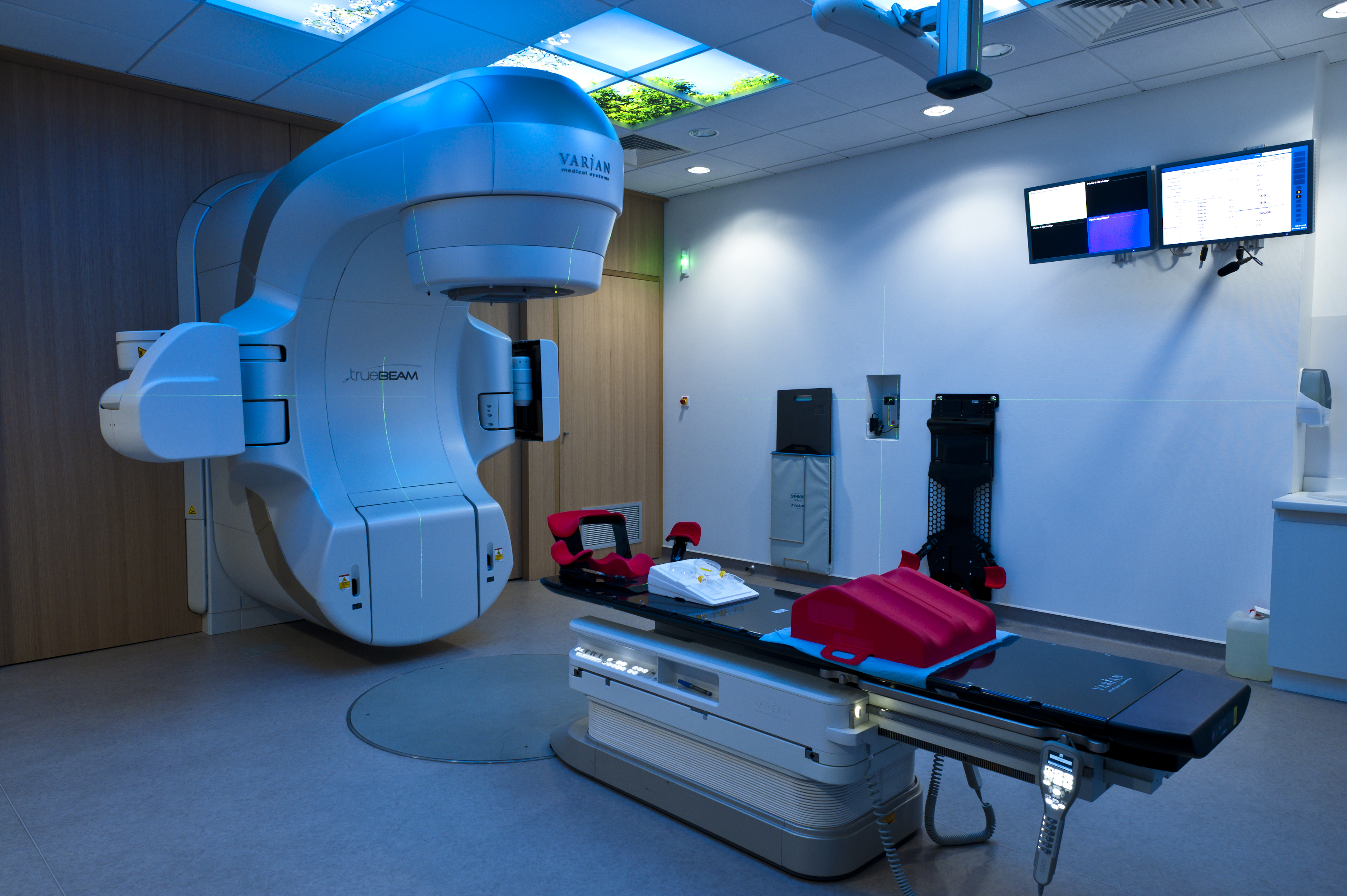
Plateforme de radiothérapie clinique du (CRLC) centre régional de lutte contre le cancer. Val d’Aurelle-Paul Lamarque, Montpellier. Inserm/ P Latron
Installed in the treatment room with an isocentric arm – with which it is possible to orientate the beam around the patient depending on all effects – this cutting edge technology will enable the indications for protontherapy to be extended even further.
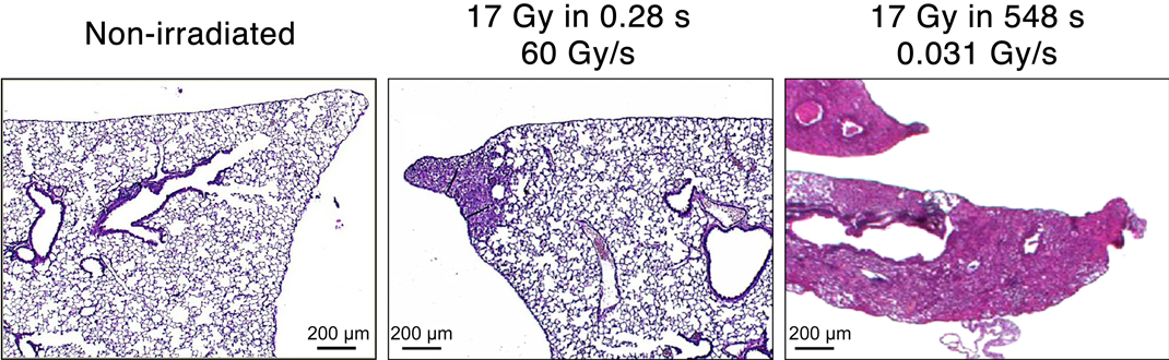
Images of tissue sections
Effect of irradiation with 17 Gy administered on 0.28 s on healthy lung tissue, equivalent to a dose-rate of 60 Gy/s (middle picture) and in 548 s, equivalent to a dose-rate of 0.031 Gy/s (right picture). Tissue irradiated with a very high dose-rate looks the same as non-irradiated tissue, while tissue irradiated at low dose-rate is completely altered.