Researcher Contact
Mickael TanterChercheur Inserm en imagerie ultrasons
Unité Inserm 979 "Physique des ondes pour la médecine"
01 80 96 30 68
rf.icpse@retnat.leakcim
Hirac Gurden
Chercheur CNRS en neurosciences
01 69 15 71 90
rf.3p2ni.cnmi@nedrug
A new ultrasound imaging technique has provided the first ever in vivo visualization of activity in the piriform cortex of rats during odor perception. This deep-seated brain structure plays an important role in olfaction, and was inaccessible to functional imaging until now. This work also sheds new light on the still poorly known functioning of the olfactory system, and notably how information is processed in the brain. This study is the result of a collaboration between the team led by Mickael Tanter at the Institut Langevin (CNRS/INSERM/ESPCI ParisTech/UPMC/Université Paris Diderot) and that led by Hirac Gurden in the Laboratoire Imagerie et Modélisation en Neurobiologie et Cancérologie (CNRS/Université Paris-Sud/Université Paris Diderot). Their findings are published in NeuroImage dated July 15, 2014.
How can the perception of the senses help represent the external environment? How, for example, does the brain process food or perfume related olfactory data? Although the organization of the olfactory system is well known it is similar in organisms ranging from insects to mammals its functioning remains unclear. To answer these questions, the scientists focused on the two brain structures that act as major olfactory relays: the olfactory bulb and the piriform cortex. In the rat, the olfactory bulb is located between the eyes, just behind the nasal bone. The piriform cortex, meanwhile, is deep seated in the brain of rodents, which made it impossible to obtain any functional images in a living animal until now.
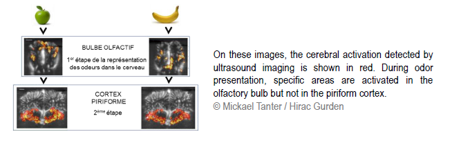
Yet the neurofunctional ultrasound imaging technique developed by Mickael Tanter’s team, called fUS (functional Ultrasound), allows the monitoring of neuronal activity in the piriform cortex. It is based on the transmission of ultrasonic plane waves into the brain tissue. After data processing, the echoes returned by the structures crossed by these waves can provide images with unequalled spatial and temporal resolution: 80 micrometers and a few tens of milliseconds. The contrast on these images is due to variations in the brain’s blood flow. Indeed, the activity of nerve cells requires an input of energy: it is therefore coupled to an influx of blood into the zone concerned. By recording volume variations in the blood vessels irrigating the different brain structures, it is therefore possible to determine the location of activated neurons.
Several imaging techniques, such as MRI, are already based on the link between blood volume and neuronal activity. But fUS offers advantages in terms of cost, ease of use and resolution. Furthermore, it provides easier access to the deepest structures that are often located several centimeters beneath the cranium. The recordings performed by Hirac Gurden’s team using this technique made it possible to observe the spatial distribution of activity within the olfactory bulb. When an odor was perceived, blood volume increased in clearly defined areas: each odor thus corresponded to a specific pattern of activated neurons.
In addition to these findings, and for the first time, the images revealed an absence of spatial distribution in the piriform cortex. At this level, two different odors triggered the same activation throughout the region. The cellular mechanisms responsible for the disappearance of a spatial signature are not yet clearlydefined, but these findings lead to the formulation of several hypotheses. The piriform cortex could be a structure that serves not only to process olfactory stimuli but rather to integrate and memorize different types of data. By making abstraction of the strict odor induced patterns, it would be possible to make associations and achieve a global concept. For example, based on the perception of the hundreds of odorant molecules found in coffee, the piriform cortex would be able to recognize a single odor, that of coffee.
This work opens new perspectives for both imaging and neurobiology. The researchers will now be focusing on the effects of learning on cortical activity in order to elucidate its role and the specificities of the olfactory system.