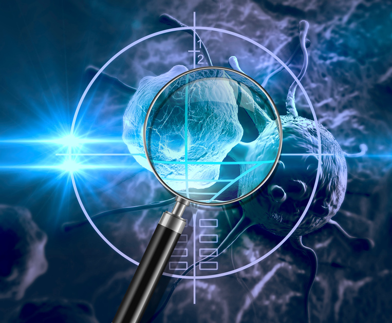
 Neural organoid with immune environment magnified twice on the left, 20 times on the right: green macrophages, red and blue neural progenitor cells (fluorescence microscopy). © Gustave Roussy
French, Singaporean and British researchers, led by Prof. Florent Ginhoux, head of a research team at Gustave Roussy/Inserm, have succeeded in demonstrating in a neuronal organoid the role of the brain’s immune environment in its formation and development. The development of these three-dimensional structures integrating neuronal cells and the immune environment is, to date, one of the most complete in vitro models of the human brain. This work is published in Nature.
At Gustave Roussy, these organoids are used to model the development of childhood brain cancers, to understand their mechanisms and discover new avenues of treatment.
“Although microglial cells, immune cells derived from the evolution (differentiation) of primitive macrophages present in the embryonic brain, are known to contribute to multiple aspects of brain development and function, their precise role remains poorly understood and little studied”, says Prof. Florent Ginhoux, director of a research team at Gustave Roussy/Inserm and Senior Principal Investigatorat A*STAR’s Singapore Immunology Network (SIgN).
The use of neuronal organoids to study their functions is one of the avenues currently favored by research.
An organoid is a 3D structure grown in the laboratory which reproduces certain morphological and functional characteristics of a human body organ or tissue. In research, these cell-cultured pseudo-organisms are a new biological model in full development in various fields, notably neurology; most studies of neuron formation (neurogenesis) are based on animal models.
With their 3D structure, the function and properties of these organoids are close to those of a real organ, but not quite as advanced. They measure just one millimeter and have no thoughts, consciousness or emotions. By generating neural organoids from human induced pluripotent stem cells (iPS cells), it is possible to model some of the key features of early human brain development. “However, current approaches do not include microglial cells,“ explains Prof. Florent Ginhoux.
The international team of researchers led by Prof. Florent Ginhoux has succeeded in producing a new type of model: neuronal organoids with microglia, by cultivating together organoids and primitive-type macrophages, all generated from the same culture of iPS induced stem cells.
Organoids and primitive macrophages are prepared separately. It takes around 25 days to obtain them. The macrophages are then placed in contact with the organoids for a further 15 to 20 days.
In the model developed by the researchers, macrophages colonized the organoids. In this 3D environment, in contact with immature neuronal cells, they differentiated into microglial cells expressing the genes and functions specific to this cell type. These microglial cells proved capable of controlling the differentiation of neuronal precursors (so-called neuronal progenitor cells), thus limiting their multiplication (proliferation), while promoting synapse creation (synaptogenesis) and axon growth (axonogenesis), two key elements in the transmission of the nerve message from neuron to neuron.
A discovery within a discovery
Prof. Florent Ginhoux’s team also observed that the organoids’ microglial cells contain high levels of perilipin-2, a molecule belonging to a family of proteins that coat lipids – including cholesterol – in droplets, enabling them to be stored in and exported from the cells. Armed with these perilipin-2-laden droplets, microglial cells facilitate cholesterol transport to the organoids. The neural progenitor cells that absorb this cholesterol undergo metabolic reprogramming as they differentiate into nerve cells.
The approach developed by Prof. Florent Ginhoux and his colleagues has significantly advanced the complexification of organoid models by integrating microglial cells. This progress is illustrated by the discovery of a key lipid-mediated pathway between microglia and neural progenitor cells, essential for the synthesis of new neurons.
“With microglia cells incorporated, the neural organoids we have succeeded in generating are a new, more complete 3D model, closer to reality. We know that the immune system plays a fundamental role in the development of cancers, so at Gustave Roussy we’re going to use them to better understand and discover the mechanisms that regulate the development of pediatric brain tumors“, concludes Prof. Florent Ginhoux.
This work has been supported by the Gustave Roussy Foundation’s “Curing childhood cancer in the 21st century” campaign.
These contents could be interesting :

iPSC-microglia promote brain organoid maturation via cholesterol transfer
Nature, Advanced publication on ligne the 1st November 2023
https://doi.org/10.1038/s41586-023-06713-1
Dong Shin Park1,2,*, Tatsuya Kozaki1,*, Satish Kumar Tiwari1, Marco Moreira3, Ahad Khalilnezhad1,2, Federico Torta4,5, Nicolas Olivié6, Chung Hwee Thiam2, Oniko Liani1, Aymeric Silvin1,3, Wint Wint Phoo7, Liang Gao4,5, Alexander Triebl4,5, Wai Kin Tham5, Leticia Gonçalves3, Wan Ting Kong1,3, Sethi Raman1, Xiao Meng Zhang1, Garett Dunsmore3, Charles Antoine Dutertre1,3, Salanne Lee1, Jia Min Ong1, Akhila Balachander1, Shabnam Khalilnezhad1,8, Josephine Lum1, Kaibo Duan1, Ze Ming Lim1, Leonard Tan1, Ivy Low1, Kagistia Hana Utami9, Xin Yi Yeo10, Sylvaine Di Tommaso11, Jean-William Dupuy12, Balazs Varga13, Ragnhildur Thora Karadottir13, Mufeeda Changaramvally Madathummal14, Isabelle Bonne2, Benoit Malleret1,2,14, Zainab Yasin Binte1, Ngan Wei Da1, Yingrou Tan1, Wei Jie Wong15, Jinqiu Zhang9, Jinmiao Chen1, Radoslaw M. Sobota7, Shanshan W. Howland1, Lai Guan Ng1, Frédéric Saltel11, David Castel16, Jacques Grill16, Veronique Minard3, Salvatore Albani8, Jerry K.Y. Chan17, Morgane Sonia Thion6, Jung Sangyong10, Markus R. Wenk4,5, Mahmoud A. Pouladi9, Claudia Pasqualini3, Veronique Angeli2, Olivier NF. Cexus5,10,18 and Florent Ginhoux1,2,3,8,15,#
1 Singapore Immunology Network (SIgN), Agency for Science, Technology and Research, Singapore 138648, Singapore,
2 Department of Microbiology and Immunology, Yong Loo Lin School of Medicine, National University of Singapore, Singapore 117545, Singapore,
3 INSERM U1015, Gustave Roussy Cancer Campus, Villejuif 94800, France
4 Department of Biochemistry, Yong Loo Lin School of Medicine, National University of Singapore, Singapore 117596, Singapore,
5 Singapore Lipidomics Incubator (SLING), Life Sciences Institute, National University of Singapore,
Singapore 119228, Singapore,
6 Institut de Biologie de l’Ecole Normale Supérieure (IBENS), Ecole Normale Supérieure, CNRS, INSERM, PSL Research University, Paris 75005, France,
7 Functional Proteomics Laboratory, SingMass National Laboratory, Institute of Molecular and Cell Biology, Agency for Science, Technology and Research, 61Biopolis Drive, #07-03, Singapore 138673, Singapore,
8 Translational Immunology Institute, SingHealth Duke-NUS Academic Medical Centre, Singapore 169856, Singapore,
9 Translational Laboratory in Genetic Medicine (TLGM), Agency for Science, Technology and Research, Singapore 138648, Singapore,
10 Institute of Molecular and Cell Biology (IMCB), Agency for Science, Technology and Research, Singapore 138673, Singapore,
11 Oncoprot Platform, TBM-Core US 005, F-33000 Bordeaux, France,
12 Bordeaux Protéome, University of Bordeaux, 33076, Bordeaux, France,
13 Wellcome Trust-Medical Research Council Cambridge Stem Cell Institute and Department of Veterinary Medicine, University of Cambridge, Cambridge, CB2 0AW, UK.,
14 A*STAR Microscopy Platform Electron Microscopy, Research Support Centre, Agency for Science, Technology and Research (A*STAR), Singapore 138673, Singapore,
15 Shanghai Institute of Immunology, Department of Immunology and Microbiology, Shanghai Jiao Tong University School of Medicine, Shanghai 200025, China,
16 INSERM U981, Molecular Predictors and New Targets in Oncology & Département de Cancérologie de l’Enfant et de l’Adolescent, Gustave Roussy, Université Paris-Saclay, Villejuif 94805, France,
17 Department of Reproductive Medicine, KK Women’s and Children’s Hospital, Singapore 229899, Singapore,
18 School of Biosciences, Faculty of Health and Medical Sciences, University of Surrey, Guildford, GU2 7XH, United Kingdom