

Researcher Contact
Valérie Gabelica
Directrice de recherche Inserm
U 1212 – Laboratoire Acides nucléiques : régulations naturelle et artificielle (ARNA)
E-mail : rf.mresni@acilebag.eirelav
Tel : 05 40 00 29 40
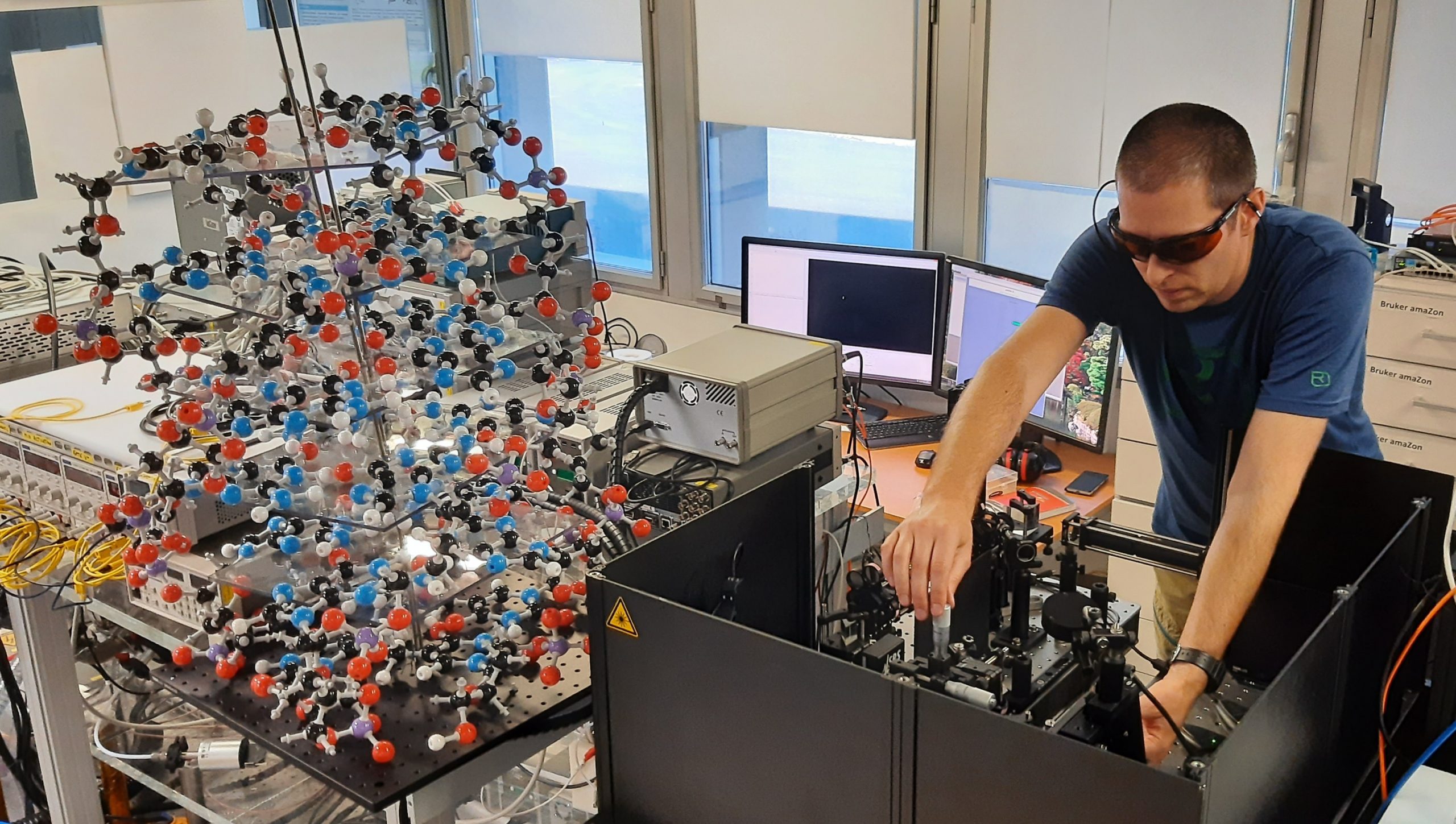
To better characterize molecular interactions, researchers must also look at molecular chirality. © Valérie Gabelica
Understanding the three-dimensional structure of DNA and RNA and how they interact with other molecules is necessary for the advancement of biomedical research and drug development. A team led by Inserm researcher Valérie Gabelica at the Nucleic acids: natural and artificial regulation laboratory (ARNA, Inserm/CNRS/Université de Bordeaux)[1] has developed an innovative method pairing mass spectrometry with circularly polarized light, enabling better characterization of these different molecular interactions. This new technique is described in a study published in the journal Science.
A technique widely used in physics and biology laboratories, and also in forensic science to analyze chemical and biological samples, mass spectrometry measures the masses of each molecule in a sample, thereby providing information on how they interact and associate with each other.
Inserm researcher Valérie Gabelica and her team at the ARNA laboratory (Inserm/CNRS/Université de Bordeaux)1 are studying how short DNA and RNA sequences fold in three dimensions and interact with other molecules. To do this, they use mass spectrometry, which gives them valuable insights into how DNA and RNA associate structurally with proteins or pharmaceutical molecules, for example.
Given that measuring molecular weight provides no information on chirality, the use of another technique, circularly polarized light[2], is necessary to study the 3D structure of molecules.
In a study published in Science, the researcher and her team describe a novel tool for studying chirality and how DNA and molecules assemble: a “2-in-1” method pairing a mass spectrometer and a laser that produces circularly polarized light.
Therapeutic prospects
By pushing back hard on the frontiers of mass spectrometry and facilitating the study of molecule chirality, this research is particularly innovative and opens up very broad prospects in the field of biomedical research.
[1] The IECB research support unit (CNRS/Université de Bordeaux/Inserm) also participated in this work.
[2]Light is a wave. It is described by signals that oscillate in space and time: electric and magnetic fields. These two fields have a direction, the magnetic field is perpendicular to the electric field. When light is circularly polarized, the electric field changes orientation along the beam and describes a spiral. The magnetic field is always perpendicular to the electric field
Valérie Gabelica
Directrice de recherche Inserm
U 1212 – Laboratoire Acides nucléiques : régulations naturelle et artificielle (ARNA)
E-mail : rf.mresni@acilebag.eirelav
Tel : 05 40 00 29 40
Mass-resolved electronic circular dichroism ion spectroscopy
Steven Daly1, Frédéric Rosu2, Valérie Gabelica1
1 Université de Bordeaux, Inserm & CNRS, Laboratoire Acides Nucléiques : Régulations Naturelle et Artificielle (ARNA, U1212, UMR5320), IECB, 33607 Pessac, France.
2 Université de Bordeaux, CNRS & Inserm, Institut Européen de Chimie et Biologie (IECB, UMS3033, US001), 33607, Pessac, France.