Menu

Osteoarthritis and metabolic disease: a “joint” therapeutic target?

© Fotolia

Osteoarthritis is an incapacitating condition with diverse etiologies, the most recently described of which is metabolic syndrome (MetS). For the first time, researchers from the “Molecular Engineering and Articular Physiopathology” (Cnrs/Université de Lorraine) and “Acute and chronic cardiovascular failure” (Inserm/Université de Lorraine) research laboratories in Nancy, France, have described the spontaneous development of this type of degenerative joint disease in a mouse model of MetS. Inserm researchers Hervé Kempf and Anne Pizard, together with their co-workers, have characterized the existence of osteoarthritic lesions of the tibiofemoral joint of the knee in the presence of metabolic disorders.

This new experimental model of metabolic-associated osteoarthritis has also made it possible to demonstrate that chronic treatment with a mineralocorticoid receptor antagonist (MCRA) improves joint impairment. This drug is already used in heart failure and has been proposed as possibly being more effective in obese patients.

This research has been the subject of a letter published in Annals of the Rheumatic Diseases.

For the researchers, “these results will make it possible to propose metabolic-associated osteoarthritis as a potential new indication for MCRA therapy”, a hypothesis that they intend to test in the clinical setting very soon.

Inserm Transfert has filed a patent application for this research.

Rare genetic obesity: promising results from a clinical trial

Pesée

(c) Fotolia


POMC (proopiomelanocortin) deficiency obesity is an orphan disease which has been described in 50 patients to date, although it is estimated to affect 100–500 people worldwide. Although there is currently no specific treatment, a publication in NEJM reports promising results from a phase 2 clinical trial that led to considerable weight loss in female patients. The study involves collaboration between clinical teams from the Charité University Hospital in Berlin, Pitié-Salpétrière Hospital (ICAN – Assistance Publique–Hôpitaux de Paris (AP-HP)), Inserm, Pierre and Marie Curie University, and an American company.

POMC is a hormone that acts in the brain to control hunger and weight. A deficiency in affected individuals leads, from early childhood, to severe obesity associated with endocrine abnormalities (deficiency in cortisol and sex hormones), red hair and very fair skin.

In this study, two female patients were treated with setmelanotide, a powerful agonist of the melanocortin receptor MC4R, involved in the pathway on which POMC acts. With this treatment (one injection per day), the first patient lost 51 kg in 42 weeks, and the second, 20 kg in 12 weeks. The very high insulin levels observed before treatment decreased considerably, and the clinicians observed a reduction in hunger, which caused great relief to the patients.

Setmelanotide thus appears to be an effective treatment for reducing weight and restoring a normal appetite to individuals with POMC deficiency. While these patients are currently continuing their treatment, the investigators are planning clinical trials using setmelanotide in the treatment of other obesity-related genetic disorders, such as Prader Willi syndrome, which is associated with reduced POMC activity and abnormalities in the leptin receptor.

Red blood cell fragments attack blood vessels

A new study published in the journal Blood shows that red blood cells can turn against blood vessels by releasing aggressive fragments that contribute to the development of cardiovascular diseases.Blood Cells : erythrocyte, thrombocyte, leukocyte

©Fotolia

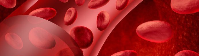
There are approximately 5 million red blood cells in every microlitre of blood. Their stock is constantly renewed, and each one is discreetly removed every three months. But abnormal red blood cells persist in the bloodstream in some diseases. They ultimately rupture and release their contents, a process referred to as intravascular haemolysis.

Does the red blood cell disappear then? Not quite…

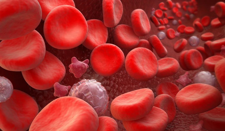
Researchers led by Olivier Blanc-Brude, a CNRS (French National Centre for Scientific Research) research fellow at the Inserm Paris Cardiovascular Center and Paris Descartes University, have just shown that at the exact moment of haemolysis, the red blood cells do not disappear: they actually release a large number of fragments, known as microparticles. And these fragments are not harmless. These observations were made using red blood cells from patients with sickle cell disease, the most common genetic disorder in France. Their rigidity leads to a considerable degree of haemolysis, which is therefore easier to study.

Red blood cells contain haemoglobin, the protein that carries blood gases and enables us to breathe. As a result, the red blood cell fragments also contain haemoglobin, but in a degraded and toxic form: the iron previously contained in the haemoglobin becomes exposed on the surface of these thousands of red blood cell microparticles.

It is this feature that makes the microparticles dangerous.

Effectively, the red blood cell particles act as carriers, and deposit the toxic substances on the cells lining the blood vessels. This transfer of haem and iron from the red blood cells to the vascular lining interferes with dilation, limiting the blood supply to the tissues that need it. This also leads to oxidative stress and promotes obstruction of the blood vessels.

In patients with sickle cell disease, red blood cell fragments may be partly responsible for very painful vaso-occlusive crises. These microparticles also probably contribute to the many lesions caused by chronic lack of oxygen, e.g. in the kidneys.

These innovative results make it possible to anticipate new therapeutic possibilities for preventing and treating vaso-occlusive crises, as well as for preventing some more common cardiovascular diseases.

These new therapies could target various components of the red blood cell microparticles, e.g. membrane components, such as phosphatidylserine, or free haem. Haemopexin is a molecule naturally found in the bloodstream, and which has the role of binding to free haem to eliminate it. It might represent a therapeutic opportunity, i.e. a haemopexin supplement might help to neutralise free haem carried by red blood cell microparticles.

These results are the subject of a patent filed by Inserm Transfert.

A vaccine against house dust mite allergy?

House dust mite allergy affects over half of asthma patients. The major allergen, Der p 2, is recognised by nearly 90% of allergic patients. Continuous exposure to this allergen leads to a type of asthma characterised by bronchial constriction and inflammation of the airways, which in the most severe cases can lead to considerable disability and sometimes serious attacks and death.

“Desensitisation” is the only current treatment for respiratory allergies. It involves exposing patients to the allergen, usually by the sublingual route, so that their immune system gradually generates protective cells and antibodies. Thanks to technological advances and genetic engineering, so-called hypoallergenic peptides, derived from allergens and used as a vaccine, represent a new direction for immunotherapy, especially for house dust mite asthma. This strategy has the three-fold advantage of being effective in a short time, offering prolonged protection, and reducing the allergic side-effects of desensitisation.

In this work, carried out by researchers from Inserm and the French National Institute for Agricultural Research (INRA), in collaboration with researchers from the University of Vienna, Austria, a fragment of Der p 2 was generated by genetic engineering, and its protective effect against house dust mite asthma was then tested in a mouse model of asthma.

To accomplish this, mice were vaccinated with this peptide on 2 occasions: before and during allergic sensitisation to house dust mites. The researchers then measured bronchial obstruction in these mice in response to metacholin (a drug that induces bronchoconstriction), and demonstrated that vaccination with this peptide resulted in normal bronchial reactivity in asthmatic mice.

Furthermore, vaccination with this peptide induces anti-inflammatory activity, by reducing the chemical messengers that attract eosinophils and neutrophils to the bronchi and lungs, while reducing the T-lymphocyte activation and immunoglobulin E secretion responsible for the allergic reaction.

For the researchers, the protective role of vaccination, using a hypoallergenic peptide, is a very attractive therapeutic option for stemming the progression of asthma, especially in children at risk. Additional studies in animals are necessary before the first therapeutic trials in humans can be planned.

The peptide used is the subject of a patent filed by Inserm Transfert.

Fatty plaques in the arteries associated with dementia in older subjects

The carotid arteries are essential for supplying blood to the brain. In a study published online in the journal Alzheimer’s & Dementia, researchers from Inserm, in collaboration with those involved in the Three Cities Study (3C), have focused on the relationships between atherosclerosis and the risk of dementia in older subjects.

Atherosclerosis is characterised by the deposition of plaque essentially composed of lipids (known as atheroma) on the walls of arteries. These plaques can be highly unstable and cause a stroke when they become detached. Previous studies have shown a link between cardiovascular risk factors and the occurrence of dementia. Atherosclerosis, which reflects lifelong exposure to these factors, can be measured noninvasively by ultrasound of the carotid arteries, making it possible to detect the presence of atheroma plaques.

In this study, researchers from the “Cardiovascular Epidemiology and Sudden Death”[1] and “Hormones and Cardiovascular Disease”[2] teams analysed data from the Three Cities (3C) French multicentre cohort. The association between carotid atherosclerosis and the occurrence of dementia after 7 years of monitoring was measured in 6,025 men and women aged from 65 to 86 years, who were initially free from dementia and non-institutionalised.

“Subjects showing carotid plaques at enrolment had a nearly 2-fold higher risk of dementia than those free of carotid atheroma,” explains Matthieu Plichart, who co-authored this work.

For the first time, the study examines the utility of measuring carotid atherosclerosis. The researchers suggest that taking carotid plaques into account might be useful in predicting the individual risk of developing these “mixed/vascular” dementias, i.e. those resulting from a combination of vascular lesions in the brain and neurodegenerative lesions. They are the second most important cause of dementia, the leading cause being Alzheimer’s disease, which is a purely neurodegenerative process.

[1] Unit 970 PARCC (Paris Cardiovascular Research Center [Inserm/Paris Descartes University])
[2] Inserm Unit 1018 “Centre for Research in Epidemiology and Population Health” (Inserm/Paris-Sud University)

Diabetes: fasting in the wrong place at the wrong time

During fasting, in order to cope with nutrient depletion, cells break down their own constituents, a process known as autophagy.

Roméo Ricci’s team at the Institute of Genetics and Molecular and Cellular Biology (CNRS/Inserm/University of Strasbourg) has shown that, during fasting, the pancreatic beta cells, which are responsible for insulin secretion, do not compensate for nutrient deficiencies by the “classic” autophagy mechanism, which is completely suppressed, but by breaking down insulin secretory granules.

ricci

© IGBMC. When protein kinase D1 (PKD1) acts (A), insulin, produced by the Golgi bodies in the beta cells, is released by the granules (in yellow, in the diagram and electron micrograph, inset). During fasting (B) and PKD1 inactivation, the insulin granules in the beta cells fuse with the lysosomes (in purple) containing the enzymes needed to degrade insulin; at the same time, mTOR activation suppresses autophagy.

In type 2 diabetes, insulin secretion by the pancreatic beta cells should be increased to cope with excessively high glucose levels. However, in this disease, the body reacts as though it were fasting. The mechanism discovered by Roméo Ricci’s team, by working against the mechanism of insulin secretion and autophagy, could be involved in the impaired functioning of pancreatic beta cells in type 2 diabetes. These results are published in the 20 February 2015 issue of Science.

Using “good cholesterol” as a therapeutic vector for treating the lungs

One of the major challenges for therapeutic science is to optimise the accurate distribution of drugs in the affected organs. Targeted delivery methods are needed to accomplish this.

In a new study published in The American Journal of Respiratory Cell and Molecular Biology, Olivier Meilhac’s team (Inserm Unit 1148, the “Laboratory for Vascular Translational Science,” in collaboration with Inserm Unit 1152, “Physiopathology and Epidemiology of Respiratory Diseases”) addressed the transport capability of HDLs (high density lipoproteins, commonly known as “good cholesterol”). These naturally occurring nanoparticles carry cholesterol, but may also be loaded with drugs or other protective molecules. Their study shows that intravenously injected HDLs help to convey a therapeutic drug, alpha-1 antitrypsin, to the lungs, to reduce pulmonary emphysema.

Pulmonary emphysema is characterised by progressive destruction of the pulmonary alveoli, which ultimately leads to respiratory failure. The cause: inflammatory cells that secrete an enzyme, elastase, which is involved in the development of these lesions. This mechanism is especially involved in patients with a genetic defect in another enzyme, alpha-1 antitrypsin (AAT), a natural inhibitor of elastase. This frequent genetic disorder affects 1 in 3,000 people, and is responsible for approximately 1% of emphysema cases.

The treatment currently offered to patients with emphysema who are deficient in AAT involves intravenous administration of AAT, with the challenge mentioned earlier, namely: how to reach the lungs efficiently? This work shows that in mice, loading AAT onto HDLs improves its delivery to the lungs, where it can limit elastase-induced damage, compared with the standard treatment.

Pulmonary emphysema is present to different degrees in patients with chronic obstructive pulmonary disease (COPD), a very common (5-10% of the adult population) and serious disease (16,000 deaths per year), which particularly affects smokers. The researchers believe that these “therapeutic HDLs” may be a useful avenue for improving the treatment of COPD.

The satiety mechanism described and restored in mice

Leptin is a hormone secreted by adipose cells, sending the satiety signal to the brain. Sometimes, certain overweight or obese individuals develop resistance to this hormone, which is no longer able to deliver its message. A team led by Vincent Prévot, Inserm Research Director at the Jean Pierre Aubert Research Centre in Lille, has used obese mice to show how the pathway transporting leptin to the brain can be reactivated.

Obesity acquired by a diet containing too much fat is associated with high and abnormal levels of circulating leptin. These circulating concentrations reflect the inability of leptin to reach the brain. So it remains ‘trapped’ in the bloodstream. In this study, published in Cell Metabolism, the researchers succeeded in finding the front door for this hormone to enter the brain and restored opening of this ‘cellular door’ in obese mice.

shcéma

Leptin transport via the tanycytes © Inserm / Vincent Prévot

The research team was first able to describe the mechanism by which leptin passes through this cellular door: tanycytes (see diagram opposite: cells shown in yellow). These cells capture circulating leptin from blood vessels that, at this site, are specially-adapted to allow its passage (step 1). As it passes though the tanycyte, captured leptin activates an ‘ERK’ signalling pathway (step 2) that triggers its release into the cerebrospinal fluid (step 3). Leptin then activates brain regions that convey its anorexigenic action (step 4).

In cases of obesity. the researchers observed that the tanycyte is able to capture the hormone but can no longer release it into the brain. It develops what they call ‘leptin-resistance’ associated with the change in the activity of its receptor, ‘LepR’. The researchers then tested mice with a pharmacological treatment enabling leptin to reach the CSF.

Our study shows that transport of leptin through the tanycyte can be restored in obese mice using a pharmacological treatment by activating the ERK signalling pathway in this cell“, explains Vincent Prévot, Inserm Research Director.

This treatment allows leptin to activate neurones and finally restore the sensitivity of mice to this hormone by gradually restoring a balanced diet. 

 “Altered leptin transport by tanycytes could therefore constitute the first physiopathological event leading to central hormone resistance in overweight or obese individuals” he concludes.

Better dosing of “good cholesterol” for greater efficacy

The measurement of cholesterol levels in an organism during medical analyses has become routine in the prevention of cardiovascular diseases. In reality, the cholesterol levels are the result of a balance in the human body between “good” (HDL) cholesterol and “bad” (LDL) cholesterol. If this balance is threatened and tilts too far towards “bad cholesterol”, the risk of cardiovascular diseases increases. Conversely, a high HDL-cholesterol level has a protective effect against cardiovascular diseases.

artere

crédit : ©Fotolia

Although researchers thought they could easily recognise HDL particles, there were, in fact, a heterogeneous mixture of extremely complex particles that were hard to study.

Very recent work has come to the conclusion that it is not a matter of having a high level of HDL-cholesterol to be protected against cardiovascular diseases, the HDL particles also need to be functional and thus capable of exercising their protective action on the body (elimination of excess cholesterol as well as anti-inflammatory, anti-oxydant, anti-thrombotic and cytoprotective properties). For example, in diabetic patients, the HDL particles are dysfunctional because their composition is altered in comparison with those in healthy individuals.

Inserm researchers (UMR Inserm/ University of Toulouse III – Paul Sabatier 1048 “Institute of metabolic and cardiovascular diseases” and 1027 “ Epidemiology and public health analyses: risks, chronic conditions and disabilities”) discuss in The journal of lipid research the discovery of a new blood marker (IF1) associated with HDL-cholesterol and that takes account of the correct “functioning” of HDL particles.

The analysis performed on 648 patients suffering from cardiovascular disease (aged between 45 and 74) and 669 people in a control group showed that the detection of IF1 in the blood reveals the presence of functional HDL particles. The amount present would make it possible to improve early diagnosis of cardiovascular disease and thus optimise existing or future therapeutic strategies designed to increase the amount of good cholesterol.

This work was the subject of an international patent in 2013 involving the early diagnosis of cardiovascular disease (patent filed by INSERM-transfert).

Breathe…inhale…run

Salbutamol is a classic asthma treatment. Its administration via inhalation stimulates the beta-2 receptors of the bronchial walls, dilating the bronchiae and thus allowing for greater respiratory capacity.

Its  use as a doping product is currently under review. An Inserm team headed by Samuel Vergès (Inserm Unit 1042 “Hypoxia and cardiovascular and respiratory physiopathology”) attempted to discover whether its inhalation could increase contractility in certain peripheral muscles, reduce fatigue and improve recovery after exercise for endurance athletes.

Eleven non-asthmatic athletes, presenting with high levels of endurance, were recruited for comparison in a two-level double-blind dosage of salbutamol (200 and 800 µg), administered through inhalation, in comparison with a placebo prior to a muscle strain test of the leg muscles.

The athletes then performed a progressive exercise routine consisting in stages of 10 intermittent contractions of increasing intensity of their quadriceps muscles. They were required at the outset to contract their muscles at about 20% of maximum capacity, then to increase the contraction strength by 10% and in 10% stages until exhaustion (when the subject was no longer capable of producing the level of strength demanded of him).

The mechanical and electromyographical responses to magnetic stimulation of the femoral nerve were recorded during and after the voluntary muscular contractions. These records make it possible to assess neuromuscular fatigue after each stage of the strength test and after 10 minutes and 30 minutes of recovery.

According to the results obtained by the Inserm researchers, the muscle responses did not change with the inhalation of salbutamol. 

Nevertheless, the total number of contractions performed before attaining muscular exhaustion increased significantly with Salbutamol intake

(an average of 72 contractions using the placebo as against 78 with 200 µg and 82 contractions with 800 µg).

For the researchers, although the muscle responses remained unchanged, supra-therapeutic  inhaled doses (i.e. doses above any therapeutic dose prescribed by a doctor) of ß2-agonists increased quadriceps muscle endurance during progressive strain and could have an ergogenic (doping) effect in an athlete. More studies are needed to clarify the underlying mechanisms.

photo coureur sport

©fotolia

fermer