Menu

Transplantation chemotherapy eliminates regenerative capacity of brain’s innate immune cells

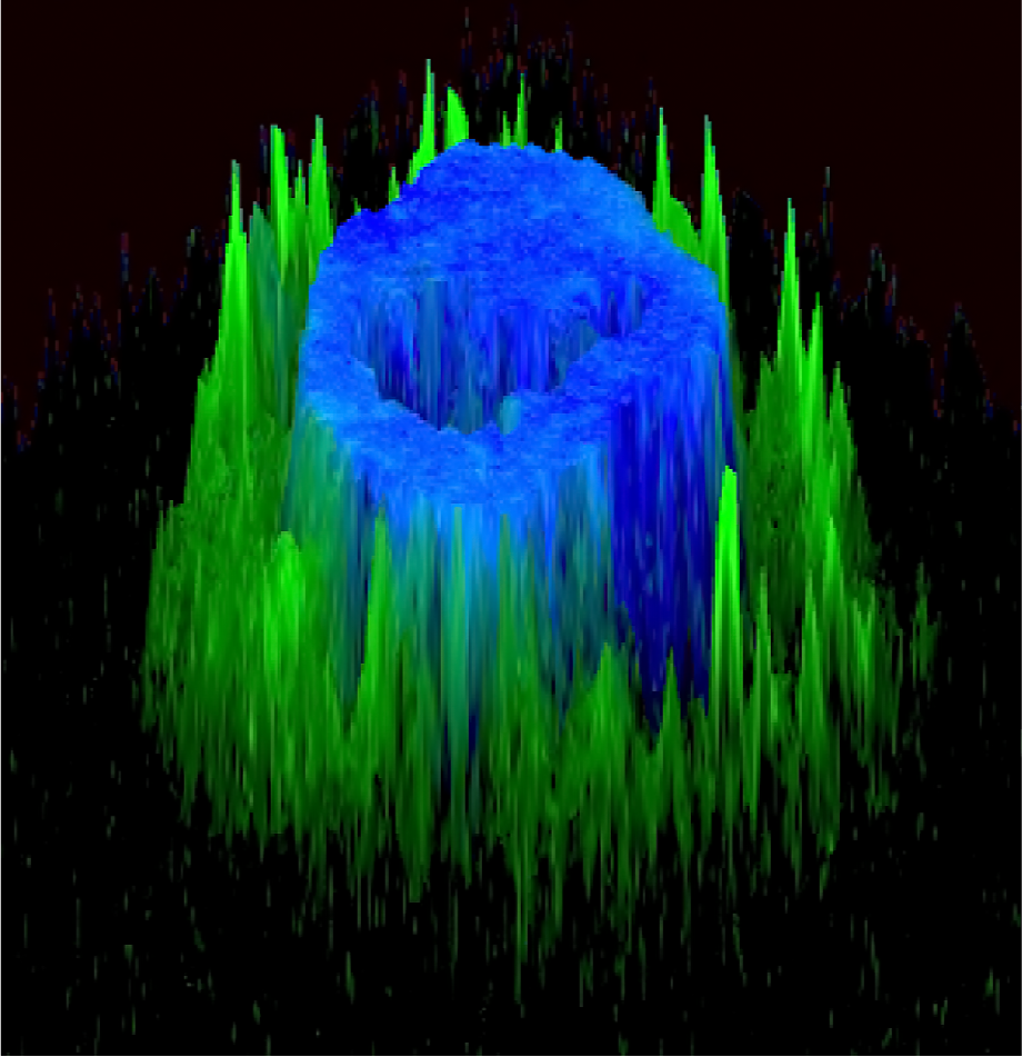
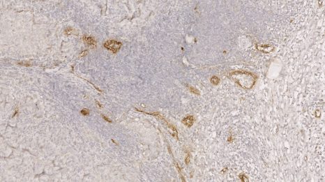
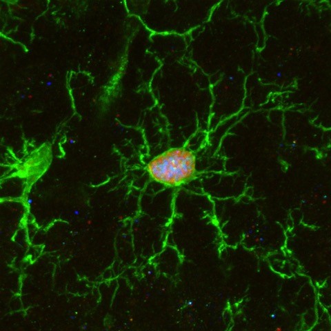
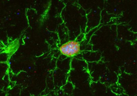
Brain microglia (green) initiating expression of cell division marker (red), but unable divide due to co-expression of a senescence marker (blue), due to the chemotherapy treatment (busulfan). © K. Sailor/ PM Lledo, Institut Pasteur.

Annually over 50,000 bone marrow transplantations occur worldwide as a therapy for multiple cancerous and non-cancerous diseases. Yet, how this procedure gives rise to bone marrow-derived cells that engraft the brain, despite being absent in the normal brain, remains unknown. In the present study, scientists from the Institut Pasteur, the CNRS and the Paris Brain Institute (Inserm) discovered how the host’s microglia, the brain’s innate immune cells, are replaced by bone marrow-derived macrophages. The key discovery was that transplantation chemotherapy eliminated the microglia’s regenerative capacity, gradually causing the engraftment of macrophages to replace microglia, providing a potential mechanism for future cell-based therapies to treat central nervous system diseases. This finding will be published in Nature Medicine on February 21, 2022.

Progressive demyelination of the central nervous system leads to devastating neurologic symptoms and premature death. As the number of cases diagnosed through pregnancy and newborn screening is increasing, more efficient therapeutic strategies are required. Recently, the successful use of gene therapy to correct disease-causing gene mutations in bone marrow stem cells, and their subsequent autologous transplantation into patients, has been clinically used for treating white matter diseases, while eliminating the need for finding matching donors. Clinical studies showed that pre-transplantation ablation of host bone marrow stem cells using the chemotherapy agent busulfan allows for efficient engraftment and tolerance to gene-modified cells, yet the mechanism involved was largely known.

Using an animal model, neuroscientists from the Institut Pasteur (CNRS) and the Paris Brain Institute (Inserm) have demonstrated the broad impact of busulfan chemotherapy used for bone marrow transplant therapy on various cell populations of the host brain.

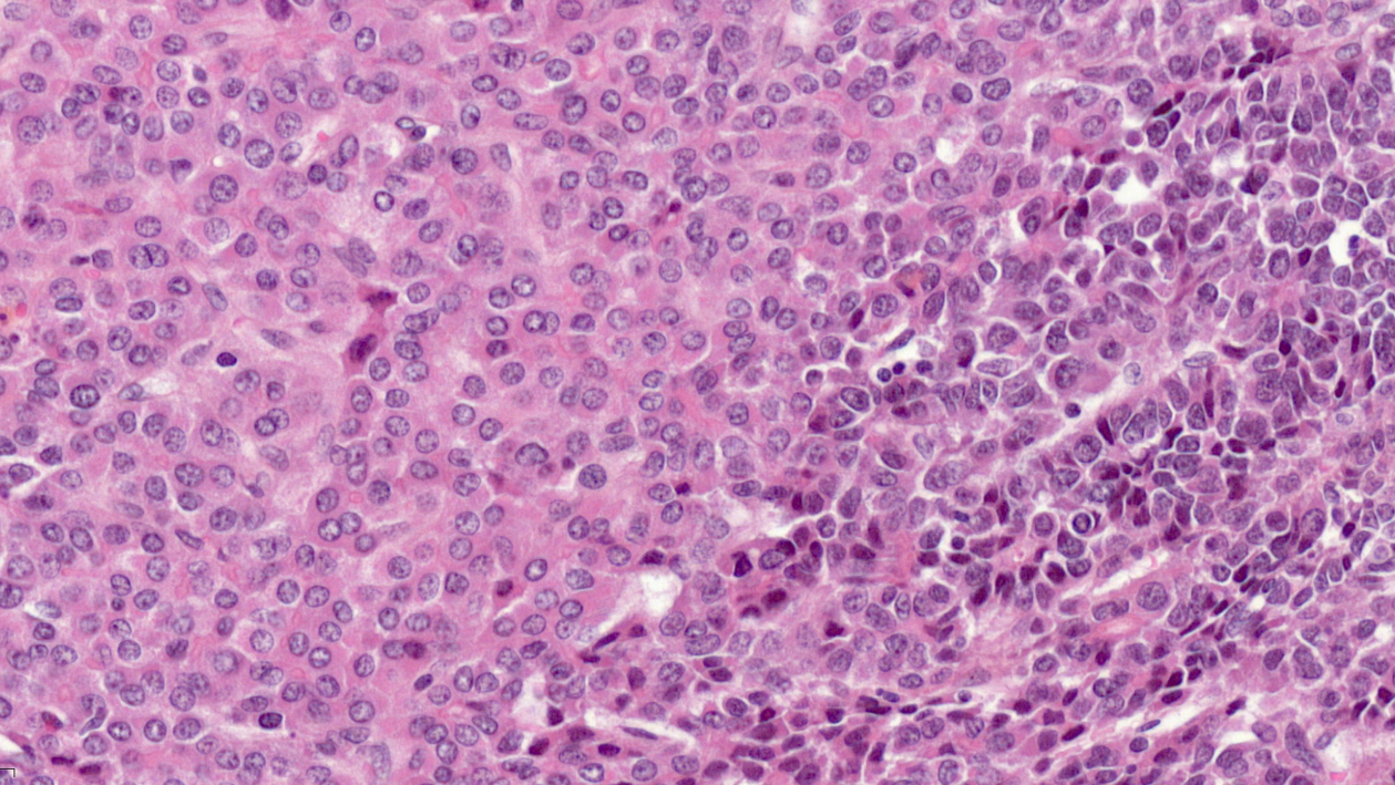
Microglia are tissue-resident brain immune cells that are instrumental for maintaining healthy brain physiology in normal and diseased states. These cells exhibit a robust self-renewal capacity and gradually turnover throughout life. However, in this study, Sailor and colleagues show that following pre-transplantation busulfan chemotherapy, microglia completely lose their regenerative capacity, becoming senescent, which thereby allowed the engraftment of bone marrow-derived macrophages into the brain. They also show these macrophages to become resident with similar surveillant dynamics as the microglia they replaced.

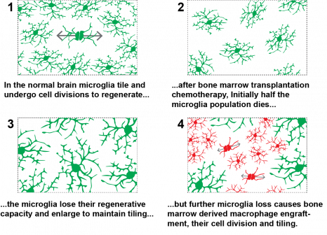
They demonstrate that busulfan administration temporarily depleted oligodendrocyte precursor cells and, importantly, caused the complete and permanent ablation of all adult neurogenesis, which may explain cognitive deficits in patients after busulfan chemotherapy, i.e. the “chemo brain”. Microglia that were eliminated because of busulfan chemotherapy, coupled with their loss of regenerative capacity, leave empty niches in the brain which are replaced by engrafted bone marrow-derived macrophages. These macrophages assumed the morphology and cellular process dynamics like normal microglia.

This study is highly relevant since for the first time it shows a mechanism dependent on microglia senescence, with the loss of their regenerative capacity, to explain how macrophages are permissively engrafted into the brain after bone marrow transplantation.

Furthermore, this work shows multiple off-target effects of bone marrow transplantation chemotherapy, which may be important for understanding post-chemotherapy cognitive issues. Understanding the mechanism of peripheral macrophage engraftment is imperative to develop further cell-based gene therapy strategies for central nervous system diseases.

“Microglial cells play an essential role in brain function and in the pathophysiology of many severe neurological diseases, both genetic and complex, such as multiple sclerosis and Alzheimer’s disease. Understanding the fate of these cells after the transplant process is essential both to clarify the consequences of chemotherapy and to develop new therapeutic strategies for serious neurodegenerative diseases,” said Nathalie Cartier, research director at Inserm and member of the NeuroGenCell team at the Brain Institute (ICM), and last co-author of the study.

“This study sheds light for the first time on a mechanism explaining how stem cell-derived macrophages enter the brain after bone marrow cell transplantation. This better understanding is essential to develop new strategies for gene and cell therapy applied to diseases of the central nervous system,” said Pierre-Marie Lledo, research director at the CNRS and head of the Perception and Memory Unit at the Institut Pasteur, and last co-author of the study.

“We show bone marrow transplantation chemotherapy to cause microglia, the brain’s resident immune cells, to lose their regenerative capacity. The microglia were unable to sustain their population which allowed bone marrow derived cells to replace them. This demonstrates how bone marrow transplantation is an effective therapy for certain neurological diseases and provides a strategy for cellular gene therapy in the central nervous system,” said Kurt Sailor, research fellow at the Institut Pasteur Unit Perception and Memory in Paris, and first author of the study.

A Novel Immunotherapy Approach Redirects Epstein-Barr Antibodies toward Disease-Causing Cells

Microscopic visualization of a cancerous cell (nucleus in blue) treated with bi-modular fusion proteins (BMFPs) to which EBV antibodies (green) bind. © Jean-Philippe Semblat and Arnaud Chêne – JRU1134 (Inserm/Université de Paris)


Monoclonal antibody therapy can be very effective in treating numerous illnesses, such as cancers, chronic inflammatory conditions, and infectious diseases. However, it is costly and uses molecules that are complicated to produce. Therefore, it is essential to identify new therapeutic alternatives so that as many patients as possible get the treatments they need. That is why researchers from Inserm, Université de Paris, Sorbonne Université and CNRS1 have designed and tested a new immunotherapy approach that uses pre-existing antibodies directed against the Epstein-Barr virus – part of the herpes family of viruses and present in over 95% of the world’s population – in order to target and destroy pathogenic (disease-causing) cells. Their findings have recently been published in a study in Science Advances.

The principle is to redirect a pre-existing immune response against the Epstein-Barr virus (EBV) to target cells that we wish to destroy. The Epstein-Barr virus – which belongs to the family of herpes viruses – is transmitted predominantly through saliva, and over 95% of the world’s population is infected with it.

The vast majority of people have no symptoms, and the virus has the ability to persist chronically in infected individuals under the effective control of the immune system. As a result, EBV antibodies circulate in these people throughout their lives.

Developing a therapeutic tool based on the recruitment of these pre-existing EBV antibodies is of major interest in redirecting this immune response against target cells that are predefined according to the disease in question. This immunotherapy may be applicable in a very large number of patients due to the presence of EBV antibodies in almost everyone.

A promising new system

The researchers have developed specific proteins called bi-modular fusion proteins (BMFPs). These possess a domain that will bind specifically to an antigen expressed on the surface of the target cell to be destroyed. This domain is also fused to the Epstein-Barr virus EBV-P18 antigen against which IgG2 antibodies are already present in the patient. The recruitment of these antibodies on the surface of the target cells treated with BMFPs will then activate the body’s immune defenses, leading to the destruction of the target cells.

The researchers first tested this system in vitro using several target cells and showed that it effectively triggers different immune system mechanisms capable of eliminating target cells.

The BMFPs were then engineered to target an antigen expressed on the surface of tumor cells and were tested in an animal model of cancer. The results are promising because the treatment led to a significant increase in survival as well as the complete remission of cancer in some animals.

“These results position BMFPs as novel therapeutic molecules that may be useful in the treatment of multiple diseases. It is a highly versatile system, since it is easy to change the binding module and as such the antigen targeted in order to adapt the treatment to many diseases, in oncology, infectious diseases, and also autoimmune conditions,” explains Arnaud Chêne, Inserm Research Officer and last author of the study.

“BMFPs are much easier and faster to produce than whole monoclonal antibodies and do not require the use of sophisticated engineering to optimize their functions, thereby reducing costs and opening up access to a broader spectrum of patients,” adds Jean-Luc Teillaud, Emeritus Research Director at Inserm.

“Pending clinical trials in a variety of diseases ranging from cancer to malaria, the technology has already led to the filing of two patents.” explains Benoît Gamain, Research Director at CNRS.


1 Two laboratories took part in this research: Integrated Red Blood Cell Biology (U1134 Inserm/Université de Paris) and Center for Immunology and Microbial Infection (U1135 Inserm/Sorbonne Université/CNRS).

2 IgGs represent the main type of antibody found in the blood and are involved in the secondary immune response

Paris Saclay Cancer Cluster : une ambition mondiale pour l’oncologie française.

Cancer Treatment: Identification of the Blood Vessels That Allow Killer Lymphocytes to Access and Destroy Tumors

lymphocytes

Microscopic visualization of lymphocytes (in green) infiltrating a tumor HEV (in red) during combination anti-PD-1 plus anti-CTLA-4 immunotherapy. The white arrow indicates a lymphocyte that is leaving the bloodstream and entering the tumor (in black). © Elisabeth Bellard and Jean-Philippe Girard – IPBS (CNRS/UT3 Paul Sabatier)

Immunotherapy, a therapeutic strategy aimed at increasing the activity of the immune system in order to recognize and destroy cancer cells, has revolutionized cancer treatment over the past decade. A better understanding of how this therapeutic approach works, and more particularly how killer lymphocytes access tumors during immunotherapy, could improve the efficacy of the treatments. The team of Jean-Philippe Girard, Inserm Research Director at the Institute of Pharmacology and Structural Biology (French National Center for Scientific Research [CNRS]/Université Toulouse III – Paul Sabatier), in collaboration with Gustave Roussy, has recently discovered the essential role played in this process by specific blood vessels known as tumor-associated HEVs. For the first time, the scientists were able to film the lymphocytes infiltrating the walls of the HEV vessels to enter the tumors. What is more, the researchers have shown in animal models that increasing the proportion of HEV vessels in a tumor improves the efficacy of the immunotherapy and leads to the eradication of the tumors. Finally, they found that the likelihood of recovery of patients with metastatic melanoma (skin cancer) and treated with immunotherapy is increased when a large number of HEV vessels are present in tumors. The findings of this study have been published in the February 3, 2022 issue of Cancer Cell[1] 

Immunotherapy with therapeutic antibodies represents a real revolution in cancer treatment. In particular, it is used to cure certain patients with metastatic melanoma (skin cancer), who would previously not have survived. Unfortunately, immunotherapy is not effective in all patients or on all cancers. A better understanding of the mechanism of action of the treatment could improve it and make it effective in a larger number of patients. 

Killer lymphocytes – white cells present in the blood – are capable of eradicating cancer cells. It is essential that many of these killer cells have access to tumors in order to defend the body against cancer. The Toulouse team has lifted the veil on the mechanisms that allow killer lymphocytes to penetrate tumors in order to destroy them – either spontaneously or following immunotherapy with anti-PD-1 plus anti-CTLA-4 antibodies.

The scientists have discovered that the HEVs, very specific blood vessels known as high endothelial venules, constitute the major gateway of lymphocytes to tumors. Using sophisticated microscopy techniques, the researchers were able to film the passage of lymphocytes from the blood to the tumor in animal models. For the first time, they were able to visualize, directly and in real time, the lymphocytes in the process of infiltrating the walls of the HEV vessels in order to access the cancer cells in the tumor. “We thought that HEV vessels played an important role in the entry of lymphocytes into the tumor, but we were surprised to see that they were virtually the only gateway,” says Jean-Philippe Girard, Inserm Research Director and last author of the study.

The researchers then observed in their models that the presence of a large number of killer lymphocytes in tumors is associated with the presence of a large number of HEV vessels. What is more, they provided proof of concept that increasing the proportion of HEV vessels in a tumor improves the efficacy of combination anti-PD-1 plus anti-CTLA-4 immunotherapy and leads to tumor eradication.

Labeling of HEV vessels (brown) on a tumor section from a patient with metastatic melanoma and treated with immunotherapy. © Jean-Philippe Girard – IPBS (CNRS/UT3 Paul Sabatier)

Finally, in collaboration with Caroline Robert’s team at Gustave Roussy[2], the scientists studied patients with metastatic melanoma. They discovered that the presence of a large number of HEV vessels in tumors is associated with a better response to combination anti-PD-1 plus anti-CTLA-4 immunotherapy.

The next step for the researchers will be to develop treatments to increase the proportion of HEV vessels in tumors, in order to improve the efficacy of immunotherapy, allowing massive recruitment of killer lymphocytes to eradicate cancer cells.

“Our research could improve immunotherapy treatment in the longer term for patients with metastatic melanoma and other types of solid tumors. It also has prognostic implications, as clinicians can now look at the HEV vessels to predict a patient’s response to immunotherapy, concludes Girard.


[1] This study was funded by Fondation ARC, the Foundation for Medical Research (FRM), the French National Cancer Institute (INCa), the French National Research Agency (ANR), and Labex TOUCAN

[2] And also team leader at Inserm U981

Myeloid Leukemia: Understanding Treatment Resistance to Move Towards Personalized Medicine

mitochondries © Adobe Stock

The patients who best respond to the dual therapy in this study have a “Mitoscore signature” associated with strong mitochondrial activity. © Adobe Stock


While the care and treatment of acute myeloid leukemia (AML) have greatly improved in recent years, overall survival remains low. Resistance to the various treatments continues to present a major clinical challenge. Using animal models, and also by working with patients themselves, scientists from Inserm, CNRS and Université Toulouse III – Paul Sabatier at the Cancer Research Center of Toulouse have identified a new biomarker predictive of response to dual therapy (chemotherapy plus targeted therapy) used in AML, as well as resistance mechanisms behind relapses. The findings of this research have been published in Nature Cancer.

Leukemia groups several types of blood cancer that affect nearly 10,000 people each year in France. These include acute myeloid leukemia (AML), which affects the hematopoietic cells[1] in the bone marrow.

For a long time, intensive chemotherapy has been the treatment of choice for patients. Although the majority respond favorably and go into remission, overall survival in the longer term remains low, with certain resistant cancer cells persisting in the body following chemotherapy and leading to relapse.

In recent years, the development of targeted therapies has improved the treatment and response of patients, prolonging survival a little – particularly in elderly people ineligible for chemotherapy. However, even with these therapies, relapse remains a major issue. Understanding the mechanisms underlying resistance to leukemia treatments and finding a way to resolve them are a central focus of the work of Inserm researcher Jean-Emmanuel Sarry and his team at the Cancer Research Center of Toulouse (Inserm/CNRS/Université de Toulouse III – Paul Sabatier).

While most scientists working on the subject are more interested in the genetic mechanisms associated with resistance, Sarry’s team is studying the non-genetic mechanisms in order to understand why some patients are more likely to relapse.

Identification of a “Mitoscore signature”

In their new study, the researchers looked at a recently approved dual therapy (conventional chemotherapy combined with a new targeted therapy), which is increasingly used in the treatment of AML.

Using patient transcriptomes (i.e. all messenger RNA derived from genome expression), they show that people who respond best to the dual therapy and who have prolonged survival have a specific biomarker – a “Mitoscore signature” – that is associated with a high level of mitochondrial activity[2]. “In other words, this strong Mitoscore signature, which reflects a high level of mitochondrial activity, is predictive of an improved response to these treatments,” explains Sarry.

Finally, thanks to single-cell sequencing[3]of residual disease[4] following this dual therapy, the researchers observed a particular remodeling of mitochondrial function allowing cancer cells to adapt to therapies and induce the patient’s relapse. In mice, the team also showed that treatment based on a molecule that inhibits the action of the mitochondria makes it possible to block this mitochondrial function remodeling, prevent relapse, and prolong the animals’ survival.

“The objective is now to test this Mitoscore signature on very large cohorts in order to validate its utility. Ultimately, the idea would be to use this biomarker to improve patient follow-up and offer more personalized therapies – by giving dual therapy, possibly also with the mitochondrial inhibitor, for those likely to benefit from it. This research could therefore have a real clinical impact in the years to come,” explains Sarry.


1 Hematopoietic stem cells are made by the bone marrow and develop into the various blood cells: red cells, white cells, and platelets. Source INCa

2 Mitochondria are intracellular organelles whose role is to provide the cells with the energy they need. They therefore play a central role in cellular energy metabolism.

3 Single-cell sequencing is a set of molecular biology techniques used to analyze genetic information at single-cell level, using next generation sequencing technologies.

4 Residual disease is the persistence in tissue of malignant cells below the detection limit of conventional techniques.

Covid-19 : les confinements ont eu un impact délétère sur la sévérité du cancer colorectal métastatique

Demonstration of the major role of mutations in the PIK3CA gene in sporadic cavernomas

Brain scan, X-ray

Brain scan, X-ray© Adobe Stock


Teams from Inserm, CNRS, AP-HP and Sorbonne University, grouped together within the Brain Institute at Pitié-Salpêtrière AP-HP hospital and coordinated by Dr Matthieu Peyre and Prof. Michel Kalamarides, studied the presence of mutations in the PIK3CA genes in cavernomas. This work was published on September 09, 2021 in the New England Journal of Medicine .

Cavernomas are low-flow cerebrovascular malformations that consist of abnormally enlarged capillary cavities with no visible brain parenchyma between the dilated vascular cavities; this condition affects 1 in 200 to 250 people. Although it is characterized mainly by bleeding visible on MRI but not causing any clinical symptoms, cavernomas can lead to seizures and hemorrhagic strokes with significant neurological complications, especially when localized in the brainstem.

Cavernomas can occur in isolation or as part of a familial genetic disease. Mutations occurring in a family context concern the CCM genes in 80% of cases . The genetics of sporadic cavernomas, which represent up to 90% of cases, are however poorly understood.

In order to study meningeal tumorigenesis and meningiomas (the most common tumor of the central nervous system of which they are experts), Dr Peyre and Pr Kalamarides have generated two new genetically modified murine models of meningiomas by activating mutation of PIK3CA and AKT1 genes in the PI3K-AKT-mTOR pathway.

The unexpected observation of typical cavernomas identical to human lesions prompted them to investigate the possible involvement of PIK3CA and AKT1 mutations in sporadic human cavernomas. They identified 39% mutations in the PIK3CA gene in a series of 88 sporadic cavernomas. Moreover, their results shed new light on the cell of potential origin of the cerebral cavernous malformations which was until now considered to be of endothelial lineage. They have in fact shown that it is in fact the PGDS-positive pericytes which in their models are at the origin of cavernomas by disorganization of the neurovascular unit.

Their results may provide a better understanding of the biology of sporadic cavernous cerebral malformations by highlighting the major role of PIK3CA mutations in them, rather than that of CCM genes , initially considered to be predominant.

This result, which was corroborated by a preclinical model, opens up new perspectives, yet to be validated, for the development of targeted therapies for the treatment of sporadic human PIK3CA mutated cavernomas which are refractory to surgery and radiotherapy or radiosurgery. and lead to frequent complications. PIK3CA inhibitors have indeed shown promising results in patients with CLOVES syndrome (PIK3CA overgrowth syndrome) as well as in patients with a wide range of tumors.

Creation of a patient-centered biocluster in oncology

Visuel_BioCluster_ParisSaclay

BioCluster©ParisSaclay

Sanofi, Gustave Roussy, Inserm, Institut Polytechnique de Paris and the University of Paris-Saclay are committed to developing personalized medicine in France through a patient-centered oncology cluster – the Paris Saclay Cancer Cluster.

Following the announcements made by French President Emmanuel Macron at the Strategic Council for Health Industries, Sanofi, Gustave Roussy, Inserm, Institut Polytechnique de Paris and the University of Paris-Saclay have announced plans to create the Paris Saclay Cancer Cluster, a center bringing together key players in oncology innovation. This project, which is unique in Europe, will bring together the best scientific, human and technological expertise to shape the future of personalized medicine and accelerate the discovery of new customized cancer treatments. Within ten years, the objective is to be able to offer rapid diagnosis at the patient’s bedside, including disease modeling and the construction of an individualized and personalized therapy.

Oncology is a field where the medical needs of patients remain largely unmet and where innovations must lead to improved diagnosis, treatment and survival.  Ensuring that France and Europe are leaders in oncology innovation in 2030 is a key challenge. France has world-renowned strengths in oncology (quality of academic research with a number of publications that ranks France 2nd in the world, hospitals, industrial companies, venture capital funds, incubators); these assets must lead to the emergence of therapeutic and diagnostic solutions that will transform the lives of patients and enable the emergence of a truly global innovation ecosystem.

The Paris Saclay Cancer Cluster project aims to build on this high-potential ecosystem by bringing together the key players in oncology innovation (patients, hospitals, universities, pharma companies, investors, national research organizations, patient associations and public authorities) to develop the most effective synergies. With strong political support, the project will lead to the creation of a Prospective Oncology Center in the Paris region.

The Paris Saclay Cancer Cluster would stand out with:

  • The co-location and multi-disciplinary nature of the five founding players, which will promote the conversion of fundamental research into concrete and transforming applications for the benefit of patients,
  • Its ability to generate massive economic impact (creation of several thousand direct jobs, patents, several billion euros in fundraising, etc.),
  • The choice to support the most ambitious projects, and the proposal of a “unique, facilitating place” with secure access to the anonymized data of over 100,000 patients,
  • The mobilization of skills, services and infrastructures to develop the future of personalized medicine and the emergence of leading companies in oncology in the region. These research projects would be conducted by new joint public-private teams, in addition to existing units, for example those of Inserm/UP-Saclay at Gustave Roussy,
  • A solid foundation for the re-emergence of European therapeutic sovereignty by locating R&D and production capacities for new cancer therapies and diagnostics in Europe (with the ambition of five French unicorns in oncology),
  • The will to open up and attract many players in the world of oncology, beyond the founding members, very quickly.


The Paris Saclay Cancer Cluster intends to be both unique and complementary to existing facilities through its research strategy, which integrates all dimensions – clinical, fundamental, academic, industrial, transdisciplinary, etc. – on a single theme, located in a single location, as close as possible to patients. The ambition of this major project is to enable France and Europe to become world leaders in cancer research,” said Professor Jean-Charles Soria, CEO of Gustave Roussy.

“Improving the care of cancer patients is fundamental for Sanofi. Facing an opponent as challenging as cancer, I am delighted that public and private players bringing together the best medical, academic, and scientific expertise, join forces to advance research and create a European dynamic. The convergence of biology and medicine with data science and artificial intelligence offer major opportunities to accelerate therapeutic innovation and create future leading companies in oncology, positioning France at the forefront of innovation in Europe and the world,” said Paul Hudson, Chief Executive Officer of Sanofi.

I am delighted with this strong impetus that will combine the best of academic research with ambitious industrial development and will fully contribute to scientific advances in the fight against cancer,” said Dr. Gilles Bloch, President and CEO of Inserm.

The University of Paris-Saclay is very pleased to be a founding member of the Paris Saclay Cancer Cluster, which is part of a will to respond with our industrial partners to one of the major challenges of our time. This common trajectory with our faculties of medicine and pharmacy irrigates training, research and innovation in cancerology. The University’s high disciplinary level in this field is enriched by interfaces in AI, data sciences, applied mathematics and engineering, also at the highest world level,” said Sylvie Retailleau, President of Université Paris-Saclay.

By participating in the creation of the Paris Saclay Cancer Cluster, Institut Polytechnique de Paris affirms its strong commitment to a major challenge facing our society, cancer research, by contributing its skills in AI, data science and engineering. This union of all the public and private players around a great ambition, allowing us to bring innovations in this field to scale, will help our country achieve its therapeutic sovereignty,” said Eric Labaye, President of Institut Polytechnique de Paris.

Next steps

After a preparation and scoping phase, the first projects of the cluster should start by the end of 2021. They will focus on identifying new therapeutic targets based on a large collection of patient samples. Beforehand, a legal structure that will host the Paris Saclay Cancer Cluster will be created and its first employees, including its Managing Director, will be recruited, while the technological platforms, data and first training courses will be accessible (via the Cluster) from 2021/2022. The founders are already meeting with various players in the fields of oncology, data and AI, who could join the cluster in the near future.

From 2023/2024, the opening of the Prospective Oncology Center on a site close to Gustave Roussy will mark a real acceleration for the cluster: the objective will be to select at least ten new projects per year.

After 2025, the cluster will enter a phase of sustainability and expansion, notably through the emergence of projects focused on the creation and optimization of new treatments and drugs and their development (accelerated and facilitated by artificial intelligence), on the invention of new administration systems to provide patients with personalized treatment, and on the deployment of innovative treatment methods directly at their bedside, while disseminating these models so that they benefit all patients regardless of where they are treated in the territories.

Cancers du foie de l’enfant : plasticité tumorale et résistance à la chimiothérapie

Certains cocktails de pesticides favoriseraient le risque de cancer du sein chez les femmes ménopausées

fermer