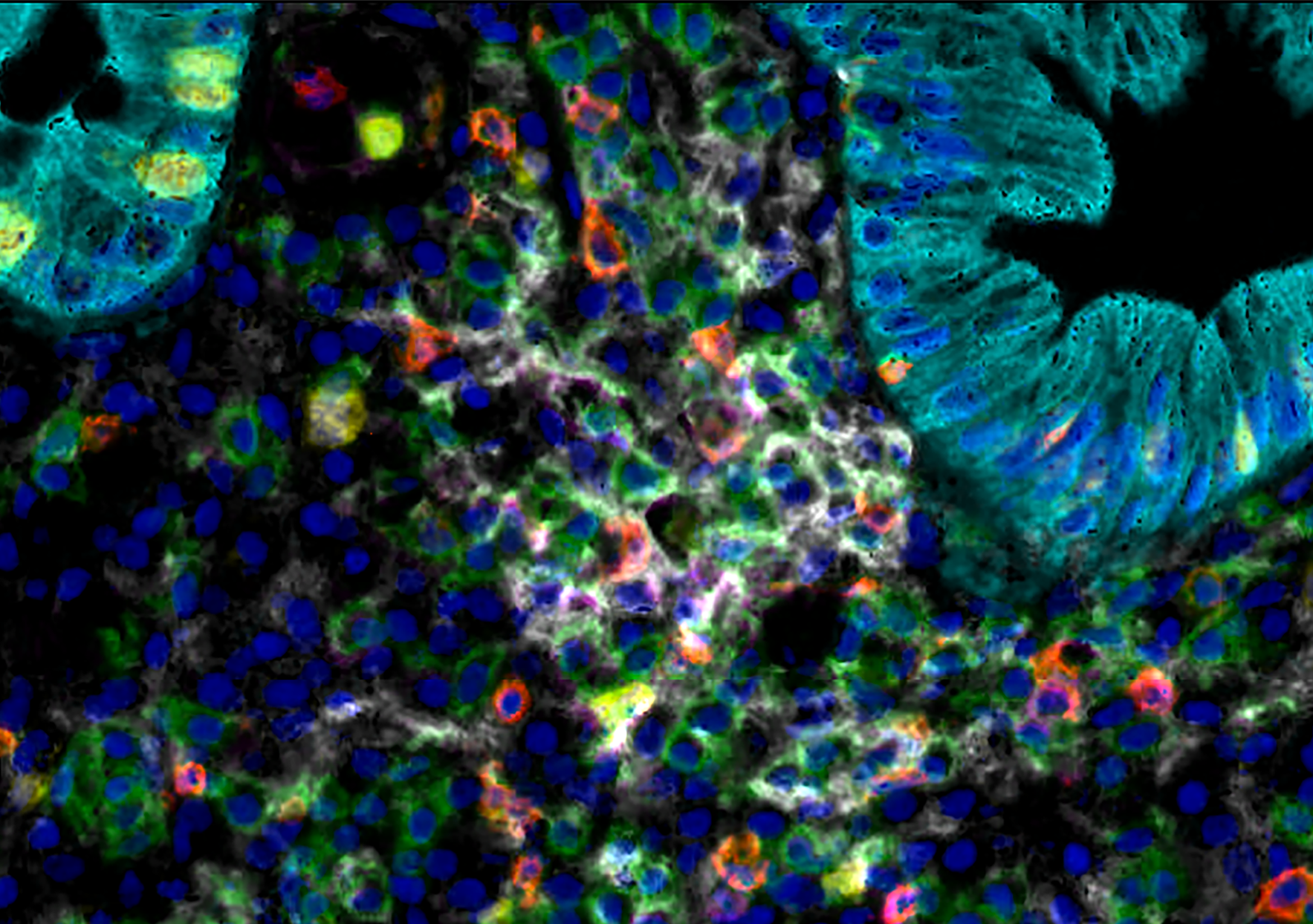
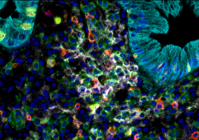
 Microscopic image of a section of a precancerous colorectal lesion, colored using multiplex immunofluorescence: cytokeratin, delineating intestinal epithelial cells, appears in cyan, cell nuclei appear in blue. Particularly active immune surveillance is visible at the center: several types of T lymphocytes stand out in green, red, purple, and white. © Morgand Erwan and Galon Jérôme/Inserm
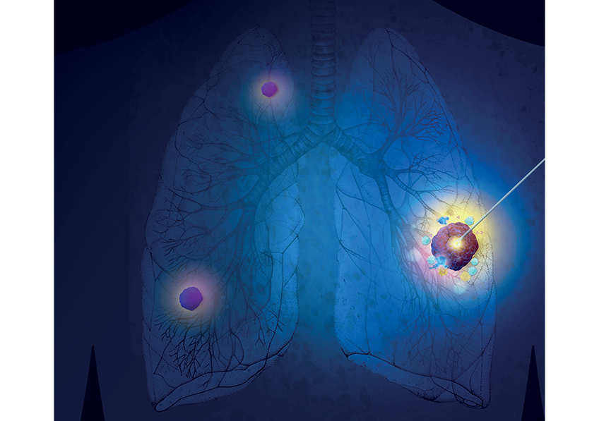
The prevention of colorectal cancer relies on the early detection and removal of precancerous lesions, which can significantly reduce the risk of progression to invasive cancer. However, the immune mechanisms involved in these lesions remain poorly understood. A research team from Inserm, Sorbonne University, and Paris Cité University has identified an association between immune activity in the microenvironment[1] of precancerous lesions, the frequency of polyp development, and the risk of colorectal cancer. These findings, published in Science Translational Medicine, pave the way for the identification of new biomarkers that could lead to the development of innovative immunotherapies.
Colorectal cancer develops in the epithelial cells of the intestinal lining from lesions known as precancerous lesions. Those that protrude and are easily visible are called polyps. Initially benign, these can gradually progress to a cancerous state. Early detection and preventive removal can significantly reduce the risk of developing invasive cancer and the associated mortality. However, the frequency of polyp formation varies from one individual to another, sometimes without any identified risk factors, and follow-up recommendations after an initial diagnosis are not always tailored to each patient’s specific risk.
Gaining a better understanding of the mechanisms underlying carcinogenesis (i.e., how a normal cell in the intestinal lining becomes cancerous) and enabling earlier intervention are therefore essential to reducing mortality due to these cancers.
Previous findings have shown that the prognosis—whether in terms of survival or response to treatment—for patients with colorectal cancer is associated with the composition of immune cells and molecules in the tumor microenvironment. Since cancers are often detected at advanced stages, this aspect remains understudied in precancerous lesions. However, recent studies have shown that the immune system is also capable of recognizing and eliminating tumor cells as early as the precancerous stage.
A research team led by Jérôme Galon, Inserm research director at the Cordeliers Research Center (Inserm/Sorbonne University/ Université Paris Cité) compared the microenvironment of a total of 258 precancerous lesions in 69 patients—none showing any risk factors for accelerated polyp development—based on the frequency of polyp occurrence.
The researchers thus identified distinct microenvironments whose specific immune profiles were associated with the frequency of polyp development. In particular, lesions in patients with a low frequency of polyp development exhibited a microenvironment indicative of enhanced immune surveillance[2] compared to those in patients with a higher frequency.
This immune surveillance was reflected in particular by a strong presence of antitumor immune cells, as well as a greater number and more mature stage of tertiary lymphoid structures. These clusters of immune cells form within the lesion itself to enable a local immune response and are associated with a better prognosis in many cancers.
“Our in-depth analyses of changes in the microenvironment also show that this ‘enhanced’ immune profile can develop very early on, as soon as the first polyp appears, and could enable the immune surveillance of emerging cancers,” adds Jérôme Galon.
Another major finding: lesions in patients with a low incidence of polyps exhibited gene activity profiles characteristic of particularly active antitumor immune surveillance, notably including increased expression of non-coding RNAs[3].
Conversely, precancerous lesions in patients with higher rates of polyp formation contained few of these non-coding RNAs. Their gene activity profiles were also characteristic of advanced carcinogenesis, even though the lesions appeared benign.
The frequency of polyp development and the risk of colorectal cancer thus appeared to be strongly associated with the immune profile of the lesions, which was itself associated with the presence of non-coding RNA.
“These observations suggest that these non-coding RNAs play a dual role: both in maintaining gene regulatory mechanisms and in orchestrating early antitumor immune surveillance at the local level,” explains Jérôme Galon. “Our hypothesis—which still needs to be verified experimentally—is that these RNAs could facilitate the recognition and targeting of precancerous cells by the immune system, thereby helping to limit the development of polyps and their progression to invasive cancer.”
The results of this study represent a significant advance in our understanding of the mechanisms of carcinogenesis. They point to the need to explore the role of non-coding RNAs in gene regulation and immunity in colorectal lesions.
“Identifying new biological markers of the progression of these lesions could enable the development of new personalized prevention and monitoring strategies, such as targeted vaccine immunotherapies, and allow for very early intervention to counteract the carcinogenesis leading to colorectal cancer,” concludes Jérôme Galon.
This work is part of the Darvac research project, led by Jérôme Galon and supported by Impact Santé, a funding program for high-risk health research led by Inserm as part of France 2030.
Impact Santé’s mission is to identify and support innovative approaches with the potential to transform biomedical research and medical practices.
With €2.9 million in funding from the program, Darvac is known as an “acceleration” project: it is based on a sufficiently solid scientific foundation to already be pursuing breakthrough research. It aims to develop vaccines that offer early and innovative protection against non-viral cancers.
To learn more about the Impact Santé program: https://presse.inserm.fr/impact-sante-30-millions-deuros-confies-a-linserm-pour-faire-avancer-la-recherche-en-sante/70177/
To learn more about the Darvac project, watch the project presentation video.
[1] The tumour microenvironment comprises all the cells or biological components (blood vessels, immune cells, fibroblasts, signalling molecules, extracellular matrix) located around cancer cells and which interact closely with them.
[2] Immune surveillance refers to the immune system’s ability to detect and eliminate, early and effectively, cells which, by accumulating mutations, have the potential to become cancerous.
[3] When DNA is transcribed into RNA, part of the resulting RNA—coding RNA—is translated to produce proteins. The other part, non-coding RNA, has regulatory functions that control gene expression (which genes are expressed and to what extent). In certain circumstances, non-coding RNA can also be translated into small protein fragments, known as peptides, which may be specific to tumour tissue. These peptides act as antigens, facilitating the detection of tumour cells by the immune system.


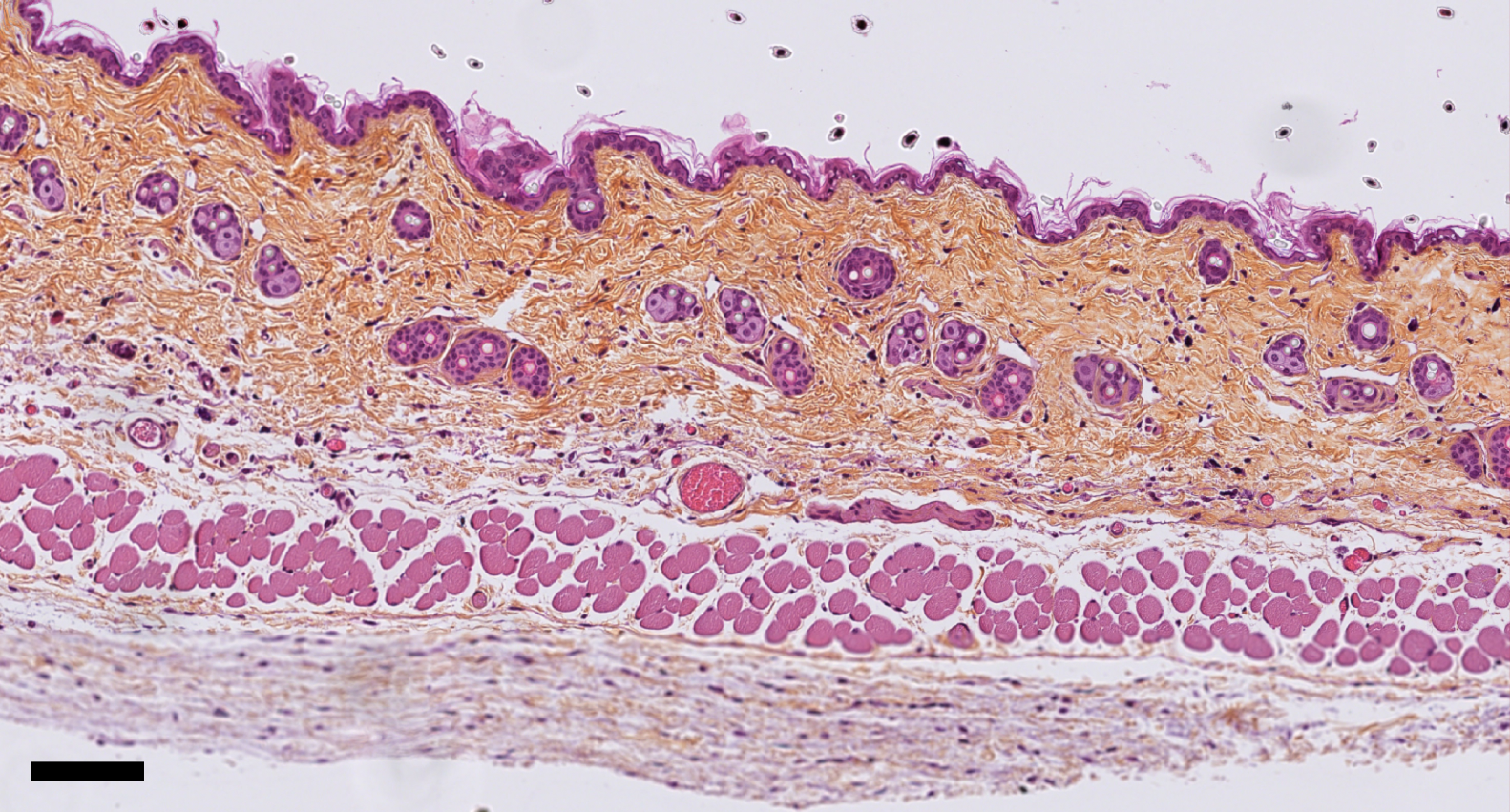
 Mouse skin structure after stretching, using histological staining. The scale bar corresponds to 100 micrometers. © Darawan Tabtim-On and Renaud Leclère – Experimental Pathology Platform, Institut Curie