Menu

Microbiome Influences Brain’s Immune Cells in a Sex and Age-dependent manner

©AdobeStock

A joint study conducted by Inserm researchers from IBENS (Institute of Biology of the Ecole Normale Supérieure – Inserm/CNRS/ENS) in Paris and researchers from SIgN (Singapore Immunology Network, A*STAR) in Singapore reveals a hitherto undiscovered impact of microbiota on immune brain cells, occurring from fetal stages. These cells, called microglia, which are known players in brain development and functioning, differentially respond to microbiome perturbations in male or female mice. These results are published in Cell.

Microglia are immune cells that respond to traumatic injury or inflammatory signals to protect the brain, acting as sensors of various environmental signals. In addition to their role as immune sentinels, microglia have also been show to regulate several steps of brain wiring and functioning. Consistently, microglia dysfunction has been linked with the etiology of several neurodegenerative diseases and neurodevelopmental disorders, including Schizophrenia or Autism Spectrum Disorders. Microglia therefore play major roles in brain circuits and could constitute an entry point for environmental signals.

To test this hypothesis, Morgane Thion and Sonia Garel, Inserm researchers, together with their associates, used a multidisciplinary approach involving germ-free mice, which lack all the microbiome, and adult mice treated with antibiotics, which acutely destroys the gut flora. Through a combination of global genomic analyses and histological studies, researchers have shown that microglia are profoundly affected by microbiota disruption, already from prenatal stages. Strikingly, the impact of the microbiome on microglia depended on the sexual identity and the age: microglia of males were perturbed prenatally whereas those of females were affected in adulthood. This surprising sexual dimorphism echoes the fact that many neurodevelopmental disorders have a higher incidence in men, whereas autoimmune diseases are more prevalent in women.

While the underlying mechanisms and consequences remain to be discovered, this study reveals a key role of microglia at the brain/environment interface and shows that males and females have distinct susceptibilities time windows to microbiome alterations. For the authors, these elements should be systematically taken into consideration at preclinical and clinical level.

An Edible Mushroom With Potential to Fight Human Genetic Diseases

Credits @ MNHN/CNRS – Christine Bailly

Could a common mushroom help fight certain genetic diseases? Although surprising, this is indeed the new discovery made by French scientists from Inserm, the French National Museum of Natural History, the CNRS, Université de Lille, and the Institut Pasteur de Lille[1]. By examining numerous extracts, the scientists thus evidenced that the mushroom, Lepista inversa, acted significantly on three isolated cell lines taken from patients with cystic fibrosis. This research was published in .Plos One

[1] Mechanisms of Tumorigenesis and Target Therapies Laboratory (CNRS, Université de Lille, Institut Pasteur de Lille), and Microorganism Communication and Adaptation Molecule Laboratory (MNHN, CNRS)

Approximately 10% of patients with rare genetic diseases such as cystic fibrosis or Duchenne muscular dystrophy (or more common genetic diseases, such as certain types of cancer) have a nonsense mutation, i.e. of a change in DNA sequence. This mutation is evidenced by the presence of a “stop codon” which does not code for a known amino acid and prematurely stops the synthesis of proteins from mutated genes. As a result, the proteins obtained are truncated and do not function properly. Unable to fulfill their role in the body, they have familiar harmful consequences: bronchial obstruction and inability to breathe in cystic fibrosis, and muscular degeneration in muscular dystrophy.

Several strategies are now being developed to correct the consequences of a nonsense mutation. One of the most promising avenues is translational readthrough. This involves the cellular machinery continuing protein synthesis despite the presence of a “stop codon” in the DNA. For this purpose, when the RNA is transformed into protein, “decoy” molecules located in the environment very close to the cellular machinery may catch it unawares and enable a complete protein to be produced, as if there was nothing amiss. Nevertheless, the molecules capable of playing this role and identified to date display very limited efficacy and/or considerable toxicity.

@ Taken from Médecine sciences https://doi.org/10.1051/medsci/2012282018


ATG            STOP                       DNA

                                                  Transcription

AUG                                            mRNA

                                                   Translation

mRNA degradation by the NMD pathway

Termination                              Readthrough inducers

Truncated protein                       Full-length protein


Figure 1. Therapeutic strategy in stop codon diseases. A nonsense mutation in a gene (DNA) causes the appearance of a premature stop codon on the mRNA which is translated by the ribosome. This triggers the synthesis of a truncated protein and possibly the degradation of this mRNA by the NMD pathway (nonsense mediated mRNA decay), which specifically recognises mRNAs containing a premature stop codon. Certain compounds, by inducing passage through the premature stop codon (an event known as “readthrough”), enable the synthesis of a full-length protein, which could bring therapeutic benefit to patients with a nonsense mutation.

By combining their expertise and using a screening system on the chemical-extract libraries at the French National Museum of Natural History, two teams of scientists[1] succeeded in demonstrating that the extract of a mushroom, Lepista inversa or Clitocybe inversa, is capable of very effectively restoring the expression of human genes presenting nonsense mutations on cells in culture.

Thanks to the partnership between the two research laboratories, the CHU de Lille, Hospices Civils de Lyon, Cochin Hospital, and the charity Vaincre la Mucoviscidose, researchers were also able to evidence significant activity on cells from patients with cystic fibrosis[2].

“Given that only 5% of functional proteins need to be restored in cystic fibrosis to have an impact on the consequences of the disease, this research is extremely encouraging,” according to the authors who point out that this strategy also has the advantage of not affecting the patients’ genetic inheritance.

“This discovery brings hope as this mushroom is edible, although it is not particularly prized for its taste; it is also very common – it grows in the Ile-de-France region, and in various regions of France and Europe,” explains Fabrice Lejeune, Inserm researcher and the last author of this research. “There is still a long way to go before we can develop a genuine therapeutic strategy,” he adds. “We still need to find a way to purify the molecules of interest present in this extract, and then carry out in vivo tests to verify their long-term efficacy and the absence of toxicity.”

This multidisciplinary study also demonstrates the value of the extract collection kept in the extract bank at the museum for teams of biologists and chemists working in the health field.

[1] Mechanisms of Tumorigenesis and Target Therapies Laboratory (CNRS, Université de Lille, Institut Pasteur de Lille), and Microorganism Communication and Adaptation Molecule Laboratory (MNHN, CNRS)


A crucial enzyme finally revealed

© L. Peris /GIN

After 40 years of research, researchers at the CEA, the CNRS, the University of Grenoble-Alps, the University of Montpellier and the Inserm have finally identified the enzyme responsible for the tubulin cycle. Surprisingly, it is not one enzyme but two which control the cycle of this essential component of the cytoskeletal structure. This work opens up new prospects for the improved understanding of the role of tubulin, changes in the cycle of which are associated with cancers, cardiac diseases and neural disorders. These results were published on 16th November 2017 in the review Science.

A collaborative international project involving researchers from the CEA (French Atomic Energy Commission), the CNRS (National Centre for Scientific Research), the Inserm (French National Institute of Health and Medical Research), the University of Grenoble-Alps, the University of Montpellier and the University of Stanford[1] has identified an enzyme, Tubulin CarboxyPeptidase (TCP), which is responsible for the biochemical transformation of cellular microtubules, or detyrosination. Detyrosination is a biological reaction for the removal of the terminal amino acid tyrosine[2] from tubulin α, a constituent of microtubules. After four decades of research, biologists have succeeded in isolating this protein by purification, and have gone on to provide evidence of its cellular activity.

Microtubules contribute to essential cellular functions

Microtubules are dynamic fibres which are present in all cells. Formed by the combination of two proteins (tubulin α et tubulin β), microtubules assume numerous functions. They separate the chromosomes which are to be contained in the two daughter cells resulting from cell division, they contribute to the polarity of cells, morphology and cellular migration. They form “rails” upon which cellular constituents, such as proteins or RNA strands, are transported.

These cellular functions are regulated by the existence of “signals” which are present on the surface of microtubules. These signals are biochemical modifications to amino acids (described as post-translational modifications, as they take place after protein synthesis), executed by various enzymes which, in this case, modify the tubulins.

 The enzyme TCP, identified after 40 years of mystery

The activity of one of these enzymes was identified for the first time in 1977 by Argentine researchers, who named it “TCP” (Tubulin CarboxyPeptidase). The function of this enzyme, which had never been identified previously (its size and sequence were unknown) is the removal of the terminal amino acid, a tyrosine, from the end of tubulin α. This is the detyrosination reaction. A reverse enzyme, ligase TTL, is responsible for resetting this tyrosine in its place. This is tyrosination. This detyrosination/tyrosination cycle is vital for the cell and the organism. Massive (abnormal) detyrosination is observed in a number of severe cancers and cardiac diseases.

The identification and characteristic definition of TCP was therefore a major objective for understanding the physiological function of the detyrosination of tubulin α and evaluating the consequences of its inhibition.

In order to isolate TCP, researchers have monitored its activity, employing conventional biochemical techniques, and have involved chemists from the University of Stanford, who have developed a small inhibitor molecule for its activity. This molecule has been used as bait to “reel in” the desired enzyme.

Tubulin detyrosination/tyrosination cycle

Microtubules are fibres which are present in all cells, comprised of a stack of α/β tubulins. Tubulin carries a tyrosine (Y) at its end, which is alternately removed and replaced by two enzymes, thereby modifying the surface of microtubules. TCP (which is represented by a saw comprised of two elements, VASH/SVBP) is responsible for detyrosination. TTL (represented by a tube of glue) resets tyrosine on the tubulin. This cycle is essential to the various functions of microtubules in cells (division, migration, etc.) and is vital for the organism. © C. Bosc, GIN


SVBPSVBP
VASH1,2VASH1,2
scie TCPTCP saw
detyrosinationdetyrosination
tubuline tyrosinéetyrosinated tubulin
tubuline détyrosinéedetyrosinated tubulin
tyrosinationtyrosination
colle TTLTTL glue


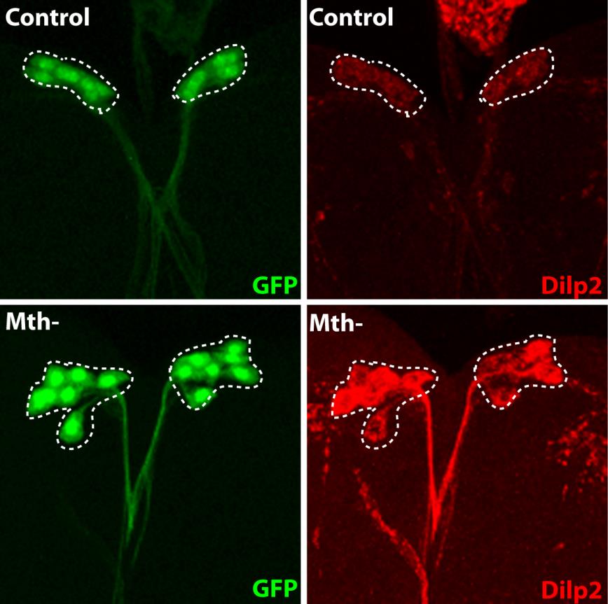
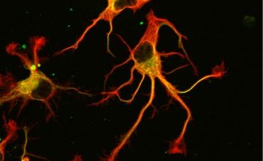
Ultimately, not one, but two enzymes have been discovered. The latter, named VASH1 and VASH2, were already known to scientists, but it was not known that these were enzymes associated with the cytoskeleton. Researchers have demonstrated that, provided they are associated with a partner protein called SVBP, VASH1 and VASH2 are capable of the detyrosination of tubulin α. To demonstrate this, researchers have inhibited the expression of the former (or that of their partner SVBP) in neurons. They then observed a very strong decline in the rate of detyrosination of tubulin α, together with anomalies in the morphology of neurons (see Figure). Researchers went further, demonstrating that these enzymes are also involved in the development of the cerebral cortex.

Prospects for the fight against cancer

Thus, forty years after the conduct of the first work on the detyrosination of tubulin α, the enzymes responsible have been revealed. Scientists are now hoping that, by modulating the effectiveness of TCP and improving their knowledge of the detyrosination/tyrosination cycle, they can advance the fight against certain cancers, and achieve progress in the understanding of cerebral and cardiac functions.

ContrôleControl
VASH1 et VASH2 réduitesVASH1 and VASH2 reduced
SVBP réduiteSVBP reduced
Tubuline deTyrosinée / Tubuline TyrosinéeDetyrosinated tubulin / Tyrosinated tubulin

Photographs of the alteration of neurons associated with a reduction in the expression of TCP enzymes (VASH/SVBP). From left to right: control neuron, neurons in which the expression of VASH1 and VASH2 is reduced, neurons in which the expression of SVBP is reduced. Neurons with a reduced enzyme show a delay in development, together with morphological anomalies.

[1] The following institutes are involved: Grenoble Institute of Neurosciences, GIN (Inserm/Univ. Grenoble-Alps); Institute of Biosciences and Biotechnologies of Grenoble, BIG (Inserm/CEA/Univ. Grenoble-Alps); Institute of Advanced Biosciences, IAB (Inserm/CNRS/Univ. Grenoble-Alps), Department of Pathology, Stanford University School of Medicine (Stanford, USA), Institute of Human Genetics, IGH (CNRS/Univ. of Montpellier), Montpellier Centre of Cell Biology Research, CRBM (CNRS/Univ. of Montpellier).

[2] Tyrosine is one of the 22 constituent amino acids in proteins.

Gene therapy: first results in children with Sanfilippo B syndrome

©Fotolia

On July 13, 2017, the journal Lancet Neurology published the results of a gene therapy trial conducted in four children with Sanfilippo type B syndrome (also known as MPS IIIB). This trial is the achievement of a two-decade partnership with financial support of AFM-Téléthon and the cooperation of the charity “Vaincre les Maladies Lysosomales” (VML). After monitoring of the treated children for 30 months, Dr. Jean-Michel Heard, from the Institut Pasteur and Inserm, and Professors Marc Tardieu and Michel Zérah, from the Paris public hospital administration (AP-HP) and the Paris-Sud and Paris Descartes Universities, conclude that the treatment was well tolerated and associated with neurocognitive benefits for the patients.

Sanfilippo syndrome is a rare genetic disease which affects approximately one in every 100,000 children. It alters brain development after birth and leads to brain degeneration several years later. The first symptoms of the disease are hyperactivity and delayed cognitive acquisition, which are usually noticed when children are around two-years old. A genetic anomaly prevents the production of an enzyme needed to breaking down mucopolysaccharides. Mucopolysaccharides are large macromolecules that help the neurons develop effective connections in young children during learning. Incomplete degradation and accumulation are toxic for brain cells. The disease progressively leads to a state of severe and multiple impairments and to premature death within periods of 5 to 10 years.

The challenge to treat Sanfilippo syndrome lies in the design of a method to supply the missing enzyme to the brain as early as possible after birth.  The therapeutic trial conducted by the Institut Pasteur at the Bicêtre Hospital (AP-HP) used gene therapy for that purpose. A gene therapy vector (AAV2/5) capable of inducing the production of the missing enzyme by brain cells was injected at several sites in the brain and cerebellum of affected children. The specific aim of this phase I/II trial was the assessment of tolerance to the surgical procedure and to the candidate drug delivered by gene therapy.

The clinical study initiated in 2013 was preceded by more than ten years of preclinical studies in animals naturally affected by the disease. The researchers administered the treatment for the first time to four children aged between 1.5 and 4 years (20, 26, 30, and 53 months). No particular clinical, radiological or biological side effects associated with the treatment were observed within 30 months of administration, indicating that it was well tolerated.

Within one month of treatment and throughout the 30 months of the trial, researchers detected the previously missing enzyme in the cerebrospinal fluid of the four treated children. Moreover, very careful and regular neurocognitive monitoring revealed positive impact on cognitive acquisition and behavioral development, which were more pronounced in the youngest treated child.

The encouraging findings of this phase I/II clinical trial suggest that treatment could be proposed to patients with Sanfilippo syndrome in the future.  To reach that goal, the next step would consist in a large and multicentric phase III clinical trial, if an appropriate partner is found.

Discovery of a new mechanism involved in the migration of cancer cells

A team of young researchers under the supervision of Guillaume Montagnac, Inserm research leader at Gustave Roussy, in collaboration with the Institut Curie and the Institut de Myologie (Myology Institute), has discovered a new mechanism which facilitates cell migration. On the surface of its membrane, the cell develops multiple small hooks which help it to attach to fibres outside the cell and move along them. This action helps us to understand better how a cell escapes from the tumour mass and moves around the body to form a new focus. This research is published in the 16th June issue of the American journal Science.

Cell migration is a normal process which is essential to life. In oncology it is involved in the formation of new metastases.

“Up till the present, we knew that the cell relied on certain structures to anchor itself within its environment. We have now identified new cell structures known as ‘clathrin-coated pits’, already known to be important for other cell functions. The cancer cell uses them as hooks to attach to other structures in order to move around, These novel structures underlie some 50% of cell adhesion to surrounding structures,” declared  Guillaume Montagnac, Leader of the ATIP-Avenir team, attached to Inserm Unit 1170, Normal and abnormal haematopoiesis”, at Gustave Roussy.  

Recognised in 1964, these clathrin pits are small invaginations of the cell membrane which allow it to renew itself or to help molecules to enter the cells. The cell uses them particularly to supply itself with nutritional material (iron, cholesterol, etc.).

Using fluorescence methods, the researchers succeeded in demonstrating with an aggressive human breast cancer line, known for its marked propensity to metastasise, that the clathrin pits adhere to collagen fibres and surround them. The pit squeezes the fibre, so strengthening its hold and allowing it to move.  

“Our Gustave Roussy team is one of the few with an interest in cell membrane dynamics when the cell is placed in 3D matrices under conditions close to normal ones. By studying these clathrin pits in 3D we were able to see the phenomenon when we were not expecting it,” concluded Guillaume Montagnac.

A breast cancer cell with actin (engine of migration) in red, the clathrin pits (cell hooks) in green and collagen fibres in blue

Neuronal Self-Defense Against Alzheimer’s Disease

Neurons

© Fotolia

It is known that IGF-1 (insulin-like growth factor) is needed for development and also plays a role throughout the body’s life. Previously, the team led by Martin Holzenberger (Inserm/UPMC Unit 938, Saint-Antoine Research Center) has shown that this hormone is involved in longevity and in Alzheimer’s disease. The team has recently conducted further research on IGF-1 and the response of neurons to this kind of neurodegeneration. These new results have been published in Brain.

Secreted by the liver and stimulated by growth hormone, IGF-1 (insulin-like growth factor) is able to stimulate the growth and maturation of bone and other organs, regulate energy metabolism, and control the aging of the whole body. In its previous work, the Holzenberger team had demonstrated in mice that when the number of IGF-1 receptors present in the neurons was reduced by genetic mutation, the level of IGF-1 in the blood decreased and the mice had a longer lifespan.

In this new study, published in the journal Brain, Martin Holzenberger and Saba Aïd have conducted further research on IGF and Alzheimer-type neurodegeneration. These researchers first show that inhibiting IGF-1 receptors in the neurons of mice led to a much later presentation in their brains of the signs of lesions typical of Alzheimer’s disease, in particular amyloid plaques and neuroinflammation. Reduced cognitive impairment was also observed in the same mice. Importantly, the team has shown that suppression of the IGF receptor leads to a series of neuroprotective effects. This confirms their previous results concerning prolonged lifespan.

This new study reveals a self-defense system used by the neurons when they suffer the kind of harmful attack typical of Alzheimer’s disease. In fact, the gene families activated in Alzheimer neurons and in the neurons deprived of the IGF-1 receptor are essentially the same. This suggests that, in the early stages of the disorder, a neuron faced with an Alzheimer-type disease is able to instigate a process of self-defense of its own accord (this is called an endogenous response). This endogenous response is not however sufficient over the long-term in a brain affected by Alzheimer’s, and effective protection against the disease requires total suppression of IGF-1 receptors in the neurons. It is not yet known at what point this neuronal response ceases to be effective against the disease.

These results enable a better understanding of the mechanisms of Alzheimer-type neurodegeneration, a disease that affects nearly one million people in France. This work is crucial, suggesting a paradigm shift concerning the role of IGF-1 in the progression of age-related neurodegenerative diseases: it is not stimulation, but rather long-term blocking of IGF signaling that would improve neuronal function and neuroprotection.

Ultimately, this work will lead to the development of new therapeutic and preventive targets in the fight against Alzheimer’s disease. However, the researchers emphasize that there is still a long way to go. “We cannot inhibit the IGF-1 receptor throughout the entire body because this hormone is essential for other cells. However, specifically targeting the neurons is a possibility. In any case, we have to better understand how to benefit from the good effects of IGF while preventing its less beneficial effects. “, concludes Martin Holzenberger.

We’re all a bit Neanderthal… or are we?

neanderthal

A study conducted by Inserm researchers at the Research Institute for Environmental and Occupational Health (Irset)[1] has shown that natural selection has “purged” our bodies of many of the traces of our ancient Neanderthal and Denisovan cousins in the genes responsible for the genetic mixing essential to reproduction. The researchers have shown that the genes expressed during meiosis in the cells that produce gametes (reproductive cells) are strongly deficient in genetic variations of Neanderthal origin that were the result of the interbreeding between Homo sapiens and Homo neanderthalensis. These results have been published in Molecular Biology and Evolution.

For decades, a question has preoccupied paleontologists regarding our now-extinct cousins, the Neanderthals and Denisovans:  what was the nature of the interactions between modern humans (Homo sapiens) and the other species of the Homo genus ?

Well, hundreds of thousands of years ago, a succession of human migrations from Africa to the other continents led to the coexistence in Eurasia of Homo sapiens with various other now-extinct species of the Homo genus. In 2014, the sequencing of a Neanderthal genome was made possible by the discovery of bone fragments containing DNA. With the very recent emergence of paleogenomics, it has been established that 1 to 3% of the genome of present-day Eurasians is inherited from the Neanderthals, whereas 3 to 6% of that of Oceanians is inherited from other ancestral cousins, the Denisovans. The women and men that populate our planet today are the result of many interbreeding events that have enabled human populations to expand thanks to the acquisition of characteristics favorable to climatic and environmental adaptations.

However, a surprising particularity recently came to light: the genetic variations inherited from interbreeding with these extinct species are not evenly distributed on the chromosomes. As such, Prof.  David Reich’s team demonstrated that these “archaic” genetic variations are present only to a very minor extent on the genes expressed specifically in the testis of modern humans.

Hence the key question studied by the researchers in Rennes: within the testis and ovary, to which specific functions are these genes, deficient in Neanderthal and Denisovan genetic variations, assigned ?

To answer that question, researchers from Inserm compared the genes present in the different cell types of the testis (germ line cells, Sertoli cells, Leydig cells, etc.).

The results obtained show that it is only those genes expressed specifically during meiosis, the process responsible for genetic mixing, that are highly deficient in ancestral alleles of Neanderthal and Denisovan origin. The conclusions were the same when the germ cells present in human fetal ovaries were studied. Since meiosis is a unique and fundamental process of spermatogenesis and oogenesis, natural selection has therefore “purged” our gene pool of the genetic variations that could have adversely affected its progression and thus prove harmful to the continuation of our species.

For the study’s coordinators Frédéric Chalmel and Bernard Jégou, this shows that “while interbreeding between modern humans and these extinct hominins has enabled us to acquire new adaptive traits important for our survival, it probably had a negative impact on the fertility of the initial hybrids. That is certainly why the genes involved in meiosis, a particularly sensitive biological process, have been purged of genetically archaic variations.  This is the first paleo-fertility study and it is likely to reveal evolutionary processes involved in certain present-day cases of infertility.”

[1]  Research Institute for Environmental and Occupational Health; Inserm; EHESP School of Public Health, Université de Rennes 1.

Communication between neurons implicated in autism spectrum disorders and intellectual disabilities

vignettecp-web

© Fotolia

An international collaborative study coordinated by Frédéric Laumonnier (Unit 930 “Imaging and Brain” Inserm/University of Tours) and Yann Hérault of the Institute of Genetics and Molecular and Cellular Biology (Inserm/ CNRS/ University of Strasbourg) provides new and original findings on the pathophysiological role of the contact areas between neurons in certain brain disorders. The study reveals that mutation of one of the genes involved in intellectual disability and autism spectrum disorder leads to dysfunction of the synapses, which are essential for neuronal communication. The research was published on April 18, 2017, in Molecular Psychiatry.

Autism spectrum disorder (ASD) and intellectual disability (ID) are neurodevelopmental disorders that generally emerge when a child’s brain is developing and often persist into adulthood. Behavioral disorders and inabilities to communicate and establish social interactions are observed in people with ASD. In addition, those with ID present difficulties with comprehension, memory, and learning. While the origins of these disorders remain poorly understood, we now know that a significant proportion are associated with genetic mutations.

During the brain development process, synapse formation is essential for brain functions such as memory and learning. Synapses are the points of contact between neurons which enable neurons to connect with each other and propagate information.  Some synapses are inhibitory and others excitatory, to enable the establishment of functional neuronal networks. However, mutations of the so-called PTCHD1 (Patched Domain containing 1) gene, which is located on the X chromosome and enables the expression of a protein potentially involved in synaptic functioning, have recently been identified in boys with the aforementioned disorders. These mutations stop the gene from expressing itself.

In order to validate the involvement of PTCHD1 gene mutations in ASD and ID, Hérault and his co-workers created a mouse model that was deficient for the PTCHD1 gene. In these animals, they observed major memory deficits and significant symptoms of hyperactivity, thus confirming the gene’s involvement in ASD and ID. Parallel studies by Laumonnier’s team showed a presence of the PTCHD1 protein in the excitatory synapses and also detected changes in the same mice’s synapses.

These changes to synaptic structure and activity in the excitatory neuronal networks were found to be particularly significant in a central brain region known as the hippocampus. This region plays a major role in cognitive processes, particularly those involving memory and the formation of new memories.

Genetic abnormalities impacting the structure or functioning of these synapses constitute a pathophysiological target in ASD and ID. In this context, this research defines a new “synaptic disease” caused by a PTCHD1 gene mutation. This dysfunction emerges during the development of the central nervous system and is associated with ID and ASD. Understanding of the pathophysiological mechanisms that underlie these neurodevelopmental disorders, particularly through the study of model organisms, is essential to improve therapeutic strategies.

A warning on taking ibuprofen during pregnancy

fotolia_4518648 Suivi médical grossesse

©fotolia

A new study conducted by Inserm researchers at Irset (Institute of Research in Environmental and Occupational Health)[1] shows that ibuprofen is liable to cause disruptions in the hormone system in the human foetal testis, with possible implications for the development of the male urogenital tract. This drug suppresses the production of various testicular hormones, including testosterone, which controls the primary and secondary sex characteristics and the descent of the testes. These effects are obtained at doses similar to the standard dosage. These results are published in Scientific Reports.

Ibuprofen, which can be obtained without prescription, is one of the drugs most commonly consumed by pregnant women. Although nearly one woman in ten reports having taken ibuprofen during her pregnancy, studies indicate that in reality up to 3 in 10 have self-medicated with it.

Epidemiological studies conducted in recent years have shown a link between taking analgesics during pregnancy and the occurrence of adverse effects in the child (low birthweight, asthma, premature birth etc.). Other research combining epidemiology, in utero experimentation in rats and ex vivo on rat and human organs, undertaken at Irset in collaboration with Danish researchers from the University of Copenhagen, showed that paracetamol and aspirin could disrupt the endocrine system of the foetal testis, resulting in an increased risk of failure of the testes to descend (cryptorchidism). Only the effects of ibuprofen had not yet been tested.

To do that, the Irset researchers – with the support of colleagues at Rennes University Hospital and the University of Copenhagen, researchers from Laberca (Laboratory for the Study of Residues and Contaminants in Food) in Nantes, and Scots colleagues from MRC Edinburgh – conducted two series of tests to study the effects of ibuprofen on the human foetal testis. In the first series of studies, the testes were cultured; in the second, they were grafted onto mice[2]. The effects of ibuprofen were studied over periods corresponding to the 1st and 2nd trimesters of pregnancy.

When testes corresponding to the 1st trimester of pregnancy were exposed to ibuprofen, there was a sharp drop in testosterone production by the Leydig cells. During the same period (up to 12 weeks of development), the researchers observed for the first time that ibuprofen also affected the production of antimüllerian hormone by the Sertoli cells. This hormone plays a key role in genital tract masculinisation.

Moreover, expression of the genes needed for the germ cells, the progenitors of spermatozoa, to function is considerably reduced in the presence of ibuprofen.

Finally, production of prostaglandin E2 (known to be produced by the testes, and known to be involved in many biological processes) and the corresponding genes are also inhibited by the presence of ibuprofen at the same developmental age.

All these effects are observed very early in the first trimester, and none of them is found in tests conducted during the second trimester.

For Bernard Jégou, Inserm Research Director and coordinator of this study, and Séverine Mazaud-Guittot, Inserm Research Fellow, the conclusions of this work, which was supported by the French National Agency of Medicine and Health Products Safety (ANSM), are to be taken seriously: “There is a well-defined window of sensitivity during the 1st trimester of foetal development during which ibuprofen seems to present a risk for the future genital and reproductive system of the child. All the indications point to the need for great prudence regarding the use of this drug during the 1st trimester of pregnancy. Furthermore, if we now take the body of available data into account, it seems that taking several analgesics during pregnancy represents an even greater danger for the hormonal balance of the male foetus.

[2] Xenografting is the transplantation of cells or organ fragments from one living organism (e.g. human cells) into the body of another species (here the mouse) in order to understand their development.

[1] Institute of Research in Environmental and Occupational Health; Inserm; French School of Public Health (EHESP), University of Rennes 1.

Breast cancer: identification of a molecular switch that controls cancer stem cells

Cancer sein Fournier

© fotolia

Some cancer cells are resistant to treatment and persist. If they are capable of proliferating again, even a very small number of these cells may be enough to reconstitute a tumour after or despite treatment. Various approaches to eliminate these “cancer stem cells” (CSCs) have been tried in recent years: targeted therapies, vaccination and tumour starvation. In an article published in the journal Cell Reports, Christophe Ginestier, Inserm Research Fellow at the Cancer Research Center of Marseille (CRCM, Aix-Marseille University/CNRS/Institut Paoli-Calmettes), and his collaborators identify a specific RNA[1] molecule that plays the role of a molecular switch that can “turn off” or “turn on” CSC proliferation in breast cancers.

Scientific data accumulated in the course of recent years have shown that tumours contain a particular population of cells with different properties. Indeed, a small number of the cells constituting a tumour have the ability, when isolated and then injected into animal models, to form a tumour identical to the original one. These cells, known as cancer stem cells (CSCs), can proliferate (and thereby renew themselves), differentiate (and thereby give rise to the different populations that make up the tumour), or even become temporarily dormant, which allows them to escape most treatments, since these mainly target dividing cells.

If the tumour is to be eliminated in such a way that it cannot grow again, the CSCs must be neutralised. The development of any new therapeutic strategy requires a better understanding of the intrinsic molecular mechanisms of CSCs. MicroRNAs have been described as regulators that can direct the “cellular destiny” of stem cells, particularly during embryogenesis. They might play a major role in CSC biology. MicroRNAs are small RNA molecules that, unlike messenger RNAs, do not act as intermediates in protein production based on information encoded by genes, but regulate the activity of other RNAs or of proteins.

Christophe Ginestier, Emmanuelle Charafe-Jauffret and their co-authors screened the full complement of microRNAs present in the genome in order to identify microRNAs capable of directing the choice for a CSC between self-renewal and differentiation. They thus observed that inactivation of one particular microRNA, known as miR-600, causes an increase in CSCs, while its overexpression reduces tumourigenicity.

They then showed that miR-600 works by acting on an enzyme needed to activate a protein (WNT) known to activate a signalling cascade involved in embryogenesis. When they inactivate miR-600, the researchers observe the expansion of CSCs. Conversely, when miR-600 production is increased, differentiation of CSCs is promoted at the expense of their proliferation: tumour progression is stopped.

This mechanism, demonstrated experimentally, clearly seems to play a role in the development of breast cancers, since the researchers were also able to show, by analysing a panel of 120 human breast tumours, that a low level of miR-600 is found to be associated with a strong activation of the WNT protein and a poor prognosis for patients whose tumours show these characteristics.

“If miR-600 is a switch for tumour aggressiveness, it may therefore constitute an excellent therapeutic target,” conclude the researchers. “Our data also tend to prove that resistance to treatment and relapse after treatment could be due to the fact that the therapies employed are not targeting the right cancer cells.”

[1] RNA: ribonucleic acid, a biological molecule present in nearly every living being. Often providing intermediate support to genes during protein synthesis, RNA can also be involved in many chemical reactions within the cell.

fermer