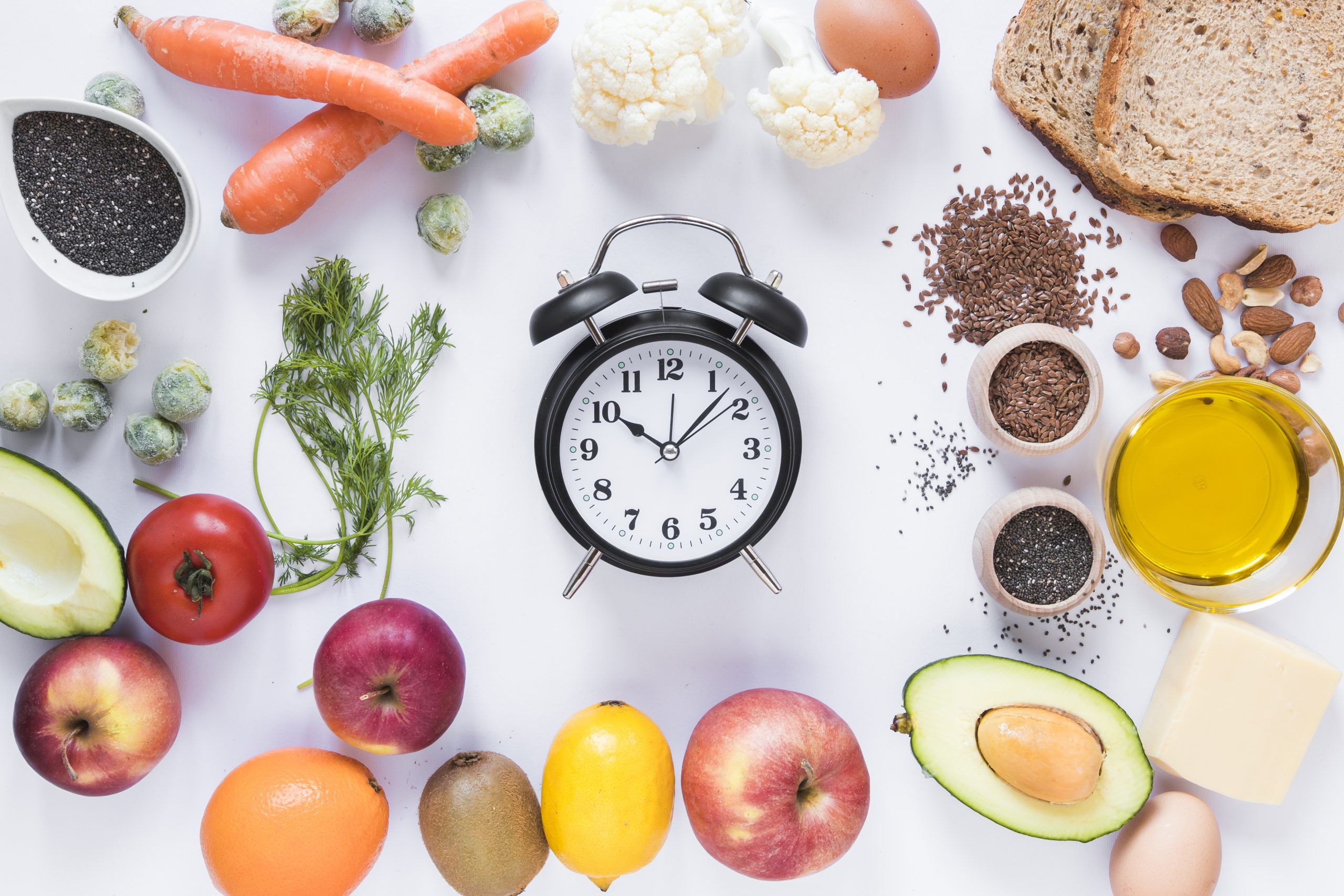
Menu

The appendix is not an unnecessary organ but is in fact correlated with a longer lifespan

koala

The appendix is an anatomical structure that can be found in a wide range of very different species, from the orangutan to the koala, beaver, and of course humans. © David Clode/Unsplash


Long considered an unnecessary organ, the appendix is now the focus of several studies that aim to better understand its role. Present in many mammals, including humans, it appears to have developed at least 16 times over the course of the evolutionary history of mammals, suggesting that its function must confer a positive selective advantage on those that have it. A new study carried out by researchers from Inserm and the French Museum of Natural History suggests that the presence of the appendix is in fact correlated with greater longevity. Their findings have been published in the Journal of Anatomy.

The appendix is a small anatomical structure of a few centimeters in size, located in the abdomen and attached to the colon, the function of which has long been poorly understood. According to the theories of Charles Darwin, the appendix was a vestigial structure, useless and devoid of function. It might even be seen as potentially dangerous to health due to the risk of inflammation of the organ. If such inflammation, known as “appendicitis,” is left untreated, it can develop into peritonitis and result in death.

Over the last few years, researchers have sought to learn more about the role of the appendix. Studies have, for example, shown that an appendectomy performed in cases of confirmed appendicitis before the age of 20 has a protective effect against the onset of a particular form of chronic inflammation of the colon and rectum: ulcerative colitis.

Researchers have also shown that the appendix is not only present in humans. It first appeared in mammals at least 80 million years ago, and over the course of evolution has developed independently multiple times in several mammalian lineages, with no obvious correlation with diet, social life, or the environment. Today it can be found in a wide range of animals: from orangutans and koalas to manatees, beavers, and platypuses. Its function has however remained a mystery, with no study reaching a definitive conclusion.

A link with mammalian longevity

The team led by Inserm researcher Eric Ogier-Denis and his colleague Michel Laurin from the French Museum of Natural History approached the question by analyzing data from 258 species of mammals, 39 with and 219 without an appendix. The scientists focused in particular on the theoretical maximum longevity (the theoretical lifespan of mammals, established based on their weight) and actual maximum longevity of the various species considered.

They have shown for the first time that the presence of the appendix is correlated with an increase in the maximum longevity observed for a species. Compared to mammals of the same weight without an appendix, mammals with an appendix have a longer lifespan.

The idea of focusing on longevity developed from our work on the relationship between appendicitis/appendectomy, ulcerative colitis and the involvement of the immune system. A more active and better educated immune system should theoretically provide greater resistance to the environment and a longer lifespan. We therefore tested this hypothesis by partnering with two internationally renowned evolutionary experts from the French Museum of Natural History. This is the first time the existence of a correlation between the presence of the appendix and a trait in the life history of mammals has been demonstrated,” explains Eric Ogier-Denis.

The team has also shown that the appendix has developed at least 16 times and has only been lost once (by the lemur Hapalemur griseus, endemic to Madagascar) during the evolutionary history of mammals, which supports the idea that

through its function this organ provides a significant positive selective advantage with regard to the laws of natural selection.

A bacterial sanctuary

The researchers believe that the most likely hypothesis to explain the link between the presence of the appendix and longevity is that the shape of the organ enables the development of a selective “bacterial sanctuary” that reduces mortality from infectious diarrhea by promoting rapid recolonization of bacterial species that are essential to the host. The presence of the appendix would therefore be associated with a decrease in mortality and thus greater longevity in mammals that have this organ.

“This does not mean that an appendectomy performed on a human to treat appendicitis has an effect on longevity. Appendicitis at a young age is clearly beneficial by strengthening the education of the immune system and enabling it to fight any subsequent infection more effectively. The treatment for appendicitis remains appendectomy and this work does not provide any evidence to suggest this treatment approach should be changed. Only an appendectomy performed in a patient without appendicitis might have harmful consequences in the context of inflammatory and infectious bowel disease,” explains Eric Ogier Denis.

This work therefore opens up clear new avenues of research for elucidating the controversial issue of the function of the appendix. Over the coming months, the researchers will build on it with field studies looking at different species of mammals to confirm the link between the appendix and longevity.

Hippurate, a metabolite derived from gut bacteria, is associated with microbiotal diversity

Microbiote

Insulin is produced by the beta cells of the pancreatic islets of Langerhans. Cells which, in type 1 diabetes, are destroyed by the immune system. In this study, the administration of hippurate improved blood glucose control and stimulated insulin secretion in animal models. © Inserm/U845/UMRS975/EndoCells SARL


Good gut microbiota function has an impact on our general physical and psychological health. Understanding how the architecture of the microbiota and the function of the bacteria that inhabit it affect the body has become a key research focus in recent years.

Within this context, researchers from Inserm and Université de Paris, in collaboration with teams from INRAE, Imperial College London and the University of Copenhagen in Denmark, have shown that hippurate, a metabolite derived from gut bacteria, is associated with microbiotal diversity. Hippurate is thought to play an important role in our cardiovascular and metabolic health, particularly by helping to regulate blood sugar. This research has been published in Gut.

For several years, the gut microbiota has been considered to play a key role in our health. Many scientific studies have highlighted the existence of a link between the diversity of the bacterial strains present and certain health parameters, particularly cardiovascular and metabolic.

The team led by Inserm researcher Dominique Gauguier focused on hippurate, a metabolite produced by the gut bacteria and that is found in urine.

The scientists combined two methods, DNA sequencing (analysis of the genetic profile) of the gut microbiota bacteria and urinary metabolomic profiling (analysis of small metabolites present in urine) in 271 individuals from a Danish cohort (the MetaHIT study).

From the data obtained, the scientists show that high levels of hippurate in urine are associated with greater gut flora diversity and increased microbiotal gene richness, two parameters that protect against cardiometabolic risk (the risk of developing cardiovascular disease and/or diabetes).

The researchers also had information about the participants’ dietary habits and body mass index (BMI). They found that in obese individuals with a diet high in saturated fat and a risk of developing cardiovascular and metabolic problems, high levels of hippurate had beneficial effects on weight and metabolic health.

schéma Gauguier eng

Figure representing the main study findings.

These findings were supplemented by a validation study in obese mice fed a fatty diet. In these animal models, the administration of hippurate improved blood glucose control and stimulated insulin secretion. “This research confirms the importance in human health of gut flora architecture and function by demonstrating the beneficial role of a metabolite produced by gut bacteria. Something we had already shown with the metabolite cresol,” emphasizes Gauguier.

The relevance of these findings is both diagnostic, as hippurate can be considered a biomarker of microbiotal diversity, and therapeutic.

One could, for example, envisage modifying the microbiota using probiotic systems to produce larger quantities of the gut bacteria that synthesize the precursors of hippurate. This would then increase hippurate levels with their attendant protective effects on cardiometabolic risk.

For the scientists, the next step is to continue their research by studying the cellular mechanisms that explain how hippurate promotes insulin secretion and blood glucose regulation.

Persistence of rituximab-resistant memory B cells contributes to relapses in adult patients with immunologic thrombocytopenia

lymphocytes B

Image of a persistent germinal center (mouse spleen, 4 months after immunization), comprising B lymphocytes or memory B cells (green), strongly interacting with follicular dendritic cells (CD35, red), and follicular T helper cells (CD4, blue). ©Inserm/Reynaud, Claude-Agnès

Teams of Prof. Matthieu Mahévas from the reference center for autoimmune cytopenias in adults and the Mondor Biomedical Research Institute (Henri-Mondor AP-HP / Inserm / Université Paris-Est Créteil), Prof. Jean- Claude Weill and Dr Claude-Agnès Reynaud at the Institut Necker-Enfants Malades (Inserm / CNRS / University of Paris) studied the presence of self-reactive memory B lymphocytes before and after treatment with rituximab in adult patients with thrombocytopenia immunologic disease (ITP), a rare autoimmune disease.

The results of this study, which is the subject of a publication in the journal Science Translational Medicine on April 14, 2021 , show in particular that a fraction of memory B lymphocytes self-reactive towards platelet antigens resist treatment with rituximab, persist in the spleen for several months and participates in relapses. The discovery of these cells could open up new therapeutic avenues.    

Patients with B cell mediated autoimmune diseases, such as immunologic thrombocytopenia (ITP), may benefit from treatment with the anti-CD20 antibody, targeting B cells, rituximab. However, a significant proportion of patients relapse after this treatment.

The teams of Prof. Matthieu Mahévas from the internal medicine department of Henri-Mondor AP-HP hospital (Prof. Godeau and Prof. Michel), from the “Transfusion and red blood cell diseases” research unit of the Mondor Research Institute Biomedical (UPEC / Inserm), Prof. Jean-Claude Weill and Dr. Claude-Agnès Reynaud from the Necker-Enfants Malades Institute (Inserm / CNRS / University of Paris), in collaboration with numerous clinicians from the National Center for Auto Cytopenias -immunes of adults (CERECAI), sought to understand why by studying the presence of memory B lymphocytes reactive towards platelets in the spleen of patients splenectomized for a relapse of immunological thrombocytopenia after treatment with rituximab.

Several innovative experimental approaches have been carried out by Dr Crickx and Chappert in order to determine the phenotype, the transcriptional program and the specificity of these B cells capable of secreting anti-platelet antibodies in the spleen of patients during relapses. This work has made it possible to demonstrate that cells newly generated after B lymphocyte reconstitution and memory cells that have resisted treatment participate in relapses.

It thus appears that these pathogenic cells, resistant to rituximab, have lost the expression of CD20 on their surface but preserved the expression of CD19, specifically expressed by B lymphocytes, which could therefore constitute a new potential therapeutic target in this disease.

The persistence of immune memory is generally studied for the benefit it provides in terms of anti-infectious protection. This work demonstrates that memory cells can also persist during periods of remission of an autoimmune disease and contribute to subsequent relapses, suggesting new avenues to be explored to promote prolonged remissions in autoimmune diseases.

This work benefited from ANR funding (Auto-Immuni-B – ANR-18-CE15-0001).

A Toxin Causing DNA Damage Found in Patients with Urinary Tract Infections

Bactérie E Coli

During a urinary tract infection, uropathogenic Escherichia coli bacteria (in red, their DNA) form bacterial communities within superficial cells of the bladder (in blue, their nuclei; in green, their cellular sugar content), here 6 hours following an infection reproduced in mice. © Inserm/Motta, Jean-Paul/Chagneau, Camille

Urinary tract infections affect over 50% of women, in some cases recurrently. E. coli bacteria are very often implicated in their development. For the first time, researchers from Inserm, Toulouse University Hospital, INRAE, Université Toulouse III – Paul Sabatier and École nationale vétérinaire de Toulouse have identified the presence of a toxin produced by these bacteria in the urine of patients, which is thought to damage bladder cell DNA. These findings pave the way for new reflection on how to refine the treatment of patients prone to recurrent urinary tract infections. The study was published in Plos Pathogens on February 25, 2021.

Every year, 150 million people are affected by urinary tract infections. These are more common in women, with more than one in two experiencing them at some point in their lives. This represents a major public health problem, especially since the frequent need for antibiotic treatment is promoting the emergence of antibiotic resistance.

Urinary tract infections develop when the urogenital area is contaminated with bacteria from the gut microbiota. Escherichia coli (E. coli) bacteria are implicated in 80% of these infections[1] and have been the subject of several years of study by Eric Oswald and his team at the Digestive Health Research Institute (Inserm/INRAE/Université de Toulouse III Paul Sabatier/École nationale vétérinaire de Toulouse) in collaboration with various research teams in Toulouse.[2]

The scientists are particularly interested in the virulence factors of these bacteria, namely their ability to infect or damage the tissues of the host. They had already shown that under certain conditions E. coli in the intestinal tract can produce a toxin, colibactin, which is associated with an increased risk of colorectal cancer. In this new study, the team analyzed the urine samples of 223 adults with a urinary tract infection linked to the presence of E. coli and who were being treated in the emergency room of Toulouse University Hospital.

They identified a biomarker reflecting the presence of colibactin produced by the E. coli in at least 25% of the urine samples collected. This is the first time that this toxin has been identified in the context of a urinary tract infection and that researchers have provided direct evidence of its production during infection in humans.

DNA damage in mice 

In an attempt to better understand and characterize the effects of colibactin in the context of urinary tract infections, the researchers turned to animal models, showing in mice that the toxin is produced during urinary tract infection with E. coli and that it induces damage to the DNA in the bladder mucosa cells.

“These experiments allow us to step outside of a highly theoretical framework and show that, during a urinary tract infection, colibactin can have a genotoxic effect: the damage to the DNA does not completely repair itself and genetic mutations can occur. While we can only speculate on the impact of these mutations at this time, it is likely that they are linked to an increased risk of bladder cancer,” clarifies Oswald.

While these findings in animal models cannot in their current state be applied to humans, the researcher and his team believe that they could nevertheless lead to more extensive and targeted monitoring of people who are prone to recurrent urinary tract infections.

In addition, a better understanding of the links between gut microbiota and recurrent urinary tract infections is considered a priority. “We could consider implementing more specific management of patients suffering from regular urinary tract infections, with systematic screening for colibactin markers in their urine.And, more proactively, propose therapeutic approaches aimed at modulating the composition of their gut microbiota, which represents the main reservoir for the E. coli implicated in these infections,” emphasizes Oswald.

More particularly, the team is working on several research projects surrounding probiotics and the intestinal reservoir to limit harmful populations of E. coli in the microbiota and promote the emergence of “good bacteria”. A domain in which they have patented with Inserm-Transfert a non-pathogenic strain of E. coli that is capable of waging “biological warfare” on uropathogenic strains.


[1] This is referred to as uropathogenic Escherichia coli (UPEC).

[2] In particular, the coordination of a French National Research Agency (ANR) project in partnership with the company VibioSphen and an Inserm team working on iron metabolism.

Discovery of a Mechanism Explaining the Beneficial Action of Cancer Drug Tamoxifen on the Cardiovascular System

Image showing the endothelial healing process in mice 3 days following carotid artery injury. © Coralie Fontaine.

A commonly used treatment in some forms of breast cancer, tamoxifen acts on the cancer cells by blocking the estrogen receptor (ER)a and thereby inhibiting their proliferation. However, the action of this drug appears to be more complex than that, with the addition of protective effects on the arteries that could reduce the risk of developing cardiovascular disease. Researchers from Inserm and Université Toulouse III – Paul Sabatier at the Institute of Cardiovascular and Metabolic Diseases have studied the effects of tamoxifen on the arteries in animal models in order to better understand its mechanism of action and refine its clinical use. Their findings have been published in the journal Circulation Research.

Following breast cancer, women are at increased risk of developing cardiovascular disease. Several studies have confirmed this association, highlighting risk factors common to both disease types as well as the toxicity of certain cancer treatments, such as chemotherapies, to the cardiovascular system. However, experimental and clinical data suggest that tamoxifen – a hormone therapy that reduces the risk of recurrence of certain forms of breast cancer[1] – also has protective effects against cardiovascular disease.

In the cancer cells, tamoxifen acts as an anti-estrogen: without eradicating the production of this hormone, it takes its place in its receptors (the ERa receptors), thereby blocking the proliferation of these cells.

However, the action of this drug is not limited to blocking the estrogen receptors of the cancer cells. In other cell types it could have the opposite effect of activating these same receptors, as estrogens do.

Different mechanisms of action

In their study, the researchers have shown that tamoxifen accelerates arterial healing by promoting the renewal of the endothelial cell layer that protects the arteries, thereby revealing a novel beneficial effect of this drug in terms of cardiovascular risk.

In order to explain this novel beneficial action of tamoxifen, the team shows that contrary to its inhibiting effect on the cancer cells, the drug mimics the action of the estrogens in the arteries, bringing about their healing.

However, the cellular mechanisms involved are thought to be different.

Whereas estradiol (the main estrogen) induces this effect by directly activating the estrogen receptors in the arterial endothelial cells, the researchers show that tamoxifen produces this same effect on the arteries by also activating the estrogen receptors, but in another cell type (the underlying smooth muscle cells).

This research therefore shines a new light on the action of tamoxifen, showing that this molecule can mimic the action of estrogens by targeting the different functions of their receptors in different cell types.

These findings could have various clinical implications, particularly because they enable a deeper understanding of the spectrum of action of this drug that is prescribed to thousands of patients in oncology.

They are also expected to generate further research on this molecule that has been in use for 40 years.

“The vision we have of tamoxifen at present is that of a hormone therapy that blocks the receptors present on the cancer cells, but this only partially explains its action. Our study emphasizes that this drug mimics estrogens by targeting pathways that are not always the ones we expect. We have revealed a protective effect on the arteries through an indirect action on the endothelial cells, but this action could also affect the immune system cells, which play a key role in the immune surveillance of tumors”, emphasize Jean-François Arnal, professor at Université Toulouse III – Paul Sabatier, and Coralie Fontaine, Inserm researcher, who have coordinated this research.


[1]So-called “hormone-dependent” cancers, for which the cancer cells express the estrogen receptor

A Disruption of the Microbiota Is Linked to the Formation of a Molecule Promoting Type 2 Diabetes

Diet plays a major role in the composition of our gut microbiota. From what we consume, the gut bacteria produce organic compounds known as metabolites, which can affect our health. © Adobe Stock

An imbalanced diet has been linked to a disruption of the gut microbiota, which promotes metabolic diseases such as diabetes. Researchers from Inserm, Sorbonne Université, Paris hospitals group AP-HP and the French National Research Institute for Agriculture, Food and Environment (INRAE) in collaboration with a Swedish team have shown, in a large European cohort, that changes in the composition of the gut microbiota lead to increased blood levels of the molecule imidazole propionate. A molecule known to render the body’s cells resistant to insulin, thereby increasing the risk of developing type 2 diabetes. Their findings have been published in Nature Communications.

Diet plays a major role in the composition of our gut microbiota. From what we consume, the gut bacteria produce organic compounds known as metabolites, which can affect our health if they are present in too large or too small a quantity in our body.

Previous studies have shown that changes in the makeup of the gut microbiota and the production of certain metabolites can directly influence the development of type 2 diabetes.

For example, they have revealed that lower levels of the bacteria that produce butyrate, a fatty acid known to improve insulin sensitivity, are linked to an increased risk of diabetes.

Other recent research suggests that an alteration of the gut microbiota disrupts the metabolism of histidine, an amino acid found in many foods, leading to increased levels of the metabolite imidazole propionate. This molecule blocks the action of insulin, preventing it from lowering blood glucose levels.

The present study published in Nature Communications confirms these initial findings in a large European cohort of 1990 participants from France, Germany and Denmark, called METACARDIS. Coordinated by Inserm, the objective of this cohort is to study the impact of changes in the gut microbiota on the onset and progression of cardiometabolic diseases and associated pathologies. “METACARDIS is a unique and valuable database in that it allows us to access very detailed characteristics of each person enrolled in the cohort with large amounts of phenotypic, metabolic, and bacterial genetic data,” emphasizes the project’s coordinator, physician Karine Clément, teacher-researcher in nutrition at Sorbonne Université.

She and her colleagues show that in the cohort, subjects with prediabetes[1] or type 2 diabetes do indeed have higher levels of imidazole propionate in their blood. The gut microbiota of these subjects is also characterized by a significant depletion of bacteria.

The researchers suggest that these alterations in the bacterial composition of the microbiota are linked to an imbalanced diet. They cause a disruption in the metabolism of histidine, which in turn leads to an increase in imidazole propionate and problems regulating blood glucose. The risk of developing type 2 diabetes then becomes higher.

“Our study suggests that people with poor diets have increased levels of imidazole propionate and that there is a clear link between the depleted composition of the microbiota, diet and type 2 diabetes. Its aim is to convey a message of prevention, emphasizing that a more varied diet can enrich the microbiota. This study also has therapeutic implications since we could envisage the future development of drugs to modify the synthesis of certain metabolites such as imidazole propionate,” explains Clément.

A number of questions continue to be raised and are expected to be elucidated in future research based on METACARDIS. In particular, the researchers want to understand how the elevation of one or more metabolites can predict, in people with diabetes, the risk of developing other complications, such as those affecting the cardiovascular system. They also want to study how increased imidazole propionate levels in people with prediabetes could increase their risk of developing type 2 diabetes earlier on in their clinical course.


This large-scale research project, based on close collaboration between several European scientific teams, has received support from the European Community (7th Framework Programme FP7-Metacardis), as well as from the Leducq Foundation.

[1] Prediabetes is a blood glucose disorder at a less advanced stage than diabetes itself. It is characterized by fasting blood glucose levels of between 1.10 g/L and 1.25 g/L (normal fasting blood glucose is below 1.10 g/L). The risk of going on to develop type 2 diabetes is increased.

Links between nutrition and the brain: how a maternal omega-3 deficiency can influence the behavioural development of offspring in animals

Animal and vegetable sources of omega-3 such as salmon, avocado, flax seeds, eggs, butter, nuts, almonds, pumpkin seeds, parsley leaves and colzal oil. © Fotolia


Omega-3 fatty acids* are essential, necessarily supplied by the diet and indispensable to brain development. Scientists from INRAE and University of Bordeaux, working in collaboration with INSERM, Laval and Toronto Universities in Canada and other partners (Harvard, Fondation Basque, etc.) have focused in particular on the impact of the maternal diet during gestation and lactation on the brain development of their offspring. They have thus shown for the first time in mice how an insufficient intake of omega-3 in the mother can alter the development of neuronal networks in the offspring, causing memory deficits. They have also deciphered the molecular mechanisms underpinning these effects. This unprecedented work, which is the result of several years of research, is published on 30 November 2020 in Nature Communications.

Essential fatty acids (omega 3 and 6) are massively incorporated in the brain of offspring via the maternal diet during gestation and lactation. Patchy scientific findings indicated that an insufficient consumption of these fatty acids by the mother during the perinatal period constitutes a risk factor for cognitive deficits in children (language, memory, learning, etc.). But what is the causal mechanism?

INRAE scientists from the Nouvelle-Aquitaine Research Centre and University of Bordeaux, and their colleagues, focused on a particular cell type in the brain: the microglial cells (or microglia) that participate in the shaping of the neuronal networks sustaining memory abilities. These brain macrophages lie at the interface between the environment and neurons.

During brain development, the microglia “sculpt” neuronal networks by “engulfing” useless synapses – the connectors between neurons – and only retaining those that are essential for satisfactory brain functioning.


The scientists focused their studies on a mouse model to determine whether maternal omega 3 status – and hence that of offspring – could exert an effect on microglia activity.

Omega 3 deficiency impacts the activity of a particular cell type in the mouse brain

The results showed for the first time that an insufficient intake of omega 3 via the maternal diet affects the activity of microglia in the developing brain; these cells adopt abnormal functioning and become hyperphagic; i.e. they lose the ability to recognise the synapses that needed to be deleted, hence “engulfing” too many of them. The neuronal network is thus poorly formed, causing deficits in the offspring memory capacities. The scientists were also able to decipher the molecular mechanisms responsible for this abnormal microglial activity.

To study this link between omega 3 intake and brain development, the scientists also developed several innovative technologies to evaluate the modification of microglial behaviour towards synapses, to analyse their lipid content, and to test different molecules in order to identify those responsible for this dysfunction and determine how it could be restored.

This work offers new perspectives for research, and studies will continue in humans in order to better understand the links between omega 3 and brain development.

In the general population, many pregnant women are deficient in omega 3, and the early identification of individuals at risk could enable preventive measures in order to counterbalance this deficiency.

* Omega 3 fatty acids are a family of essential fatty acids. This contains the fatty acids that are essential to developing satisfactory functioning of the body, but they can only be supplied by diet. They are found in numerous vegetable oils (walnut, rapeseed, linseed, etc.) and in the flesh of fatty fish.

An Immune “Signature” to Identify Diabetic Patients at Risk of Developing Severe Covid-19

Researchers have identified biomarkers in blood samples taken from diabetic patients. © Inserm/Latron, Patrice

Type 2 diabetes is a risk factor for the development of a severe form of Covid-19. Identifying the immune- and inflammatory markers associated with these severe forms of the disease in this patient population would enable earlier and more appropriate care. Researchers from Inserm, the Paris hospitals group AP-HP and Université de Paris have identified an immune signature in hospitalized diabetic patients that would make it possible to predict the risk of admission to intensive care. Their findings have been published in EMBO Molecular Medicine and supplement those of other studies published in recent months on the identification of biomarkers predictive of severe forms of Covid-19.

In the early months of the Covid-19 pandemic, type 2 diabetes was identified as a risk factor for developing a severe form of the disease and has been linked to higher mortality. Therefore, understanding why this is and identifying biomarkers to predict which diabetic patients will progress to a severe form of Covid-19 requiring intensive care constitutes a research priority in order to improve their care and increase their chances of survival.

Type 2 diabetes is characterized by chronic inflammation, related to the disruption of adipose tissue which produces lipids recognized as “danger signals” by certain cells of the immune system. The immune response is then deregulated, leading to local and then systemic inflammation.

As part of the team led by Inserm Research Director Nicolas Venteclef at Cordeliers Research Center (Inserm/Université de Paris/Sorbonne Université), researchers Fawaz Alzaid and Jean-Baptiste Julla prepared an observational study in a hospital setting. It was conducted at the University Center for the Study of Diabetes and its Complications led by Jean-François Gautier, a diabetologist researcher at Lariboisière Hospital AP-HP. The objective was to better understand the link between pre-existing inflammation in diabetes and the risk of developing a severe form of Covid-19. The scientists sought to characterize the immune and inflammatory “signatures” of diabetic patients hospitalized following infection with SARS-CoV-2 and who presented severe symptoms of the disease.

They looked at the immune response of 45 patients hospitalized with Covid-19, thirty of whom had type 2 diabetes. Among the study participants, 35% of the diabetic patients developed a severe form of the disease requiring a stay in intensive care, compared to 25% of the non-diabetic hospitalized patients.

The researchers analyzed blood samples from all of the patients. They found that those most severely affected had fewer lymphocytes (a type of white blood cell) than those who had not been in intensive care. The team observed particularly low levels of cytotoxic CD8+ lymphocytes, immune cells particularly involved in the antiviral response with important functions of recognizing and eliminating infected cells. This was observed in all of the intensive care patients, regardless of diabetic status.

However, the diabetic patients having required intensive care differed from non-diabetic patients in the same case because they also had fewer monocytes (another type of white blood cell) in their blood. Changes in the morphology of these monocytes were also observed, as these immune cells in patients with type 2 diabetes had a larger average size than those found in blood samples from non-diabetic patients.

Finally, the researchers noted an increased presence of inflammatory markers associated with the type 1 interferon pathway, powerful antiviral molecules.

“These findings have major clinical implications as they suggest that there is an immune- and inflammatory signature specific to diabetic patients at risk of developing severe Covid-19. If physicians notice a decrease in monocyte frequency and a change in their morphology, they have the possibility to identify patients who will require further follow-up and potentially a place in intensive care. This will make it possible to refine and improve care,” explains Inserm researcher Fawaz Alzaid.

This research also provides data to support ongoing clinical studies that suggest the importance of a disruption of the type 1 interferon pathway in the development of severe forms of the disease, and the potential therapeutic value of anti-interferon drugs, already highlighted in recent research involving Inserm, published in Science.

A New Therapeutic Target for Type 2 Diabetes Discovered Thanks to a Rare Disease

An image representing a 3D photo of a human adipocyte: in green the ALMS1 protein reservoir, in red a part of the cytoskeleton, and in blue the cell nucleus. © Vincent Marion

A new therapeutic target for type 2 diabetes has recently been identified by researchers from Inserm and Université de Strasbourg, in collaboration with several European hospitals. The target in question is ALMS1, a protein whose function is still poorly understood. It has come to light thanks to the study of a rare disease, Alström syndrome, which affects different organs and associates childhood obesity and type 2 diabetes. This research paves the way for the development of a new drug and has been published in Diabetes.

Obesity and type 2 diabetes are strongly intertwined. Around 80% of obese subjects develop the disease, although the reasons for this association have not yet been clearly established. In order to study the links between them, the team of Inserm researcher Vincent Marion at the Laboratory of Medical Genetics (Inserm/Université de Strasbourg) focused on Alström syndrome, an extremely rare monogenic[1] disease that affects multiple organs, and leads to both obesity and type 2 diabetes.

This disease is caused by mutations in the ALMS1 gene coding for a protein whose function is still poorly understood. “The fact that it is a monogenic disease provided a starting point for studying the complex mechanisms of type 2 diabetes,” emphasizes Marion. The team found that abnormalities in adipose tissue caused by loss of ALMS1 function led to type 2 diabetes in people with Alström syndrome. What is more, in animals, restoring the function of this protein restored blood glucose balance. The researchers have therefore identified a new therapeutic target for type 2 diabetes: the ALMS1 protein.

These findings are the result of several years of research based on different clinical and experimental approaches, carried out in vivo in subjects with Alström syndrome and in a mouse model for the disease, as well as on in vitro observations. The researchers have identified abnormalities in the structure and function of adipose tissue in people with Alström syndrome that are far more significant than those found in obese individuals with the same body mass but without the disease. In mice, these abnormalities were associated with the inability of adipocytes, which make up the adipose tissue, to absorb glucose. “By preventing adipocytes from absorbing glucose, the loss of ALMS1 function is directly responsible for type 2 diabetes, making it a very interesting therapeutic target,” explains Marion.

ALMS1 therapeutic target in diabetes

In the study published in Diabetes, the researchers wanted to evaluate the therapeutic relevance of this protein by restoring the expression of the ALMS1 gene in their mouse model. Doing so restored blood glucose balance in these animals by increasing their glucose absorption.

The researchers also worked in vitro with adipocytes from people with Alström syndrome in order to understand the underlying molecular mechanisms that explain why this protein helps restore blood glucose balance. They found that in these adipose tissue cells, the ALMS1 protein acts far downstream of an insulin-controlled molecular signal chain.

“Thanks to this research using a rare disease model, we have discovered a molecule that by itself is capable of increasing glucose absorption by the adipocytes and maintaining good blood glucose balance. This makes it a very good therapeutic target for type 2 diabetes in general, whether or not it is associated with obesity,” explains Marion.

By identifying and using a molecule capable of targeting this ALMS1 protein in subjects with type 2 diabetes, the hope is to improve diabetes control, regardless of the level of circulating insulin in these individuals. A peptide is already under development.

Preclinical animal testing is being finalized and clinical trials are expected to begin in 2021 in subjects with type 2 diabetes, whether or not they are obese. Ultimately, if this drug candidate proves to be safe and effective, it could be prescribed alone or in combination with other diabetes drugs that target other molecular mechanisms.

On the strength of these results, the researcher has founded the company ALMS Therapeutics, in order to capitalize on this discovery.

[1] Genetic disease resulting from the mutation of a single gene

For diabetic patients confined during the Covid 19-epidemic, the AP-HP, in partnership with Inserm and University of Paris, launches CoviDIAB, a national application for information, prevention and support online

©Dimitri Karastelev on Unsplash

The AP-HP, the Federation of Diabetological Hospital Services (Bichat Hospital, Professor Roussel, Cochin Hospital, Prof. Larger, Lariboisière Hospital, Professor Gautier) and the AP-HP Connected Health Responsibility Centre (Dr Boris Hansel Pr Patrick Nataf) offer this week to diabetic patients confined at home during this period of Covid-19 outbreak a national programme of information, prevention and support for their health.

“Diabetes is one of the situations at risk of severe COVID-19 infection,” recalls Professor Ronan Roussel, diabetologist at the Bichat Hospital. “According to preliminary Chinese studies, it multiplies by 2 to 4 the risk of death due to this infection and patients with diabetes are likely to question their personal risk.” To meet this expectation, the Federation of diabetology proposes, at national level, concrete assistance based on e-health.

“We have identified three immediate needs: informing in real time, answering practical questions and directing diabetic patients to appropriate care in case of symptoms” said Dr. Boris Hansel, co-director of the Connected Health Responsibility Centre at Bichat hospital.

A collaborative initiative of teams of the AP-HP, Inserm and University of Paris.

The application is based on the IRIADE platform. Diabetic patients can register themselves or through a doctor on the website www.covidiab.fr

Therefore, the diabetic patient accesses a media library updated daily. Notifications are sent every time an information useful for diabetic patients is published. In addition, through questionnaires, personalized advice are provided. Finally, questions on health status are regularly asked to refer the patient to specific treatments according to updated recommendations (GP, EMS, ER) if necessary by promoting teleconsultation. The system provides for physicians who wish it the supervision of individual alerts allowing them to work directly with their patients.

LIVE video sessions are offered to Covidiab registrants with diabetologists, hygienists and infectiologists.

The application is intended for all diabetic patients residing in France (mainland and overseas).

“The project involves teams  INSERM U1148 (LVTS connected health director Didier Letourneur)  and U1138 (IMMEDIAB, Director: Nicolas Venteclef) , AP-HP Research Foundation  and University of Paris. 

This project has also received the support of the Youtube channel PuMS: https://www.youtube.com/pumsuniv 

fermer