Menu

Direct-acting antivirals: confirmation of their short-term clinical efficacy in “real life”

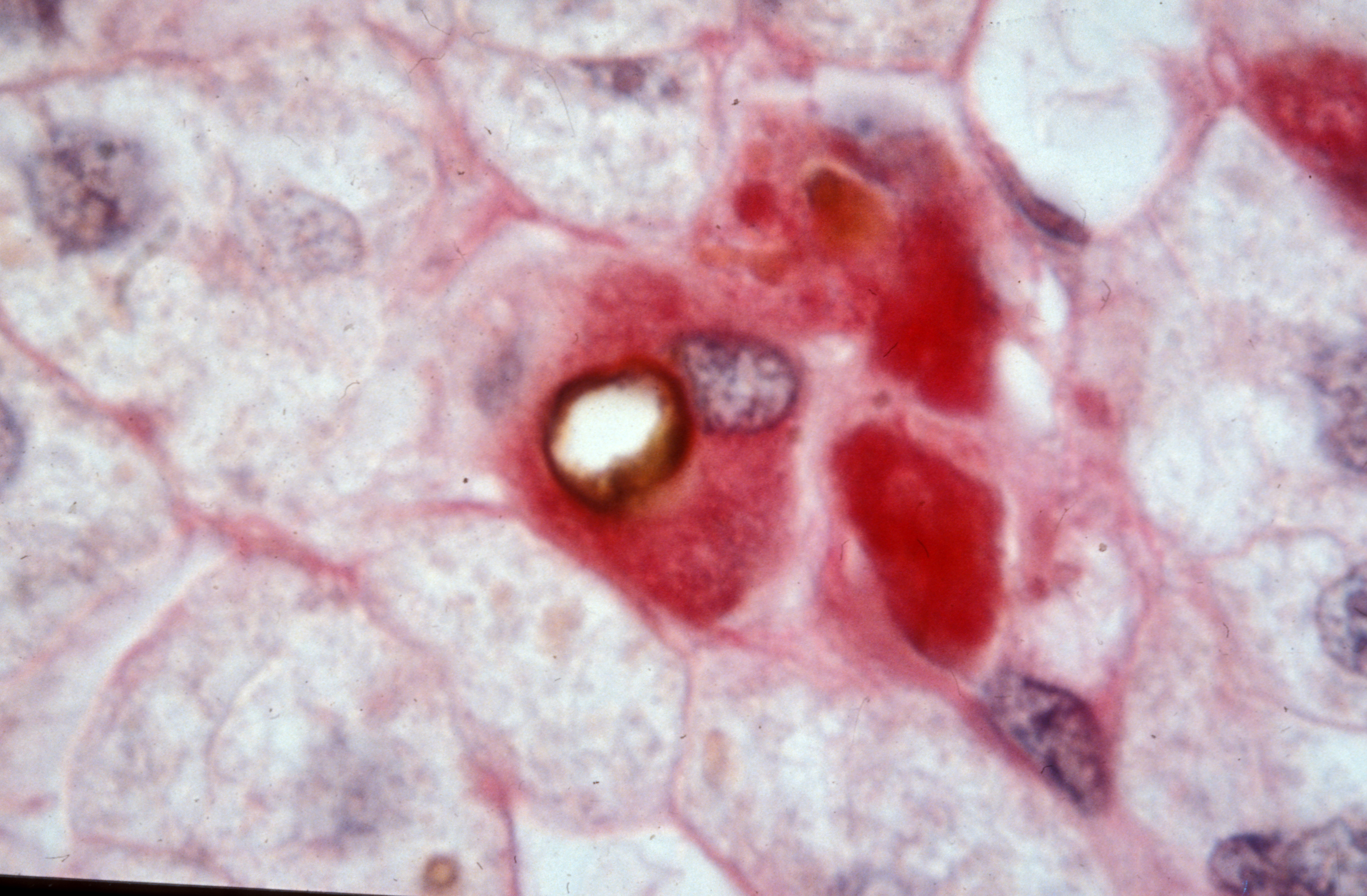
©Inserm/Jammart, Baptiste, autophagy vesicle (HCV induced)

A study published in The Lancet on 11 february 2019 shows that direct-acting antivirals have short-term clinical benefits in the treatment of hepatitis C virus infection. These results come from ANRS-funded interdisciplinary research conducted by clinicians,hepatologists, and epidemiologists of the Inserm, Sorbonne University and AP-HP and coordinated by Professors Fabrice Carrat and Stanislas Pol, and Dr Hélène Fontaine,[1] in 9895 patients of the ANRS CO 22 HEPATHER national cohort recruited in 32 centers in France.

The most recent treatments of hepatitis C virus (HCV) infection, the direct-acting antivirals (DAAs), are remarkably effective. Indeed, they eliminate the virus in almost all treated patients (95% in general) in 8 to 12 weeks. DAAs were first prescribed in France in 2014. Initially, priority was given to patients with advanced HCV infection, but from January 2017 DAA therapy was extended to all patients with chronic HCV infection.

The virologic efficacy of DAAs is well established, but until now prospective data on their clinical efficacy (ie, their impact on the progression of liver disease associated with HCV infection in real life) were scarce and related to highly selected patients or to patients from retrospective surveys. An ANRS-funded team of researchers has now compared clinical progression of HCV infection in patients receiving or not receiving DAA therapy. The researchers monitored clinical progression in “real life” in 9895 HCV-infected patients included between 2012 and 2015 in the ANRS CO22 HEPATHER cohort (see box below).

In these 9895 patients, who were followed up for a median period of 33 months(2), statistical analysis showed in the 7344 patients who received DAAs before the end of the study that this treatment was associated with reductions in mortality and in the occurrence of hepatocellular carcinoma (liver cancer). After adjustment for individual factors (age, disease staging, presence of other diseases, etc.), the patients treated with DAAs showed a 52% reduction in mortality risk and a 33% decrease in the risk of liver cancer compared with patients at a similar disease stage but not treated with DAAs.

“We could have expected these results. It seems logical that the elimination of the virus causing the damage is linked to clinical improvement,” said Prof Fabrice Carrat. “Our results show that these benefits are obtained soon after virologic control and the patients are no longer highly selected as in early trials. Our analysis reflects real-world efficacy for all patients.”    

The prolonged collection of data from these patients cured of an HCV infection will allow definition of the long-term benefit of DAA therapy and of the modalities needed for medical follow-up (How frequent should liver cancer screening be? How long after the cure? At what cost?). One of the difficulties sometimes encountered in this sort of study arises when patients who are cured are lost to follow-up. The “linking” of medical data from the patients of ANRS CO22 HEPATHER cohort to the national health data system (SNDS), which was validated by the French Data Protection Authority (CNIL) on 19 July 2018, should help researchers obtain exhaustive information on healthcare consumption by these patients over the long term.

This study was conducted in collaboration with the AFEF (French Hepatology Society) and supported by the ANR (French National Research Agency) in the framework of investments for the future and from industrial partners: Gilead, Abbvie, MSD, Janssen BMS, and Roche.

The ANRS CO22 HEPATHER cohort initiated in 2012 in collaboration with the French Association for the Study of the Liver now includes more than 21 000 patients (6500 infected by HBV, 14 600 by HCV, and 95 by both). The main purpose of this cohort, which is coordinated by Professors Fabrice Carrat and Stanislas Pol, and Dr Hélène Fontaine, is to measure the benefits and risks associated with different therapeutic modalities in the management of hepatitis B and hepatitis C and to identify their individual, virologic, environmental, and social determinants.

(1) Fabrice Carrat (Institut Pierre Louis d’Epidémiologie et de Santé Publique, Sorbonne université, Inserm UMS-20 – unité de santé publique – hôpital Saint-Antoine, AP-HP), Stanislas Pol (Département d’Hépatologie, Hôpital Cochin AP-HP; Université Paris-Descartes; Inserm, Institut Pasteur), Hélène Fontaine (Département d’Hépatologie, Hôpital Cochin, AP-HP).

(2) Median: the value that separates the higher half from the lower half of a data sample.

When HIV Uses Camouflage to Evade the Immune System

 Hybridation in situ des ARN du virus du sida (VIH) dans les cellules immunitaires infectées (HeLa CD4+). ©Inserm/Fournier, Jean-Guy, 1994

How is HIV able to escape the surveillance of the immune system within the very cells it infects? Researchers from Inserm, CNRS, Université de Montpellier and Université de Lorraine decided to take a closer look at one of these evasion mechanisms. They were able to observe the ability of HIV to “camouflage” its RNA within the infected cell using an intracellular enzyme. This research, published in Nature, provides new knowledge on the evasion mechanisms of HIV in the face of the innate immune system.

From the very first stages of a viral infection, the intracellular “radars” of the innate immune system make it possible to rapidly trigger an antiviral response through the secretion of type I interferons, proteins produced by the white blood cells to regulate and stimulate the immune response.

The Human Immunodeficiency Virus (HIV) targets the cells of the immune system and causes severe immune deficiencies responsible for AIDS. When HIV infects a cell, its genome made up of single-stranded RNA is transformed into DNA. It then inserts itself into the nucleus of the host cell where it incorporates into its genome. The success of these early stages depends on the ability of the virus to camouflage itself within the cell and pass unnoticed by its detectors, particularly those capable of recognizing the nucleic acids of its genome as foreign RNA.

Researchers from Inserm, CNRS, Université de Montpellier and Université de Lorraine decided to take a closer look at this mechanism through which HIV can evade the surveillance of the cells by using a system of camouflage. Within the cells, an enzyme called FTSJ3 is found which is capable of modifying some of the nucleic acids comprising cell RNA by adding to them a methyl group. This modification is a signature of self (set of molecules resulting from the expression of the individual’s genome, as opposed to non-self) which enables the detectors to recognize the cellular RNA as such in the human cells and to avoid its destruction by the immune system.

The research team was able to reveal that HIV recruits the FTSJ3 enzyme in order to methylate its own genomic RNA. The cellular detectors of foreign RNA then prove incapable of recognizing this duly “camouflaged” viral RNA as foreign and so cannot trigger the production of type I interferons within the cell to induce the immune response. The invisible virus is then free to transform its RNA into DNA, integrate the cell’s genome and continue the infection.

These findings represent a significant advance in the understanding of HIV infection by revealing a new evasion strategy of the virus when confronted with the innate immune system’s cellular detection system. Further elucidation of these evasion mechanisms could in the longer term enable the development of therapeutic and/or vaccine strategies aimed at modifying the virus so that it leads to the establishment of an antiviral response which, when this is early, enables the cell to implement an immune response and control the infection.

This research has received support from the European Commission, MSD Avenir, Fondation pour la Recherche Médicale and the French National Research Agency.

Cancer under pressure: visualizing the activity of the immune system on tumor development

Cancérogenèse : Surexpression de TRF2, marqué en vert, dans les vaisseaux tumoraux, marquage rouge, dans un cancer ovarien. ©Inserm/Wagner, Nicole, 2014

As tumors develop, they evolve genetically. How does the immune system act when faced with tumor cells? How does it exert pressure on the genetic diversity of cancer cells? Scientists from the Institut Pasteur and Inserm used in vivo video techniques and cell-specific staining to visualize the action of immune cells in response to the proliferation of cancer cells. The findings have been published in the journal Science Immunology on November 23, 2018.

Over time, the uncontrolled proliferation of tumor cells results in the accumulation of new mutations and changes to their genome. This gradual process creates significant genetic diversity among the cancer cells in any given patient. And although the cells in the immune system, especially T cells, are potentially able to eliminate these abnormal cells, tumor diversity can have a harmful effect, complicating the action of the immune system and rendering some therapies ineffective. Understanding this frantic race between tumor development and the immune response is key to the success of future immunotherapy techniques.

Scientists in the Dynamics of Immune Responses Unit (Institut Pasteur/Inserm), directed by Philippe Bousso, in collaboration with Ludovic Deriano, Head of the Genome Integrity, Immunity and Cancer Unit (Institut Pasteur), investigated how spontaneous immune responses to tumors influence this tumor heterogeneity. They demonstrated that the immune system can employ mechanisms to significantly reduce tumor diversity, favoring the emergence of more genetically homogeneous tumor cells.

In their study, the scientists marked each cancer cell subclone with a separate color in a mouse model. By monitoring these different colors they were therefore able to characterize the evolution of tumor heterogeneity in time and space. They were also able to observe the contacts between T cells and cancer cells and determine how some tumor cells are destroyed. Their research highlights the drastic impact the immune system can have on tumors by reducing their heterogeneity.


Visualizing the action of stained immune cells.
In this video, the tumor cells are shown in gray. The tumor-specific T-cells, in purple, come into contact with the cancer cells and destroy them. The killed cells are shown in blue. In green, the control cells circulate but do not kill the tumor cells. © Institut Pasteur / Philippe Bousso


Visualizing different clusters of cancer cell clones.
This video illustrates how tumor subclones, each marked by a different color (blue, orange and green), develop in the bone marrow. The vessels are shown in white. © Institut Pasteur / Philippe Bousso

The same impact on the heterogeneity of tumor cells has also been observed in response to immunotherapies that release the brakes on the immune system, an approach which was awarded the Nobel Prize in Physiology or Medicine this year.

This research shows that taking into account the interaction between immunotherapies and tumor heterogeneity could contribute to the development of optimum therapeutic combinations and sequences.

In addition to the organizations mentioned above, this research was funded by the Fondation de France, the French National Cancer Institute (INCa) and the European Research Council (ERC).

liver cancer and hepatitis C virus to maintain interest in screening programs cirrhotic patients cured of the virus infection with interferon or antiviral direct

Professor Pierre Nahon, the Hepatology Service of Hospital Jean Verdier, AP-HP and Professor Etienne Audureau the Public Health Service of the Henri Mondor Hospital, AP-HP with the University Paris 13, the Inserm and Sorbonne Paris Cité, reported the results of a prospective observational study within the cohort ANRS CO12 CirVir. The latter describes the evolution of patients with compensated cirrhosis due to infection by the virus of hepatitis C, enrolled in liver cancer screening programs or hepatocellular carcinoma (HCC) treated with interferon before 2014 direct antivirals ( DSA) since. The findings, published in the journal Gastroenterology in November 2018, show that if the liver cancer risk is greatly reduced after viral eradication in these patients, it still persists and justified to keep patients with viral cirrhosis C cured in screening programs. This study also confirms the benefits of virologic cure the risk of hepatic carcinogenesis regardless of the type of antiviral treatment.

The direct antiviral (DSA) directed against hepatitis C virus (HCV) have revolutionized the treatment of infected patients since they are made available in 2014 in France. Ensuring virological cure in nearly 100% of patients today at the cost of few side effects, their long-term benefit is still unknown. liver cancer risk or hepatocellular carcinoma (HCC) is the most feared complication in these patients when HCV-induced cirrhosis. Old data obtained before the era of AVD when based on very restrictive interferon treatments allowed to cure that less than 50% of patients have suggested a decreased risk of HCC in case of viral eradication.

The cohort ANRS CO12 CirVir is the oldest prospective cohort of patients infected with the hepatitis B and C supported by the ANRS [1] . Between March 2006 and December 2012, 1353 patients with cirrhosis, uncomplicated and histologically proven originating infection with the hepatitis C virus were enrolled in 39 French centers. All these patients were enrolled in HCC screening programs as recommended, with conducting a liver ultrasound every 6 months. Patients were followed until December 2016, which allows the analysis with a median decline of more than 5 years.

The researchers were able to analyze the incidence of HCC in the two eras successive therapy (interferon and DSA). The analyzes confirm that if the liver cancer risk is greatly reduced after viral eradication regardless of the type of treatment (it is divided by a factor of about 4), it nevertheless persists and justifies maintaining in screening programs patients with viral cirrhosis C cured. Data from the CirVir cohort reported in this study were also allowed to provide answers about the risk of liver cancer in AVD. The latter is indeed not increased compared to interferon era when a number of confounding factors are taken into account in the analyzes.

The cohort ANRS CO12 CirVir, by its prospective longitudinal nature, has in recent years to study the factors associated with the occurrence of major clinical events in patients with cirrhosis. Nearly twenty works based on data collected prospectively in all Hepatology Services of the territory of 10 years have been published in international journals, covering fields as wide as hepatocellular carcinoma, bacterial infections, extrahepatic cancers and cardiovascular diseases.

(1) The cohort ANRS CO12 CirVir multicenter prospective cohort of patients with viral cirrhosis B and / or C uncomplicated, was initiated in 2006 and included 1822 patients. Patient follow-up was completed in 2017 and a large part of them have now been included in the cohort ANRS CO22 HEPATHER .

Bile acid receptor controls hepatitis B virus replication

 Hépatite en voie de guérison : foyers de macrophages contenant de la bile et des pigments. ©Inserm/Hadchouel, Michelle, 1990

Researchers from CIRI – International Center for Research in Infectious Diseases in Lyon (Inserm, CNRS, ENS Lyon and Claude Bernard Lyon 1 University), supported by the ANRS, are demonstrating the link between activation of a bile acid receptor found in liver cells and the reduction in hepatitis B virus replication in mice infected with the virus. This study, coordinated by Professor Patrice André (Claude Bernard Lyon 1 University), was recently published in The FASEB Journal.

Despite the existence of an effective, well-tolerated vaccine, hepatitis B caused by infection with the hepatitis B virus (HBV) is still a global public health problem with 250 million people suffering from chronic infection, at high risk of developing cirrhosis of the liver and cancer. Current direct-acting antivirals significantly reduce the risk of complications, but are unable to control infection via the host’s immune defenses; hence, life-long treatment is required. A team of researchers supported by the ANRS has studied the role of the Farnesoid X receptor (FXR) in controlling HBV infection. FXR is a liver nuclear receptor activated by bile acids. Its main known function is to control the synthesis and excretion of bile acids in bile.

Previous research by the same research team had already suggested interdependence between bile acids and hepatitis B. An initial part of the study, conducted on in vitro cell models, published in October, was able to reveal a mechanism for this interdependence. A second part, conducted in vivo, confirms these results. The researchers were able to provide in vitro evidence that FXR is a host factor favoring HBV replication. This viral multiplication is suppressed following inhibition of FXR expression or during activation thereof via agonist molecules mimicking the action of bile acids.

The researchers then evaluated the effects of treatment with agonists with a view to activating FXR in an HBV-infected mouse model. They then observed that HBV replication was less active in the treated mice compared to the control mice. This result demonstrates, for the first time in vivo, that activation of FXR by an agonist molecule may lead to suppression of HBV replication.

Lastly, scientists also observed that young HBV-infected mice did not respond to treatment, in contrast to adult mice.

These findings suggest that the immaturity of the bile acid structures could lead to an increased risk of chronic HBV infection observed in neonates and young children.

This study introduces a new concept, namely that the role of bile acid metabolism is more far-reaching than expected and extends to the control of at least one viral infection,” points out Patrice André. “FXR agonists could offer an additional therapeutic approach to relieve the burden of life-long treatments for these HBV-injected patients.”

Inserm Transfert has filed a patent for this research. “In order to bring these advances and a new agonist, which we have identified, into the clinical sphere, we have created a start-up, EnyoPharma, which has acquired the patent license,” explains Patrice André, adding: “The results of a phase I trial will be available shortly, and a phase II trial is in the preparatory stages.”

Outbreak of multidrug-resistant tuberculosis undetected by standard tests

Fluorescence microscopy of BCG vaccine. ©Inserm/Latron, Patrice, 2017

Amid a plan announced by the United Nations to eradicate tuberculosis by 2030, a new study has revealed the emergence of multidrug-resistant strains of the disease which go undetected by WHO-endorsed tests. These findings, from an international research team co-directed by CNRS researcher Philip Supply at the Center of Infection and Immunity of Lille (CNRS/INSERM/Institut Pasteur de Lille/Université de Lille), are published in the 17 October 2018 edition of The Lancet Infectious Diseases. This follows another article, published in the 26 September edition of The New England Journal of Medicine, proposing a new algorithm to detect resistant strains of tuberculosis.

On 26 September, the United Nations announced a plan to raise $13 billion annually for the fight to eradicate tuberculosis by 2030. With 10 million new cases and 1.6 million deaths in 2017, it is the most common infectious disease in the world, ahead of HIV.

In over 450,000 new cases of antibiotic-resistant tuberculosis that likely appeared, only 25% were detected. A study by an international research team1 co-directed by Philip Supply, a CNRS researcher at the Center of Infection and Immunity of Lille (CNRS/INSERM/Institut Pasteur de Lille/Université de Lille), has underlined this serious problem of under-detection, in South Africa in particular.

The findings, published in The Lancet Infectious Diseases, show that certain South African isolates of Mycobacterium tuberculosis (the bacterium which causes the disease) carry a specific combination of mutations which make them resistant to the two primary first-line antibiotics prescribed: rifampicine and isoniazide.

This combined resistance goes undetected by the standard tests endorsed by the World Health Organization: the gene region carrying a particular mutation causing rifampicine resistance is not included in the DNA test, and the resistance to the treatment due to this mutation is not detected in cultures.

This omission leads to unsuccessful first-line treatments in patients, increased mortality and contagion, and the development of additional antibiotic resistances. Researchers especially detected the presence of mutations probably causing decreased sensitivity to bedaquiline, the newest molecule used to treat cases of multidrug-resistant (MDR) tuberculosis. These mutations appeared immediately following its launch in the country from 2013 on.

This was discovered thanks to a new MDR screen test developed by Genoscreen2 together with P. Supply. Unlike standard DNA tests, this one analyses a wide panel of target genes in the bacteria and can identify resistance to over a dozen antibiotics simultaneously. These results are obtained in as little as one to three days, compared to the weeks needed for cultures. The test will help solve the problem of under-detection of MDR tuberculosis. It will benefit from a new algorithm for the detection of resistance mutations, the effectiveness of which has been detailed in an article published in The New England Journal of Medicine by another consortium (CRyPTIC)3 in which Dr Supply and Genoscreen took part. This study was based on an analysis of 10,000 genomes, making it one of the biggest microbial genome sequencing projects conducted to date.  

[1]National Health Laboratory Service, Dr George Mukhari Tertiary Laboratory, Pretoria, Sefako Makgatho Health Sciences University, Pretoria, and Gauteng Department of Health, Hatfield, South Africa; National Reference Laboratory, Ministry of Health, Mbabane, Swaziland; Forschungszentrum Borstel and German Center for Infection Research, Borstel Site, Borstel, Germany; Institute of Tropical Medicine and University of Antwerp, Antwerp, Belgium; Université Catholique de Louvain, Brussels, and Katholieke Universiteit Leuven, Leuven, Belgium

[2]Company specialised in genomics, based at the Institut Pasteur de Lille.

[3]See http://www.crypticproject.org/wp-content/uploads/2018/09/Prediction-of-Susceptibility-to-First-Line-Tuberculosis-Drugs-by-DNA-Sequencing.pdf

NONO, “The Red Flag System” That Detects HIV

Illustration du virus HIV ©Adobestock

There is not one but several types of HIV. Although HIV-1, which is the most common, wreaks havoc in infected populations, this is not the case for HIV-2 which less frequently leads to the development of AIDS. But why does the immune system do a better job of fighting this version of the virus? Researchers from Inserm and Institut Curie looked at this question. Researchers from Inserm and Institut Curie identified the NONO protein, a detector which is more sensitive to HIV-2 and responsible for direct recognition of the virus by the immune system. This work, published in the journal Cell, provides a better understanding of the natural control of HIV and paves the way for new progress in the search for a vaccine for this virus.

AIDS develops when the immune system of an HIV-positive individual becomes unable to fight infection and becomes dramatically weak. The majority of people infected and not treated develop fatal AIDS. But in some cases AIDS does not develop in certain untreated HIV-positive individuals. This explains the existence of several forms of HIV.

Although HIV-1, which affects 25 million people, without treatment leads to AIDS in 99% of cases, this is not the case for HIV-2. This particular form of HIV is very close to HIV-1 but differs in terms of genetics. It is found mostly in West Africa and affects 1 million people. HIV-2 leads to the development of AIDS in fewer than 25% of cases, has no impact on the life expectancy of most people infected, and proves to be difficult to transmit to others. Furthermore, HIV-positive individuals with the HIV-2 form of the virus, and who also contract HIV-1, show improved resistance to the latter.

Researchers from Inserm and Institut Curie in Unit 932 Immunity and Cancer (Inserm/Institut Curie/PSL University/Paris Descartes University) studied the reasons for the immune system’s better control of HIV-2.

In 2010, this research team had already shown that dendritic cells – the immune system’s “sentinel” cells – were able to detect HIV-2 much more efficiently than HIV-1. In order for the immune response to be effective, there needs to be good immune recognition.

Based on this observation, the researchers sought to understand the molecular mechanisms involved in the dendritic cells’ recognition of HIV-2, and to find out why this recognition is effective in comparison to that of HIV-1.

They thus discovered that the NONO protein, located in the dendritic cells, acted as a detector able to recognize the internal casing (or capsid) of HIV-2 a lot better than that of HIV-1, and as a result to trigger an immune response to fight the virus.

The capsid – which surrounds the genetic material of viruses – is made up of proteins, and NONO is apparently able to recognize a specific protein pattern of the capsid of HIV-2.

This study provides a better understanding of the natural mechanisms involved in infection control by HIV. According to Nicolas Manel, Inserm researcher in charge of the study: “the next step in this research project is to understand how this detection system works at the molecular level and how this detection triggers the immune response. We are developing innovative vaccine strategies in the lab, and this discovery paves the way for the new studies needed to develop a new generation of vaccines capable of “imitating” the HIV-2 capsid, and as a result triggering an immune response in people infected with the HIV-1 virus.”

20% of reactions to radiologic contrast media are real allergies

©Adobestock

A team of Pole-Imaging Research Explorations-European Hospital Georges Pompidou AP-HP, Paris Descartes University and INSERM led by Professor Olivier Clément, and a team from Caen University Hospital and the University of Caen Normandy, led by Dr Dominique Laroche, conducted the first national prospective multicenter study on allergic reactions to contrast media in radiology. 31 centers in France bringing together radiologists investigators, allergists, anesthetists and biologists have investigated 245 cases of hypersensitivity to contrast media.

Promoted by the AP-HP, the study, funded by the Hospital Regional Program Clinical Research, 2003, shows that allergy is responsible for over 20% of hypersensitivity reactions to contrast media and recommends that patients diagnosed allergic, having a high risk of recurrence, are subject to monitoring based on skin tests performed in an allergist specializes in drug allergy.

This work was published in the journal EClinicalMedicine the Lancet in its issue of July 2018.

In radiology, patients may experience immediate hypersensitivity reactions to iodinated contrast media (for scanners) and gadolinés (for MRI) is injected them in the examination. The reactions such as hives, angioedema, bronchospasm, hypotension and anaphylactic shock. Severe reactions, rare, occur within minutes after injection and require from the imaging team a quick diagnosis and management.

For iodinated contrast agents, reactions have long been falsely labeled “iodine allergy” and mistaken reactions to seafood or skin disinfectants.

But the real allergy to contrast medium is diagnosed by elevated plasma markers tryptase and histamine in the first hour of reaction and intradermal skin tests to make between six weeks and six months after it. The few retrospective studies post on the performance of this type of skin test showed that between 13 and 65% of the responses were truly allergic in origin, according to the populations tested. However, these studies suffered from a lack of clinical data, in particular the name of the injected product, or incomplete or late tests performed, or they mixed the immediate reactions and delayed reactions.

A team of Pole-imaging research explorations-European Hospital Georges Pompidou AP-HP, Paris Descartes University and Inserm, led by Professor Olivier Clément, and a team from Caen University Hospital and the University of Caen Normandy, led by Dr Dominique Laroche studied prospectively immediate hypersensitivity reactions to iodinated products and gadolinés. This multicenter study was conducted in 31 French centers equipped to perform skin tests six weeks to six months after a reaction.

After receiving contrast media for radiology review, 245 patients with immediate reaction took a blood sample in the first hour after it to measure the levels of histamine and tryptase in their plasma. They are seen to offer six weeks after a visit to the allergist to test all existing contrast agents (10 gadolinés iodinated or 5).

Skin testing revealed three types of reactions: allergic (if positive test contrast diluted); potentially allergic (if positive test only to pure product) and nonallergic. They identified 41 patients allergic to iodine products and 10 patients allergic to gadolinés products.

The results showed that over the reaction was severe, the more allergic mechanism revealed by the skin test was frequent : 9.5% in the skin reactions; 22.9% in the moderate reactions; 52.9% in reactions involving life-threatening, and 100% when there was cardiac arrest. Similarly, the levels of histamine and tryptase plasma increased with the severity of the reaction. The presence of cardiovascular signs were also very strongly linked to allergic mechanism.

The group of potentially allergic patients showed clinical symptoms and histamine assays and tryptase intermediate between the group of patients allergic and non-allergic people. This suggests that some of them are truly allergic to the contrast material.

The teams also studied cross-reactions with other different contrast the one responsible for the reaction products: 62.7% of patients had allergic cross-reaction to one or more pure products tested.

This study shows that 21% of radiology hypersensitivity reactions are actually caused by an allergy to contrast media.

Allergic patients have a greater risk of recurrence if their is reinjected contrast agent giving a positive skin test.

Patients exhibited severe symptoms (anaphylactic or cardiovascular symptoms) should benefit from a dose of histamine and tryptase the waning of resuscitation and allergy testing in the six months to determine the allergic or not of their reaction, and especially to know which products will be shown against or authorized for future injections.

Deciphering the link between skin allergies and the gut microbiota

Crédits: AdobeStock

Over the last few years, scientists have discovered connections between gut microbiota imbalances and various diseases. Now, in a study using mice, biologists from the CNRS, INSERM, and Claude Bernard Lyon 1 University—together with colleagues from the Institut Pasteur de Lille and the NIH (USA)—have revealed a surprising relationship between a viral detection system, the composition of the gut microbiota, and the development of skin allergies.[1] Their findings, published in PNAS (September 24, 2018) suggest potential new therapies.

The number of microorganisms hosted in our digestive tracts is 10 to 100 times greater than that of all the cells that make up our bodies, and the delicately balanced ecosystem they constitute may be modified by our diet and medication. Epidemiological data of various kinds suggest a link between changes in gut microbiota composition and the development of allergic diseases, like eczema, at body sites far removed from the intestine. But an explanation for this association had been lacking until now.

At the International Center for Infectiology Research (CNRS / INSERM / Claude Bernard Lyon 1 University / ENS de Lyon)—or CIRI—a team led by two researchers from the CNRS focused their attention on mice deprived of the MAVS gene, which plays a key role in the detection of viruses by the immune system. They noted an altered gut microbiota and severe allergic skin reactions in these mice. To demonstrate a relationship between the two phenomena, the researchers transferred the altered microbiota to normal mice. The latter in turn developed severe allergic reactions, showing that the transplanted gut bacteria were responsible.

Furthermore, the biologists revealed that such modification of the gut microbiota led to greater intestinal permeability, which allowed certain intestinal bacteria to migrate to the spleen and lymph nodes and increased the severity of allergic skin reactions.

These findings shed light on the unexpected role played by an antiviral protein (MAVS) in the maintenance of gut microbiota equilibrium. By showing that changes in the gut microbiota exacerbate the allergic response in the skin, this research sets the stage for the development of new therapies. In the not so distant future, might we treat eczema, or enhance already existing treatments, by acting on the microbiota? This approach is already being investigated for other diseases, like cancer.

[1] NIH: National Institutes of Health. This research was conducted at the International Center for Infectiology Research (CNRS / INSERM / Claude Bernard Lyon 1 University / ENS de Lyon), in collaboration with the Center for Infection & Immunity of Lille (CNRS / INSERM / Lille University / Institut Pasteur de Lille) and with the participation of the Cancer Research Center of Lyon (CNRS / INSERM / Claude Bernard Lyon 1 University / Centre Léon Bérard / Hospices Civils de Lyon).

Major advances in the diagnosis and treatment of allograft rejection

Crédits: Allogreffe d’aorte abdominale décellularisée, Inserm/Allaire, Eric

Prof. Alexander Loupy, Hospital Necker Children AP-HP and Prof. Carmen Lefaucheur, the Saint-Louis Hospital AP-HP and the University Paris Diderot in the Cardiovascular Research Center (Inserm / Paris Descartes University), showed, in an article published in the journal   New England Journal of Medicine September 20, 2018, the latest advances and applications of artificial intelligence carried out in the field of transplantation, including the diagnosis and the treatment of allograft rejection.

These interdisciplinary work focused on transplant patients of heart, kidney and lung. They have helped to change the past five years, three times, the International Classification of rejection. They contribute to improving the management of transplant patients on diagnosis and treatment plans.

The transplantation has become the treatment of choice at the onset of organ impairment. 120,000 new organ transplants are performed each year worldwide, but only one million people living with a functioning graft. This finding can be explained by a lack of improvement in graft survival in recent decades and a number of available organs sometimes limited.

The rejection of the organ caused by the production of antibodies by the recipient patient is recognized as one of the main causes of failure of a transplant. A better understanding of the mechanisms of this rejection now allows to diagnose accurately and offer a personalized therapeutic approach.

A multidisciplinary approach involving clinical specialists, pathologists, immunologists transplantation, epidemiologist and statisticians, has been developed in close collaboration with Professor Xavier Jouven, head of the cardiology department of the European Hospital Georges Pompidou AP-HP and the team “cardiovascular Epidemiology and sudden death” of the cardiovascular research Center Inserm and Université Paris Descartes, to evaluate this rejection to the population level. New diagnostic categories were established and patient groups likely to lose their accelerated graft were identified and defined.

The allograft rejection may for example be detected by

> An integrative analysis of multiple biomarkers (reactive antibodies directed against the donor, inflammatory markers); 

> A detailed study of the transplanted organ (identification of gene expression and characterization of cells infiltrating the graft that may cause rejection of the short or medium / long-term allograft).

Work by a team AP-HP / Inserm / Paris Descartes, and coordinated by Professor Alexander Loupy have thus demonstrated that the ultra-fine analysis of genes expressed by cells of the heart via a new technique called “molecular microscope “identifies precisely and patients with early beginnings of heart transplant rejection. (Read more:  >> Diagnosis of transplant rejection in heart: a French team shows the interest of a new method, molecular microscope (March 2017) ). Other more recent studies have demonstrated the usefulness of algorithms to improve efficiency and performance of clinical trials in transplantation *.

Finally, the interest in this approach to artificial intelligence “machine learning” applied to transplantation was realized by obtaining two funding within the hospital research future investment program (RHU ) and the European program for research and innovation 2020.

This research thus open the way to a medicine of the future in which the mathematical algorithms will be used for daily monitoring of patients and the medical decision making. A concrete example is the development of a predictive tool for the survival of kidney transplants.

* Complement-Activating Anti-HLA Antibodies in Kidney Transplantation: Allograft Gene Expression Profiling and Response to Treatment. J Am Soc Nephrol. 2018 Feb; 29 (2): 620-635. doi: 10.1681 / ASN.2017050589. Epub 2017 Oct. 17.

fermer