Menu

When Infection Strikes, Our Brain and Immune System Join Forces

©Adobestock

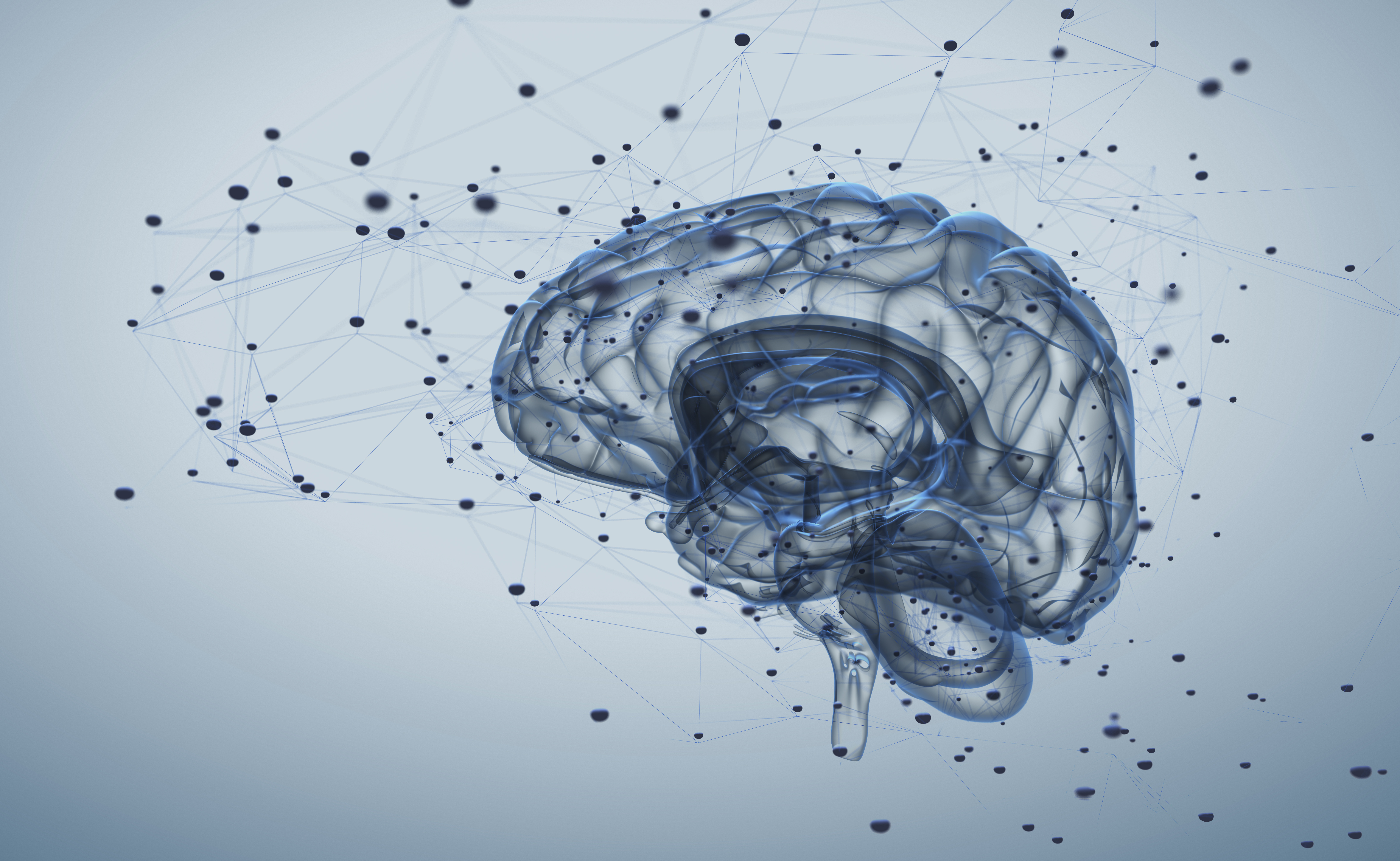
When infection strikes, what if our immune system was not alone in the fight? What if its major ally was in fact the brain? Researchers from Inserm, CNRS and Aix-Marseille University (AMU) have observed mechanisms of cooperation between the nervous system and the immune system in the response to pathogenic aggressions. This research, published in Nature Immunology, reveals the role of the brain in regulating the inflammatory reaction induced by the immune system in the event of infection and its protective effect against a potential self-destructive exacerbation of that inflammation.

In the event of infection with a virus or other pathogenic organism, the immune system springs into action to eliminate the infectious agent. The immune cells release inflammatory molecules called cytokines, which are responsible for the inflammation process necessary to fight the dissemination of pathogens in the body. There are times, however, when the inflammatory reaction is excessive and toxic to the body. It can provoke lesions in the infected organs which, when too severe, can lead to death.

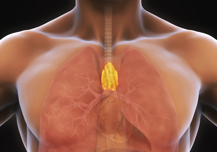
Previous studies have shown that, in the event of infection, the brain is mobilized to regulate the inflammatory reaction. When it detects the cytokines produced by the immune cells, the brain induces the blood secretion of hormones known to be negative regulators of inflammation: glucocorticoids. The properties of these hormones are widely used in medicine in many pathological conditions, but little is known about their specific mode of action.

In this context, researchers from Inserm, CNRS and Aix Marseille University (AMU) at the Center of Immunology Marseille-Luminy (CNRS/Inserm/AMU) studied, in mice, the mechanism of action of the glucocorticoids produced following activation of the brain in controlling the intensity of the inflammatory reaction caused by viral infection.

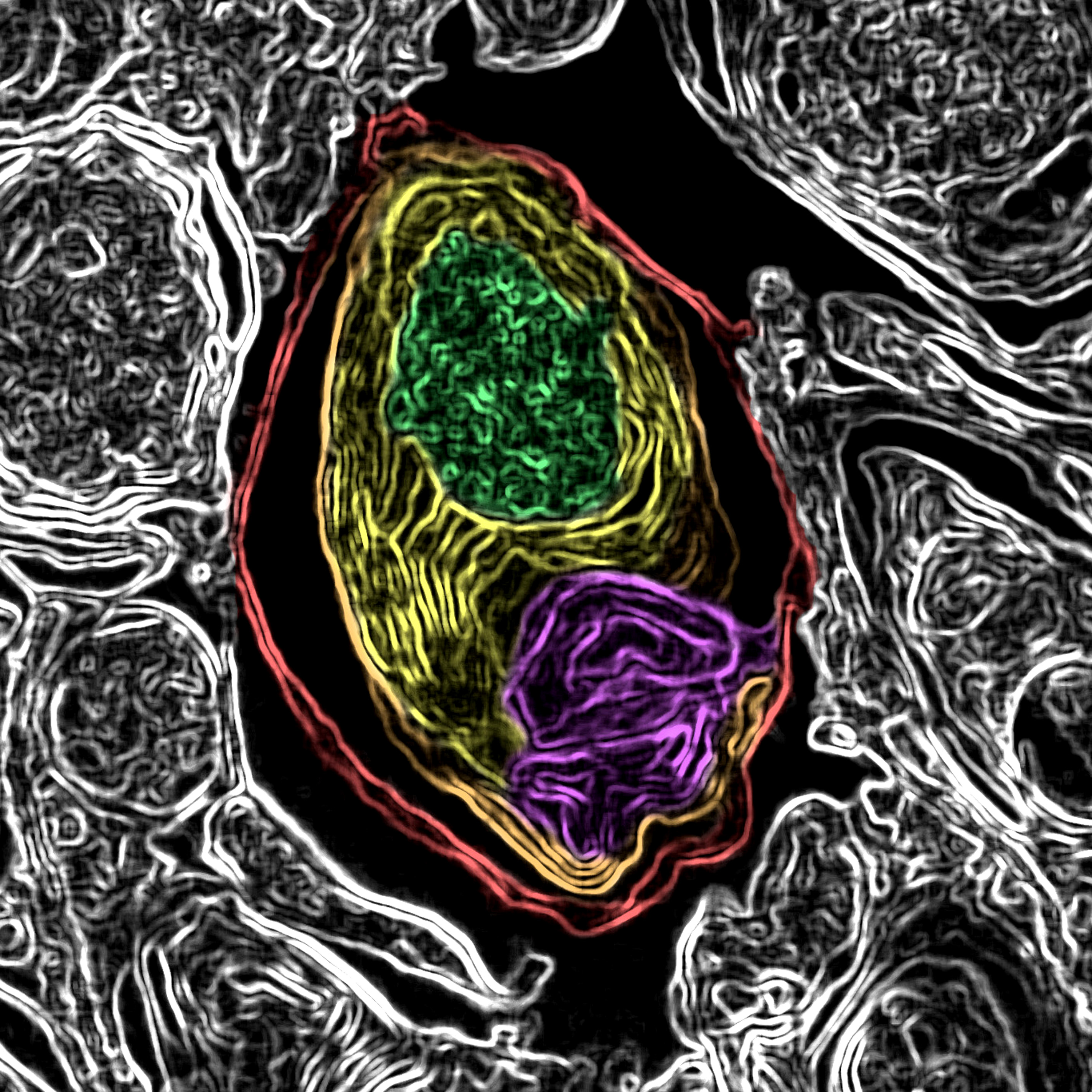
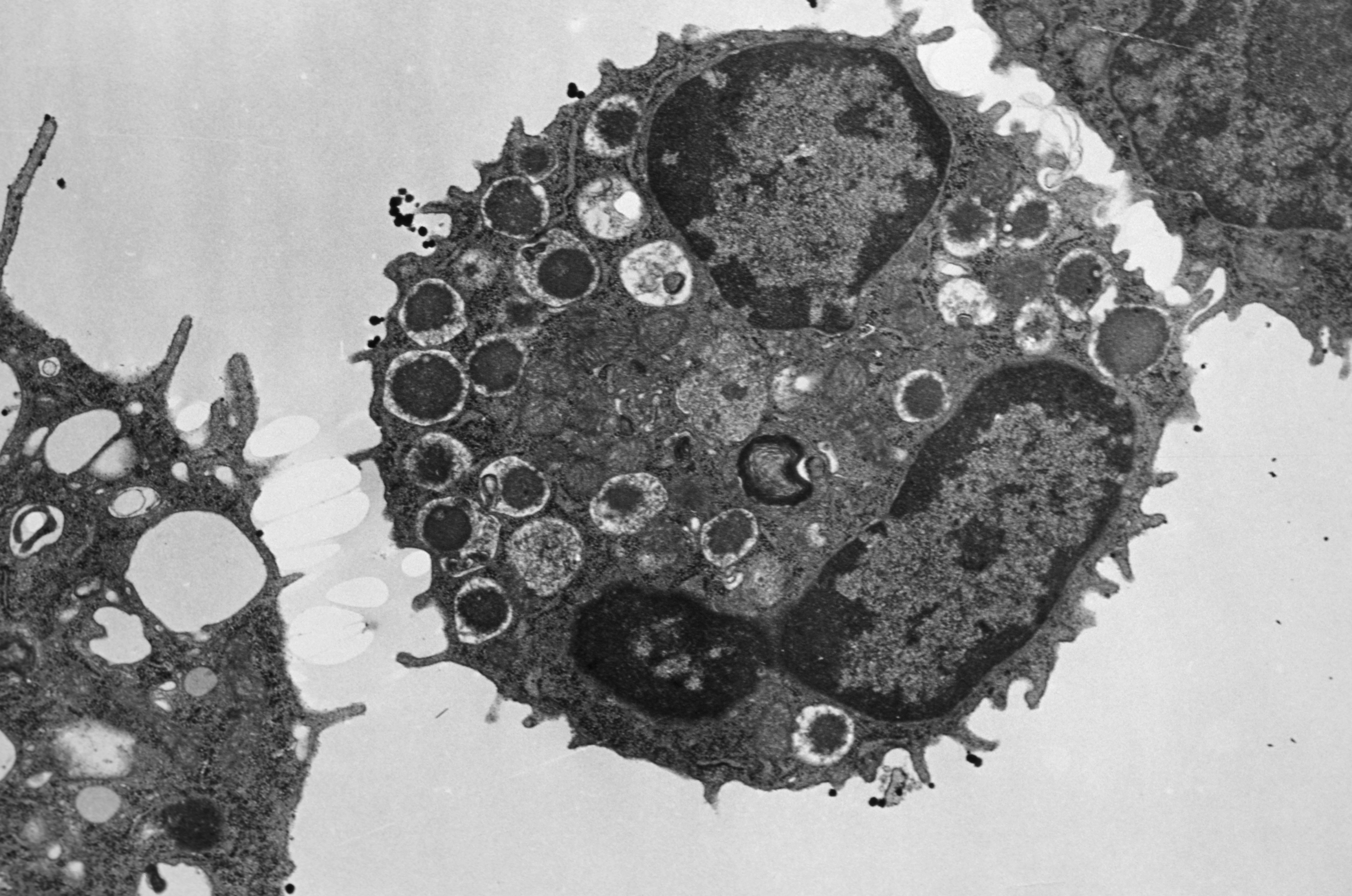
Their findings show that glucocorticoids regulate the activity of a population of immune cells which produce inflammatory cytokines and exert a major antiviral and anti-tumor action: natural killer (NK) cells.

These cells possess a receptor which is activated by the glucocorticoids produced after the infection. This activation leads to the expression on the surface of the NK cells of a molecule called PD-1, which is attracting considerable interest within the medical community and is targeted in many cancer treatments due to its inhibitory action on the activity of the immune cells that express it.

The researchers observed that mutant mice which do not express the glucocorticoid receptor in their NK cells were more likely to develop a severe hyper-inflammation reaction and die in the event of an infection. This research demonstrates that expression of the glucocorticoid receptor by the NK cells is necessary to regulate the intensity of the inflammation so that the response against the virus does not become toxic to the body. In addition, the study shows that this regulation is governed thanks to the inhibitory effect of PD-1 which, in the infectious setting, limits the production of inflammatory cytokines by the NK cells.

According to Sophie Ugolini, Inserm researcher and study director: “The most unexpected part of our discovery was that this regulation prevents the immune system from going into overdrive and destroying healthy tissues while fully maintaining its antiviral properties necessary for the effective elimination of the virus. “

This study could enable the development of new therapeutic strategies which would target this regulation pathway. Aside from infections, the researchers especially hope to explore the potential role of this regulation pathway in some cancers.

One quarter of deaths and cases of serious after-effects related to meningitis in children can be avoided by applying the immunization schedule

© Chris Benson on Unsplash 

In a context of increasing vaccine hesitancy, researchers from Inserm and pediatricians from the Nantes and Grand-Ouest university hospitals together with the Paris public hospitals (AP-HP) sound the alarm concerning the consequences of severe bacterial infections in children. In a study published in Paediatric and Perinatal Epidemiology, the researchers showed, over a period of 5 years, that 25 % of deaths and 25 % of serious after-effects occurring in children with a severe bacterial infection (primarily meningitis) could have been avoided by simply applying the immunization schedule, notably against meningococcus and pneumococcus.

Bacterial infections are common and for the most part efficiently dealt with by our natural defenses, with an occasional helping hand from antibiotics. Nevertheless, children –and more particularly babies– form a vulnerable population in the face of some severe bacterial infections (meningitis, purpura fulminans, septic shock, etc.) which can lead to serious after-effects (paralysis, sensory deficit –particularly hearing loss–, hydrocephalus, epilepsy, amputation) and even death. While vaccines against the principal bacteria responsible for these infections exist (meningococcal C and pneumococcal conjugate vaccines), defiance towards vaccination, a growing phenomenon in France, is leading to a lack of complete vaccination in some children, putting them at risk of serious after-effects or death.

That is why researchers from Inserm and pediatricians wished to determine, in children having contracted a severe bacterial infection, the proportion of those which could have been avoided had immunization taken place according to the official recommended schedule.

In France’s Grand-Ouest administrative region, all children aged 1 month to 16 years admitted to pediatric intensive care for severe bacterial infection or who died prior to admission were prospectively included from 2009 to 2014 in a study coordinated by Inserm and funded by the Ministry of Health. An infection was considered theoretically preventable by vaccination if the child had either not been vaccinated or had been incompletely[1] vaccinated and that the bacterial strains identified in his or her body were targeted by the vaccines recommended at the time of infection onset.

According to the results of this study, meningococcus and pneumococcus were the principal bacteria causing severe infections in children (65 %), responsible for 71 % of the deaths and close to half of the cases of serious after-effects, despite the introduction of meningococcal C and pneumococcal conjugate vaccines to France’s vaccine schedule in 2002 and 2009.

Only 39 % of the children were correctly vaccinated against these bacteria, meaning that 61 % had either been incompletely vaccinated or not at all. More important still, according to the Inserm study, 25 % of the deaths and 25 % of the cases of after-effects could have been prevented by complete and timely immunization according to the recommended schedule.

In France, while vaccination against the two principal bacteria responsible for these severe infections is now mandatory for all children born after January 1, 2018 (meningococcal C and pneumococcal conjugate vaccines), the majority of the deaths related to meningococcus C occurred in children over 2 years of age who had not had their catch-up vaccination. These children are not currently concerned by the obligation to get vaccinated, which is why it is fundamental to apply the current catch-up recommendations.

As such, according to the researchers, “morbidity-mortality rates related to severe vaccine-preventable infections could be reduced by one quarter with better implementation of immunization programs in France. Such information could help enhance the perception of vaccine benefits and fight vaccine hesitancy”.

[1] “Incomplete vaccination” status was defined as the receipt of fewer injections than those recommended for the age of the child, with authorized delays of 15 days for the pneumococcal primary vaccination and 1 month for the pneumococcal booster and meningococcal C vaccination (according to Gras P, 2017)

Genetic control of human thymic function: Needle in a haystack

A study conducted by a group of researchers from Paris Diderot University, INSERM and the Institut Pasteur reveals the existence of a genetic factor influencing the function of the human thymus. The results of the study, part of the Laboratories of Excellence project Milieu Intérieur coordinated by the Institut Pasteur, are published in the journal Science Translational Medicine on September 5, 2018.

Our immune system has developed different strategies to respond to external pathogens and to destroy emerging cancer cells while keeping autoimmunity in check. Among those strategies, adaptive immunity enables to keep a memory of an initial encounter, with increased recall responses after subsequent antigenic challenges. This is the basis of vaccination. Adaptive immunity is dependent on T lymphocytes present in lymphoid organs (spleen, tonsils, lymph nodes and mucosa associated lymphoid tissue) which patrol the body. Their name, « T » cells, derives from the thymus gland, the organ where they are produced.

The thymus is therefore a critical organ in health and disease, acting throughout life for the generation of new T cells. Its function and output is high in newborns and children but decreases with age, accounting in part for higher infection rates and cancer incidence in the elderly, both major public health issues in an ageing world. However, apart from age, environmental or genetic factors that may govern thymic function in humans remain unknown.

The objective of the Milieu Intérieur Consortium (http://www.milieuinterieur.fr) is to describe in a large-scale multidisciplinary approach the immune variation within the French population. The consortium enrolled 1000 healthy adults (500 men, 500 women aged 20 to 69 years) and gathered an expansive collection of biological specimens together with extensive data on medical history, vaccination and lifestyle. The scientists leveraged samples from this cohort to see how thymic output, known to decrease over time, is affected by other factors.

In addition to seeing sex-dependent differences, thymic function being higher in women of all ages, the most striking result from the study was to identify a genetic factor influencing the thymic function in the general population. A genome-wide association study revealed variants that were associated with thymic output, which was confirmed in an independent cohort of 612 individuals (MARTHA cohort), as well as in a mouse model. This common genetic variation is located in the T cell receptor locus itself and associates with marked differences in the levels of thymic function among individuals. Taking into account age, sex and this genetic variation, we could define a “biological age” of the thymus in men and women. Between individuals of the same chronological age, a difference of up to 18.5 years in “thymic age” depending on sex and genetics could be calculated. For instance a 58.5 year old women and a 40 year old man of different genotypes may have a similar “thymic age”, possibly explaining some of the differences in immune responses between healthy individuals.

This discovery may have a direct impact for the advancement of precision medicine in all situations where T cell generation is key, such as in the context of allogeneic stem cell transplantation, HIV disease or vaccine development. It would be also interesting to study the association of this genetic variation with the incidence of autoimmune diseases, which is higher in women.

2018 Ebola Epidemics: What is the Latest in Vaccine Research?

©Inserm/Delapierre, Patrick, 2018

Since July, a new outbreak of Ebola virus disease was identified in the Democratic Republic of the Congo – the second since May.  In an attempt to halt the outbreak, a new vaccination campaign has begun in the affected region of North Kivu. It is against this background that researchers from the consortium PREVAC (Partnership for Research on Ebola VACcination) take stock of advances in Ebola vaccine research in The Lancet and point to the necessity to continue clinical trials.

Since the end of July, the Democratic Republic of the Congo has been facing its second Ebola outbreak this year, following the one in May.  These outbreaks, which can be added to the one of 2017 in the same country, and the widespread outbreak that occurred in Guinea, Liberia and Sierra Leone from 2013-2016, show the extent to which the risk of Ebola re-emergence is real. While no approved treatments or vaccines exist right now, some vaccines are, however, reaching advanced stages of development. Ebola vaccine research must continue because, when combined with effective public health measures, it is an essential element in preventing and responding to future epidemics.

In 2013, Inserm and its Aviesan partners founded REACTing, a multidisciplinary consortium bringing together research groups and laboratories of excellence in order to prepare for and coordinate research to combat the health crises linked to emerging infectious diseases. It was in this context that, Inserm, the National Institute of Allergy and Infectious Diseases and the London School of Hygiene & Tropical Medicine, in collaboration with the health authorities and scientists of four Ebola-stricken countries (Guinea, Liberia, Sierra Leone and Mali) formed the international consortium PREVAC (Partnership for Research on Ebola VACcination) and launched in 2017 a large-scale clinical trial of three Ebola vaccination strategies. The objective of the trial, whose partners also include the universities of Bordeaux and Minnesota, the NGO ALIMA, and three pharmaceutical companies (Janssen Vaccines & Prevention, B.V., part of the Janssen Pharmaceutical Companies of Johnson & Johnson; Bavarian Nordic; and Merck Sharp & Dohme Corp., known as MSD outside the U.S. and Canada), is to identify the most promising vaccination strategies for protecting individuals from Ebola.[1]

Two promising vaccines regimens are being trialed as part of PREVAC, including rVSV∆G-ZEBOV-GP which has been used since August 9, 2018, in response to the new epidemic in the Democratic Republic of the Congo. The second, Ad26.ZEBOV and MVA-BN-Filo prime-boost, is also under evaluation in PREVAC. The latter is also being tested in the EBOVAC project, of which Inserm is a partner.

On August 10, 2018, in an article published in The Lancet, PREVAC researchers take stock of Ebola vaccine research. They note that some key questions remain to be studied via various vaccine strategies. Researchers are concentrating on gaining a deeper understanding of the immune response to the Ebola vaccines, the question of the rapidity and durability of the immune response (and therefore the potential protection) in vaccinated individuals, the safety and the ability of the vaccine to trigger an immune response in children, as well as the nature of the responses in people with immunodeficiencies and pregnant women.

According to the PREVAC team, various vaccine strategies and scenarios need to be researched to identify the most effective way to prevent and respond to future Ebola outbreaks.

Post-exposure vaccination, targeted preventive vaccination for people who have come into contact with infected individuals, as well as preventive vaccinations for at-risk populations, such as healthcare and frontline workers, and possibly residents of areas regularly affected by outbreaks, are some of the strategies to be investigated.

Addressing these challenges requires the pursuit of collaborative partnerships centered on Ebola research. It also requires increasing the local population’s trust in the vaccines and vaccination campaigns. Indeed, the adherence of those enrolled and the engagement of the community throughout the clinical trial process is fundamental to success.  Bringing social sciences into clinical research makes it possible to increase the population’s trust and its involvement in the trials.


[1]All the information on the PREVAC clinical trial can be found on the dedicated Inserm page:

https://www.inserm.fr/actualites-et-evenements/actualites/ebola-plus-2-000-personnes-deja-incluses-dans-essai-vaccinal-prevac

Cost-effective universal screening for hepatitis C in France

Crédits: Inserm/Jammart, Baptiste

An estimated 75 000 people in France are unaware they are infected by hepatitis C virus. An ANRS-funded study by Sylvie Deuffic-Burban, a research associate at IAME (Infection, Antimicrobials, Modeling, Evolution) (Inserm – Université Paris Diderot – Université Paris 13), and her team shows that a universal screening strategy applied to hepatitis C is cost-effective and improves life expectancy in those infected, compared with targeted screening. These modeling results will be published on 1st July 2018 in Journal of Hepatology.

It is currently recommended in Europe that screening for hepatitis C virus (HCV) should target people at high risk of infection. In France, public health data suggest that in 2014 approximately 75 000 people aged 18 to 80 were infected by HCV, but were unaware of their status. In at least one in ten cases, these people are at an advanced stage of the disease when diagnosed. Today’s treatments of HCV infection are both highly effective and well tolerated, and cure the infection in a few weeks in over 95% of cases.

In Professor Yazdan Yazdanpanah’s Inserm research team, Sylvie Deuffic-Burban has developed a mathematical model that assesses the efficacy and cost-effectiveness of different screening strategies, including universal screening.

This study applied data from a 2004 InVS seroprevalence survey to 18- to 80-year-olds in France, excluding people with diagnosed chronic HCV infection. The researchers developed their analytical model using a combination of these seroprevalence data and findings from studies of the characteristics of people infected (age, sex, stage of the disease at diagnosis, alcohol intake, etc.), the natural progression of the disease, the efficacy of treatments, the quality of life of the patients treated, and the cost of treatment of infection. The screening strategies assessed targeted the following groups: the at-risk population only, all men aged between 18 and 59, all people aged between 40 and 59, all people aged between 40 and 80, and everyone aged between 18 and 80 (universal screening).

The modeling results show that universal screening is associated with better life expectancy adjusted for quality of life than other strategies. Universal screening is cost-effective if the patients tested for HCV infection are treated rapidly after diagnosis.

Sylvie Deuffic-Burban points out that “Screening, on an individual basis, enables rapid treatment, which avoids the development of serious complications. In time, collective screening helps eliminate hepatitis C from a population that has been screened without restrictions.

The results of this ANRS-funded study therefore argue in favor of universal screening for HCV in France, followed by immediate treatment of those diagnosed with HCV infection.

Sylvie Deuffic-Burban concludes that “Although our model is unable to test the idea, the epidemiological similarities of HCV, HIV, and HBV suggest that universal and combined screening for these three viruses could be of particular interest.

A new twist on how parasites invade host cells

Crédits: Inserm/Bougdour, Alexandre

Researchers from CNRS, INSERM and the Université Grenoble Alpes have decoded the mechanisms used by the parasite Toxoplasma gondii to enter the cells of a host. Using high-resolution, high-speed imaging, they identified a unique process by which the parasite closes the ‘entry door’ it creates in order to enter and inhabit a host cell. The results of the study, at the crossroads between cell biology, parasitology and biophysics, appear in the 28 June 2018 edition of Cell Host & Microbes.

Toxoplasmosis is a widespread infection caused by the parasite Toxoplasma gondii, which multiplies within a host and irreversible tissue damage. Humans primarily become infected by eating undercooked meat and poorly washed fruits and vegetables. After infecting the digestive system, the parasite enters deep tissue in the nervous system, among other places, and remains there to develop, nearly undetected.

To do so, T. gondii implements an ingenious invasive strategy. Scientists at the Institute for Advanced Biosciences (IAB) (CNRS/INSERM/Université Grenoble Alpes) have successfully reconstructed the steps taken by the parasite to gain entry to a host cell. T. gondii injects a protein complex into the host cell membrane to form a door through which it passes in a matter of seconds. It then performs a twisting motion to close the door behind itself. This rotational force also allows it to seal itself into a vacuole, a small sac that acts as a nest, where it continues to develop at the host’s expense.

The study, pioneering in its field, unveils a stage in the invasion process that likely constitutes one of the first “signals” to Toxoplasma gondii that it can begin the intracellular phase of its cycle. The team is now focusing on understanding in detail the mechanical properties of this door that opens and closes cell membranes.

Copyright: © Isabelle Tardieux

Once it enters the cell, the parasite rotates to close the door it has opened. This movement also seals it into a vacuole, a sac where it stays and develops.

Influence of the Gut Microbiota on Tryptophan Metabolism and our Health

Crédits: Fotolia

Numerous compounds are involved in the complex interactions that exist between our body and its microbiota. One of these is the essential amino acid, tryptophan. On June 13, 2018, in the journal Cell Host and Microbe, a team from Inra, AP-HP, Sorbonne Université and Inserm gathered the most recent advances concerning the central role of tryptophan in the dialog with our gut microbiota. Data which opens opportunities for research and future therapeutic applications, particularly in the treatment of inflammatory bowel disease.

Back in 2016, a team from Inra, AP-HP, Inserm and Sorbonne Université studied the role of the CARD9 gene in susceptibility to inflammatory bowel disease (IBD)[1]. Without this gene, the mice studied were more sensitive to gut inflammation and their gut microbiota did not make effective use of tryptophan, an essential amino acid supplied only through diet whose metabolites are particularly implicated in the immune pathways. In addition, the researchers also showed that transferring the microbiota of mice without CARD9 to genetically normal mice also transmitted this susceptibility to gut inflammation. How is the microbiota capable of modulating immune response? How are genetics, immunity and gut bacteria linked? And, more specifically, what is the role of tryptophan in the dialog between the host and its microbiota?

To answer these questions, the same team two years later has gathered the most recent scientific advances on the role of tryptophan and its metabolites in the dialog with the gut microbiota. In their synthesis they address two aspects in particular: 1) the effects of the tryptophan derivatives produced directly by the microbiota bacteria, and 2) the indirect control of the host tryptophan metabolism by the gut microbiota. The researchers describe and analyze the three major pathways of tryptophan metabolism in the gut:

  • The aromatic hydrocarbons (AhR) pathway:

Tryptophan is transformed by certain microbiota bacteria into indole derivatives which are capable of activating AhR, a receptor present on immune cells and epithelial cells in the gut. By activating this receptor, the immune cells notably produce interleukin-22, which has an anti-inflammatory action and a protective role of the mucosa.

  • The serotonin pathway 

This neurotransmitter is produced from tryptophan and affects all parts of the body.  It is involved in various biological processes and in many pathologies. Yet more than 80% of the serotonin of our body is produced in the gut by specialist cells and under the influence of the microbiota.

  • The indoleamine 2,3-dioxygenase (IDO) pathway:

From tryptophan, the IDO pathway leads to the production, notably of kynurenine but also of many other metabolites which are involved in immune, metabolic and even neurological processes.

In decoding the complex balance between these various metabolic pathways, this research allows a better understanding of the development of intestinal and extra-intestinal diseases. They reveal new connections between the microbiota and our health and open opportunities for new therapies.

[1] Read the press release “Genetics and the Gut Microbiota together contribute to IBD”: http://presse.inra.fr/en/Press-releases/Genetics-and-the-gut-microbiota-together-contribute-to-IBD

Pandoravirus: giant viruses invent their own genes

Pandoravirus quercus, found in Marseille, France. Thin section, viewed via electron microscopy. Scale bar: 100 nm.  ©IGS- CNRS/AMU.

Three new members have been isolated and added to the Pandoravirus family by researchers at the Structural and Genomic Information Laboratory (CNRS/AixMarseille Université), working with partners at the Large Scale Biology Laboratory (CEA/Inserm/Université GrenobleAlpes) and at CEA-Genoscope. This strange family of viruses, with their giant genomes and many genes with no known equivalents, surprised the scientists when they were discovered a few years ago. In the 11 June 2018 edition of Nature Communications, researchers offer an explanation: pandoviruses appear to be factories for new genes – and therefore new functions. From freaks of nature to evolutionary innovators, giant viruses continue to shake branches on the tree of life!

In 2013, the discovery of two giant viruses unlike anything seen before blurred the line between the viral and cellular world. Pandoraviruses are as big as bacteria, and contain genomes that are more complex than those found in some eukaryotic organisms[1]. Their strange amphora shape and enormous, atypical genome[2] led scientists to wonder where they came from. 


The same team has since isolated three new members of the family in Marseille, continental France, Nouméa, New Caledonia, and Melbourne, Australia. With another virus found in Germany, the team compared those six known cases using different approaches. Analyses showed that despite having very similar shapes and functions, these viruses only share half of their genes coding for proteins. Usually, however, members of the same family have more genes in common.

Furthermore, these new members contain a large number of orphan genes, i.e. genes which encode proteins that have no equivalent in other living organisms (this was already the case for the two previously discovered pandoraviruses). This unexplained characteristic is at the heart of many a debate over the origin of viruses. What most surprised researchers was that the orphan genes differed from one pandoravirus to another, making it less and less likely that they were inherited from a common ancestor!

Bioinformatic analysis showed that these orphan genes exhibit features very similar to those of non-coding (or intergenic) regions in the pandoravirus genome. Findings indicate the only possible explanation for the gigantic size of pandoravirus genomes, their diversity and the large proportion of orphan genes they contain: most of these viruses’ genes may originate spontaneously and randomly in intergenic regions. In this scenario, genes “appear” in different locations from one strain to another, thus explaining their unique nature.  

If confirmed, this groundbreaking hypothesis would make these giant viruses craftsmen of genetic creativity – a central, but still poorly explained component of any understanding of the origin of life and its evolution.


[1] Organisms whose cells contain nuclei, unlike the two other kingdoms of living organisms, bacteria and archaea.

[2] Up to 2.7 million base pairs.


This research received funding from the Bettencourt Schueller Foundation, through the “Coup d’Elan Prize for French Research” awarded to Chantal Abergel in 2014.


Credit: IGS- CNRS/AMU

The search for the origin of mast cells

©Inserm/Nabarra, Bernadette, 1989

A team of researchers from CNRS, INSERM and Aix-Marseille Université (AMU) at the Centre of immunology (Marseille-Luminy (CIML), together with the Singapore Immunology Network (SIgN)1, has proven that not all of the immune system’s important mast cells are produced in bone marrow, as was previously thought. Scientists found embryonic mast cells in mice with functions that are likely to be different than the mast cells found in adults. The study appears in the June 2018 edition of Immunity.

Mast cells are immune system cells that play a role in inflammatory processes in the body. They provide an initial line of defence against certain pathogens and play a vital role in allergic reactions. Until now, research on these cells has shown that they are produced in bone marrow, as are most cells found in blood.

In this study, scientists from CNRS, INSERM and AMU identified other mast cells generated in the embryonic stage of life. Called “primitive mast cells”, these cells are produced in the yolk sac, an extra-embryonic organ known to supply nutrients to the embryo and generate certain blood cells. In mice, these primitive mast cells are generated after the 8th day of embryonic development. They then migrate to what will be the dermis and remain there until birth, gradually disappearing as the embryo begins to produce the other “definitive” mast cells.

Given that an embryo is already protected by the physical and immune barriers of its mother, researchers concur that primitive mast cells do not serve an immune-related purpose. The next step for researchers is to understand why the body produces two successive waves of mast cells with what appears to be same genetic origin but different functions. The study is also an opportunity for the scientific community to approach the subject from a different angle.

1The Singapore Immunology Network (SIgN) is part of the Agency for Science, Technology and Research of Singapore (A*STAR)

Nosocomial infections can also be caused by the Bacillus cereus bacterium

Bacillus cereus – Inserm/Leclerc, Henri, 1990 – Source : Inserm images


While Bacillus cereus is well known as a source of food infections, researchers from INRA and ANSES, working with doctors at nine French hospitals[1] including those in the Paris Public Hospital System (AP-HP), have demonstrated for the first time that this bacterium is also responsible for inter- and intra-hospital nosocomial contamination. This study, conducted in 39 patients between 2008 and 2012, also found strains of B. cereus in the hospital environment capable of causing infections that can sometimes be fatal. These results, published in PLOS ONE, suggest that more attention should be paid to these hospital infections, in order to improve patient care.

Bacillus cereus is found everywhere: in soil, in food, on almost all surfaces, on human skin, etc. In spore form, the bacterium is resistant to cooking and pasteurisation. B. cereus is the second most frequent agent responsible for foodborne infections in France and the third in Europe; characteristic symptoms are diarrhoea and vomiting. In rare but more severe cases, B. cereus may also be responsible for non-dietary clinical infections, particularly among vulnerable individuals (such as newborns and the elderly). However, the actual incidence of such clinical infections by B. cereus is unknown and there is little information available on the characteristics of the bacterial strains involved.

Over a five-year period, using clinical and epidemiological data collected from nine participating hospitals in France – including two in the Paris Public Hospital System (AP-HP) – and with the support of laboratories supervised by INSERM, researchers from INRA and ANSES conducted a study on B. cereus in the hospital to obtain an in-depth characterisation (phenotypic and genotypic) of the bacterial strains. This study was conducted in 39 mainly immunocompromised patients who had been infected by B. cereus, eight of whom died.

This work revealed nosocomial contamination by B. cereus and enabled an in-depth analysis of the genetic profile of the strains of B. cereus identified in the hospital. The same B. cereus strain was found in several patients between whom no link could be established, as well as in the hospital environment.

Eight groups of patients carrying the same strain were thus identified, with one strain infecting up to four patients. Moreover, the same B. cereus strain was identified two years apart in separate patients of the same hospital. The pathogen is therefore a source of infection for hospitalised patients, probably due to the ability of B. cereus to sporulate and/or to form biofilms.

Molecular characterisation of the strains also showed that a strain with the same genetic profile could be identified in several patients within the same hospital, as well as in different hospitals.

These results highlight the need for vigilance with regard to B. cereus in the hospital – especially in immunocompromised individuals – and for great thoroughness in cleaning and disinfection procedures. The work also offers insights on the development of diagnostic tests based on virulence factors that are able to determine whether or not the B. cereus strains present a danger to human health.

The in vitro effectiveness of the first-line antibiotic therapies recommended for treating B. cereus infections was also demonstrated. Thus, in order to improve patient care, early diagnosis of a serious B. cereus infection could enable antibiotic treatments to be adjusted, without waiting for the results of additional analyses.

[1] Micalis Institute and MaIAGE – Applied Mathematics and Computer Science, from Genomes to the Environment (INRA, AgroParisTech), ANSES, Institute for Digestive Health Research (University of Toulouse III – Paul Sabatier, INSERM, INRA, ENVT), Toulouse University Hospital (CHU), Centre for Immunology and Infectious Diseases (INSERM, UPMC), Pitié-Salpêtrière Hospital, AP-HP, Antoine-Béclère Hospital, AP-HP, Nice CHU, Strasbourg CHU, Chambéry CHU, Grenoble CHU.
fermer