Menu

Rift Valley Fever: An Example of the Fight Against Emerging Viral Diseases

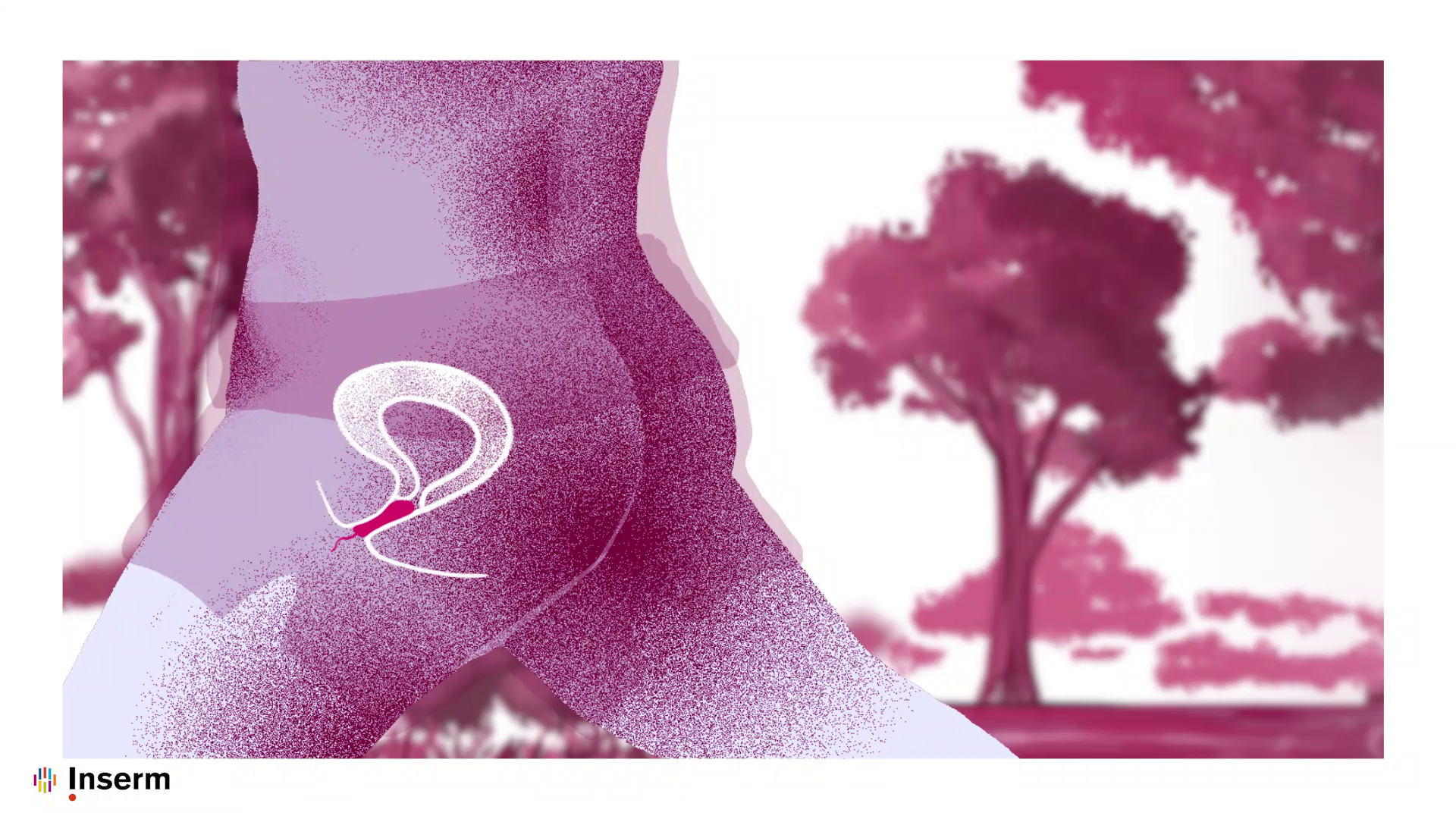
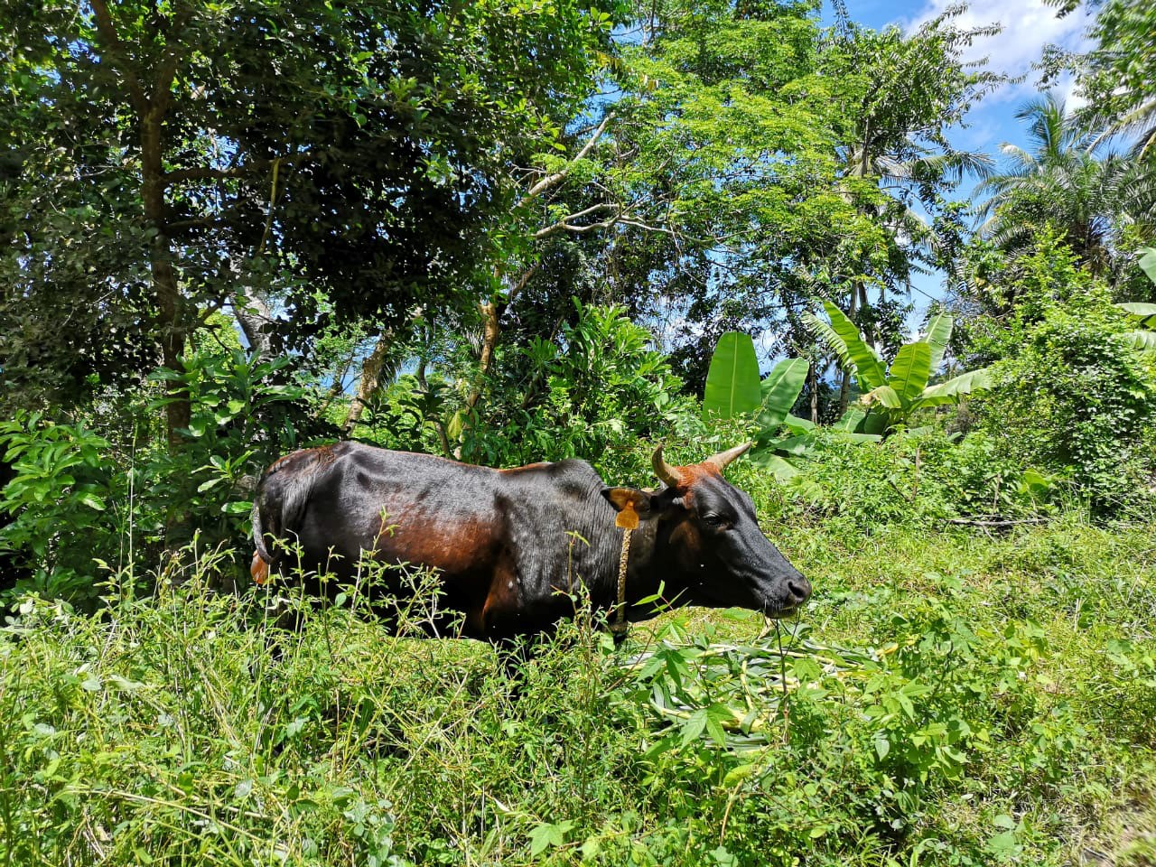
RVF is transmitted from livestock to humans. © DAAF 976

A viral disease responsible for major epidemics, occurring mainly in Africa, Rift Valley fever (RVF) is transmitted from livestock to humans. Despite being listed as a priority emerging disease by the World Health Organization (WHO) R&D Blueprint program in 2015, there had until now been little research into the dynamics of its transmission. As part of a multidisciplinary collaboration, researchers and public health professionals from Inserm, the French Public Health Agency, Cirad and the London School of Hygiene and Tropical Medicine, with the support of the REACTing consortium, have developed a mathematical model to study the dynamics of the 2018-2019 RVF epidemic in Mayotte and to quantify for the first time the beneficial impact of vaccinating livestock. This research has been published in the journal PNAS.

Against the background of the continued Covid-19 pandemic, research into emerging diseases and zoonoses – infectious diseases transmissible from animals to humans, has never been so important.

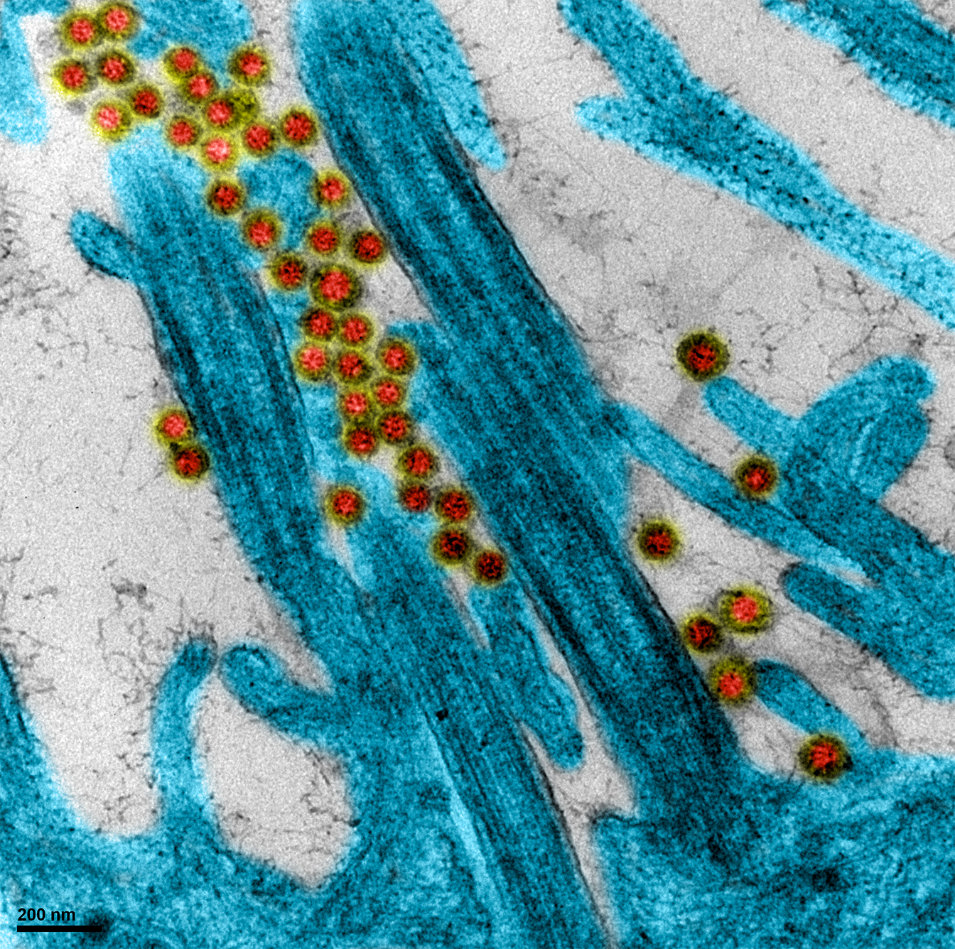
Rift Valley fever (RVF) is a viral zoonotic disease occurring primarily in some African regions, Mayotte and the Arabian Peninsula. It mainly affects livestock, causing waves of miscarriage and widespread mortality in the youngest animals. Humans can be infected through direct contact with the body fluids of contaminated animals or through the bites of infected mosquitos. To date, no human-to-human contamination has been reported. While the majority of patients develop asymptomatic or benign forms, in rare cases (1 to 3% of patients) the disease can take a more severe turn, characterized by ocular and meningeal disorders, and life-threatening hemorrhagic fever.

A major public health concern in some countries, RVF was listed by the WHO as an emerging priority disease in 2015, involving the accelerated development of its means of control. While research into vaccines for livestock is making progress, the potential impact of vaccination on the dynamics of the epidemic had never previously been evaluated.

The team, coordinated by Inserm researcher Raphaëlle Métras and her French Public Health Agency colleague Marion Subiros, looked at the 2018-2019 RVF epidemic in Mayotte. Since 2008, thanks to the implementation of two surveillance systems – one concerning the animals (helped by the Veterinary Services of Mayotte), the other humans – a large amount of high-quality RVF surveillance data has been collected. These data concern livestock seroprevalence and human epidemiology (number of human cases, sociodemographic characteristics, disease exposure criteria and geolocation).

As part of their study, the researchers and their colleagues developed a mathematical model integrating these data collected jointly by the two systems in order to reproduce the transmission dynamic of the virus during the 2018-2019 epidemic. One of the objectives was to obtain more information on how the virus passes from infected animals to humans.

What the scientists have shown is that for the first time in the context of an RVF epidemic there was a higher level of transmission to humans from mosquitos than through direct contact with infected livestock. If it is assumed that 30% of the population of Mayotte are farmers, up to 55% of human infections would have been caused by mosquito bites, versus 45% through exposure to livestock.

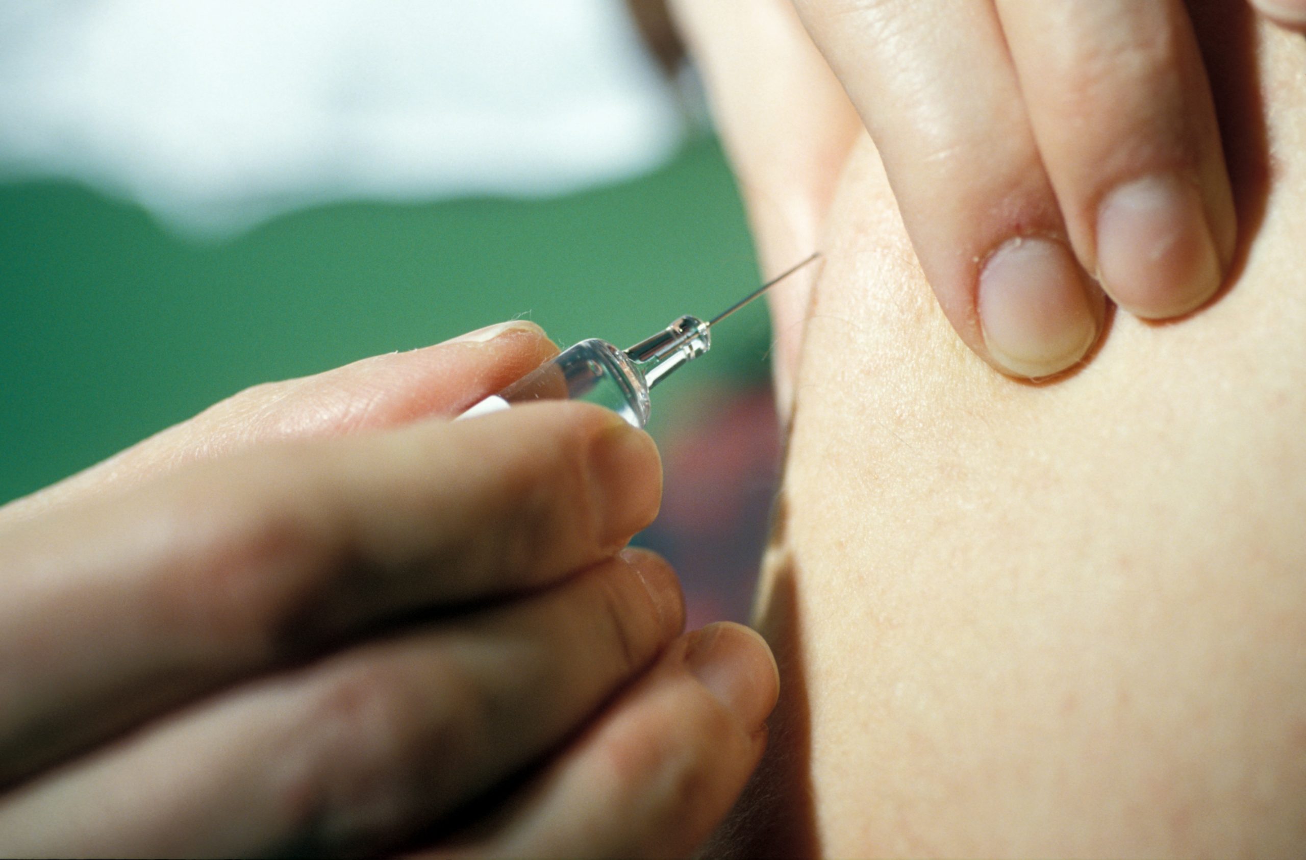
This is the first study to provide figures on the distribution of transmission through mosquito bites versus transmission through human direct contact.

The team also modeled the potential impact of vaccinating livestock to reduce the scale of the epidemic, using a suitable vaccine with the target characteristics of the WHO Blueprint program (especially vaccine efficacy). The findings of the model show that vaccinating 20% of the livestock could reduce the number of human cases by 30%. Reactive and mass vaccination campaigns in livestock would therefore be an essential measure when it comes to reducing the incidence of the disease in humans.

In a context in which zoonotic disease epidemics are emerging in succession, this research illustrates the importance of implementing a “One Health” approach, taking a systemic and unified approach to public, animal and environmental health at local, national and global levels.“The health emergency associated with the Covid-19 pandemic must force us to rethink how we see the links between human, animal and environmental health. Our research highlights the importance and added value of a multidisciplinary and integrated One Health quantitative approach in fighting zoonotic diseases. They also provide avenues for improving the surveillance of and research into emerging infectious diseases,” concludes Métras.

Prise en charge des formes sévères de Covid-19 : une méta-analyse incluant l’étude CAPE-COVID* plaide en faveur de la corticothérapie

Publication of Explore Covid-19 translationnal study findings

Coronavirus SARS-CoV-2 causing COVID-19 disease observed in close-up on the surface of a human respiratory epithelial cell.©M.Rosa-Calatrava/O.Terrier/A.Pizzorno/E.Errazuriz-cerda

New research suggests targeting C5a-C5aR1 axis could limit severe inflammatory response

Innate Pharma SA (Euronext Paris: IPH – ISIN: FR0010331421; Nasdaq: IPHA) (“Innate” or the “Company”) today announced the publication of a Nature paper entitled, “Association of COVID-19 inflammation with activation of the C5a-C5aR1 axis,” authored by Innate researchers in partnership with scientists from Hôpitaux Universitaires de Marseille AP-HM (La Timone and North Hospitals), Laveran Hospital, Aix Marseille University, the Centre d’Immunologie de Marseille-Luminy (Inserm, CNRS, AMU) and Marseille Immunopole/AP-HM immunoprofiling laboratory at La Timone Hospital. 

This Marseille-based exploratory research taskforce, named EXPLORE COVID-19, analyzed immune cells in COVID-19 patients at different stages of the disease. The goal of the study was to gain translational insights to better understand the immune response in COVID-19 patients and identify potential targets to fight the viral infection.

The study found that patients who progress towards severe COVID-19 disease, including those with severe pneumonia and acute respiratory distress syndrome (ARDS), exhibit an activation of the C5a/C5aR1 pathway. Specifically, researchers observed high levels of circulating C5a and over-activation of the C5a-dependent myeloid cell pathway, which is believed to contribute to inflammation in the lungs.

This research also focused on avdoralimab (IPH5401), a clinical-stage monoclonal antibody that blocks C5aR1 (CD88). Avdoralimab prevents C5a-induced myeloid cell recruitment and activation. Innate is currently investigating avdoralimab in oncology, which provided pharmacokinetic and safety data prior to investigation in COVID-19.

The findings published in Nature suggest that the C5a-C5aR1 axis blockade could be considered as a potential therapeutic strategy for severe respiratory disease associated with SARS-Cov-2 infection. The analysis found the C5a-C5aR1 axis blockade as a means of limiting myeloid cell infiltration at inflammatory sites and preventing the excessive lung inflammation associated with ARDS in COVID-19 patients.


“There is an urgent need to better understand COVID-19 disease progression and the associated complement cascade to help improve the prognosis of COVID-19 patients who present severe symptoms,” said Pr. Eric Vivier, PhD, Chief Scientific Officer at Innate Pharma and Professor at AP-HM, Aix-Marseille University and Centre d’Immunologie de Marseille-Luminy (Inserm/CNRS/AMU). “We are encouraged by this exploratory study, as we’re beginning to understand the impact of the immune response on the evolution of COVID-19 and pathways able to modulate this response.”    


Based on findings from this study, the Company previously announced the launch of an investigator-sponsored trial named FORCE (FOR COVID-19 Elimination). This is a randomized, double-blind Phase II clinical trial to further explore avdoralimab in COVID-19 patients with severe pneumonia, which is currently ongoing.


You can read the full publication in Nature here.


About the EXPLORE COVID-19 study:

Researchers analyzed the immune response of COVID-19 patients with no or few symptoms, patients who require oxygen, and a group of severe patients who require prolonged mechanical ventilation. The study included 82 individuals: 10 healthy controls and 72 COVID-19 patients, including 10 patients presenting few symptoms, 34 patients with pneumonia, and 28 patients with ARDS.

Avdoralimab in cancer:

Avdoralimab is a therapeutic antibody that specifically binds and blocks C5a receptor 1 (C5aR1) expressed on myeloid cells, including monocytes, macrophages and neutrophils.  It is currently in Phase I development in solid tumors, including hepatocellular carcinoma and non-small cell lung cancer.  

Avdoralimab in COVID-19:

C5a has been implicated in the pathogenesis of ARDS by promoting a proinflammatory environment, through the attraction of myeloid cells (neutrophils, monocytes and macrophages) and the stimulation of their cytokines production. Avdoralimab blocks C5aR1 and has the potential to reduce the inflammatory response in the lungs.

COVID-19: Together, Remdesivir and Diltiazem Open up New Therapeutic Avenues

SARS-CoV-2 infected human respiratory epithelium. The image shows viral clusters in the cilia of the epithelial cells, numerous highly-characteristic cytoplasmic vesicles containing large electron-dense accumulations of viral material, and numerous viruses being formed. © Manuel Rosa-Calatrava, Inserm; Olivier Terrier, CNRS; Andrés Pizzorno, Signia Therapeutics; Elisabeth Errazuriz-Cerda, UCBL1 CIQLE. VirPath (International Research Center for Infectious Diseases U1111 INSERM – JRU 5308 CNRS – ENS Lyon – UCBL1). Colorized by Noa Rosa C.


As the COVID-19 pandemic continues, finding a treatment to effectively combat the disease remains a major research challenge. Researchers from Inserm, CNRS, Université Claude Bernard Lyon 1 and ENS Lyon at the International Research Center for Infectious Diseases have developed a unique strategy for selecting, evaluating and repurposing existing drugs to assess their efficacy against SARS-CoV-2. They have also developed several highly-relevant preclinical models of infection using human respiratory epithelia of nasal and bronchial origin reconstituted in vitro. Thanks to their expertise, the researchers show that combining the Ebola treatment remdesivir with the antihypertensive diltiazem could bring significant benefit to COVID-19 patients. Their findings have been published in Cell Reports Medicine.

As part of the REACTing program coordinated by Inserm, the VirPath team led by Inserm researcher Manuel Rosa-Calatrava at the International Research Center for Infectious Diseases (Inserm/CNRS/Université Claude Bernard Lyon 1/ENS Lyon) is working on repurposing existing drugs for new therapeutic indications in viral infections.

To test the therapeutic efficacy of these molecules against COVID-19, the team began developing and characterizing experimental models of viral infection in February. This involved the in vitro reconstitution – as close as possible to human physiology – of human respiratory epithelia of nasal and bronchial origin. “We have been using these preclinical infection models, which are highly predictive of in vivo infection, for several years,” clarifies Rosa-Calatrava.

The researchers have also developed protocols for viral-genome and infectious-particle quantification. Their observations and analyses confirm and supplement current knowledge of the mechanisms of SARS-CoV-2 infection and virus-host interactions. “One particular observation from our models infected with the virus was the induced production of interleukin IL6, which is a marker of the disease’s severity,says Rosa-Calatrava.

A large number of drug candidates were evaluated using these models, including two molecules of interest: remdesivir and diltiazem, alone and in combination. Remdesivir presents antiviral activity against RNA viruses, including SARS-CoV-2. In vitro cell models, animal models, as well as several ongoing clinical trials are showing initial positive results against this virus.

Diltiazem is an antihypertensive used in the treatment of angina pectoris. It has already been characterized and repurposed by VirPath researchers to strongly stimulate the endogenous antiviral innate immune response, particularly against influenza- and respiratory viruses. The human toxicity of these two repurposed molecules has also already been evaluated, significantly reducing the time needed for their clinical development in the new SARS-CoV-2 indication.

The results of this study show a significant reduction in viral load in SARS-CoV-2 infected epithelia when treated with remdesivir. This effect is increased when diltiazem is added in combination. “By stimulating the innate immune response of the epithelia, diltiazem potentiates the effect of remdesivir and makes it possible to reduce doses. This bearing in mind that remdesivir presents some in vivo toxicity in addition to being a very costly drug,” emphasizes Rosa-Calatrava.

The team is continuing its preclinical tests with this dual therapy in animal models and hopes to launch a clinical trial as early as next winter if the positive results are confirmed.

Ebola Survivors Present Severe Immune System Abnormalities Two Years After Recovery

This study is the fruit of close collaboration between teams from Inserm, IRD, INSP, Université Paris-Est Créteil, and CERFIG in Guinea. © Aurélie Wiedemann.


Four years after the end of the Ebola epidemic in West Africa, and as it continues to wreak havoc in the Democratic Republic of Congo, the scientific community wonders about the after-effects that may remain among survivors. To learn more, researchers from Inserm and Université Paris-Est Créteil at the Institute for Vaccine Research decided to look at how their immune profiles had changed. To do this, they studied a cohort of survivors developed by Inserm, the French National Research Institute for Sustainable Development (IRD) and the Center for Training and Research in Infectious Diseases in Guinea (CERFIG) – the PostEboGui cohort. Their findings reveal the presence of abnormal levels of immune and inflammatory markers in the blood two years after developing the disease. These have been published in Nature Communications.

In the 2013-2016 Ebola outbreak in West Africa, more than 28,000 people were infected, causing over 11,000 deaths. While the long-term health impacts on survivors are still poorly understood, an increasing number of studies describe persistent clinical after-effects in these patients, such as generalized fatigue, musculoskeletal pain, and eye disorders.

To pinpoint these health problems, the research teams used data from the follow-up of cohorts of survivors, such as the Inserm PostEboGui cohort, developed with the French National Research Institute for Sustainable Development (IRD) and the Center for Training and Research in Infectious Diseases in Guinea (CERFIG). Made up of 802 former Ebola patients enrolled in several centers across Guinea, the aim of this cohort was to describe and analyze the clinical, immunological, psychological and socio-anthropological impacts of Ebola over a two-year period.

The study, published in Nature Communications and conducted by Prof. Yves Lévy with Aurélie Wiedemann at the Vaccine Research Institute (VRI, Inserm/Université Paris-Est Créteil), is one of the first to focus on the long-term immuno-inflammatory profile of Ebola survivors.

The researchers based their study on the analysis of blood samples from 35 members of the PostEboGui cohort who were recruited to participate in this study on average two years after the onset of their disease. A control group was also set up to compare their immune profiles. Each patient was seen three times for these blood samples to be taken. Samples of saliva, urine and semen were also analyzed in order to rule out the presence of the virus.

Such research was only possible thanks to the involvement of the local teams, which had been specifically trained in handling biological samples. It is therefore the fruit of close collaboration between Inserm and IRD teams and Guinean laboratory technicians and scientists at the National Institute of Public Health (INSP) and CERFIG.

Inflammation and immune markers

Analysis of the blood samples showed that even when survivors are physically recovered and no longer have any detectable virus, they still present a specific immune profile, different from that of people who have never contracted the disease.

In particular, the researchers identified the presence of immune cells known as CD4+ and CD8+ memory T cells, specific to the virus, which remained in the blood of the 35 survivors two years after the disease. In addition, there was also a higher number of cytotoxic CD8+ T cells involved in the destruction of the infected cells as well as the presence of IgG antibodies specific to the Ebola virus in these survivors.

What is more, the team showed the presence of a large amount of inflammatory markers in the blood samples (pro-inflammatory cytokines, markers of immune activation), which indicate the persistence of inflammation in Ebola survivors. Finally, this study showed that certain specific immune markers were associated with the persistence of symptoms in these patients.

These findings therefore highlight the long-term persistence of Ebola-specific immune activity and intense and chronic inflammation in these former patients, two years after being infected with the virus. “Our work underscores the importance of long-term follow-up of Ebola survivors, something that has already been emphasized in studies of clinical after-effects. It is important to see how their condition and immune profile evolve and whether they are moving towards a chronic disease,” says Wiedemann.

In the context of the ongoing Ebola epidemic in the Democratic Republic of Congo, a new cohort with an immunity component is currently being developed there, following a similar strategy to that used to implement PostEboGui. It will be an opportunity for researchers to confirm their findings on the immune profile of survivors in a larger number of patients.

Menstrual Toxic Shock Syndrome: Encourage Proper Tampon Use to Limit Risks

Inserm’s new educational video on staphylococcal toxic shock. © Camille Henry/Inserm

Staphylococcal toxic shock syndrome is linked to the presence of Staphylococcus aureus bacteria in the vaginal microbiota of some women for whom the misuse of intravaginal protection (tampons, menstrual cups, etc.) could increase the likelihood of developing it. Toxic shock is characterized by multiple symptoms that include digestive disorders, high fever, and skin rashes. In the most severe cases, it can lead to multi-organ failure and death.

Although the incidence of toxic shock remains very rare, researchers from Inserm, CNRS, Hospices Civils de Lyon, Université Claude Bernard Lyon 1 and ENS Lyon within the International Center for Research in Infectious Diseases and the National Reference Centre for Staphylococci have identified the risk factors that may in some cases favor it. In a study published in March 2020 in the journal EClinical Medicine, Prof. Gérard Lina’s team suggests simple measures to put in place for better tampon use during menstruation.


All the advice can be found in Inserm’s new educational video aimed at raising awareness and reassuring tampon users. 

Do not hesitate to share this prevention tool on your platforms and distribute it widely.

COVID-19: Vaccine Research at Inserm

The mobilization of Inserm researchers has led to major advances in SARS-CoV-2 knowledge and vaccine research.

© Inserm/Depardieu, Michel

Developing an effective and safe vaccine is one of the priority objectives in the fight to contain the COVID-19 pandemic. Since the complete sequencing of the SARS-CoV-2 genome in January 2020, research teams in France and internationally have been working tirelessly to better understand the immune response following infection and to test candidate vaccines. At Inserm, a dozen teams are involved in vaccine research projects. In particular, three initiatives have recently been selected by France’s Ministry of Higher Education, Research and Innovation on the advice of the COVID-19 Analysis, Research and Expertise Committee (CARE) and Inserm’s REACTing consortium, in order to receive special support and thus accelerate research.

Currently, more than 200 teams around the world are engaged in research projects to develop a vaccine against COVID-19. These include some thirty French groups that are members of the AVIESAN alliance or the biotech/industry ecosystem. Researchers from Inserm are also not to be outdone, since a dozen of these projects involve its units.

While the degree of progress of this research varies, some of these teams are currently in the phase of identifying the antigenic sequences of the virus that induce the specific immune response against SARS-CoV-2 and minimize the possible production of facilitator antibodies (a type of antibody that facilitates the entry of the virus into cells). In addition, some of the proposed vaccine platforms have been used previously for other candidate vaccines, including against HIV, influenza and toxoplasma, or for oncology vaccines.

The various ongoing projects can be divided into three main categories. The first is that of subunit vaccines, which do not contain live components but rather antigenic fragments of the pathogen. The second is a group of live attenuated candidate vaccines, all of which are prophylactic. The third is vaccines based on DNA or RNA coding for SARS-CoV-2 antigens.

Innovative candidate vaccines

Within this dynamic ecosystem, three projects involving Inserm units have been identified as priorities by the Ministry of Higher Education, Research and Innovation.

The first is driven by the Vaccine Research Institute (VRI), under the supervision of Inserm. The team led by Pr. Yves Lévy is involved in French COVID-19, the national cohort of patients infected with SARS-CoV-2, coordinated by Inserm’s REACTing network, in conjunction with 56 hospitals in France. Initially, using data from these patients, the researchers’ objective was to characterize the immune response in COVID-19 positive patients. Indeed, this data is essential and a prerequisite for the development of any vaccine. Based on this work, but also on their expertise in vaccine research, they are now developing a candidate vaccine in which the SARS-CoV-2 antigens would be presented by monoclonal antibodies to certain key cells of the immune system (dendritic cells). VRI has already developed several candidate vaccines based on this strategy, including against HIV (for which clinical trials will start in 2020).

The second vaccine research project, carried out at the Lille Center for Infection and Immunity by Inserm researcher Camille Locht and his team, is based on the repurposing of vectors with known activity, by integrating SARS-CoV-2 antigenic sequences. In this particular case, the chosen vector is a whooping cough vaccine. The objective is to develop a safe candidate vaccine, whose action on the body is well documented and very specific to the novel coronavirus because it incorporates carefully selected antigens.

Led by Inserm researcher Patrice Marche at the Institute for Advanced Biosciences and researcher Fabrice Navarro, head of CEA-Leti’s Microfluidic Systems and Bioengineering Laboratory, the third project also proposes an original vaccine approach against SARS- CoV- 2. It is based on an innovative delivery system involving lipid nanoparticles developed by the researchers. These highly stable and well-tolerated nanoparticles were originally created to encapsulate and transport drugs to target cells. In the fight against SARS- CoV-2, the researchers hope to encapsulate antigens of the virus to elicit a strong immune response.

Having already used this technique in HIV vaccine research, the team should quickly be able to develop this new candidate vaccine on a large scale, which represents a major advantage in making research faster and more efficient.

Developing a safe and effective vaccine against COVID-19 is a long process. Nevertheless, the mobilization of the scientific community, and in particular Inserm researchers, is enabling major advances to be made in terms of understanding the virus and the immune response to it, and in terms of setting up numerous trials to test a wide variety of vaccine strategies in record time.

Cardiac arrest outside the hospital during the peak of the COVID-19 epidemic: studies highlight pulmonary embolism as the main determinant

Cardiac arrest outside the hospital during the peak of the Covid-19 pandemic could be partly due to massive pulmonary embolism© Adobe Stock

French investigators from the Paris Medico-Legal Institute, the radiology department of the Sainte-Anne hospital / GHU Paris, the anesthesia-resuscitation department of the AP-HP hospitals Saint-Louis and Lariboisière, the University of Paris, Inserm and CNRS have hypothesized that cardiac arrests outside the hospital during the peak of the COVID-19 pandemic could be partly due to massive pulmonary embolism. The details of this work were published on May 28, 2020 in the European Journal of Heart Failure .

The Forensic Institute of Paris and the radiology department of Sainte-Anne hospital use whole-body scanners for examinations requested by the justice authorities. These scanners were compared between the two-week period corresponding to the epidemic peak (March 23 to April 7, 2020) and all of 2019. The elements sought on the scanner were the presence of signs of pulmonary infection suggestive of COVID-19, phlebitis of the lower limbs and proximal pulmonary embolism, responsible for cardiac arrest.

This study shows that requests for forensic autopsies for unexplained sudden death were 14 times more frequent during the epidemic peak than in 2019.

The vast majority of unexplained sudden deaths during the epidemic peak had lung lesions suspected of Covid-19 infection.

The age of the deceased patients ranged from 27 to 99 years. Most people died at home, some had fever and / or cough, and the majority suddenly lost contact with family or emergency services 30 minutes to a few hours before the onset of cardiac arrest.

CT analysis shows a 3 times higher frequency of proximal pulmonary embolism and phlebitis during the epidemic peak compared to all of 2019.

These results suggest that a significant proportion of the victims of sudden death during the epidemic peak were probably linked to proximal pulmonary embolism which must be quickly referred to cardiogenic shock treatment centers. This study also confirms the vital role of intensive prevention of thrombosis in patients with COVID-19 infection.

Inflammatory Bowel Disease: Immune Cells Spearheading the Cure

This microscopic image of a section of mouse intestine shows significant lesions, inflammatory signs and dysfunctional healing of the intestinal mucosa. © Sonnenberg Lab

Inflammations of the intestine affect many patients, often with reduced therapeutic prospects. The role of iron in these inflammatory processes is increasingly well documented, opening up new avenues of treatment. A collaborative study between the team led by Inserm research director Carole Peyssonnaux at Institut Cochin (Inserm/CNRS/Université de Paris) and Greg Sonnenberg’s team in New York (Weill Cornell Medicine) shows that the hormone that regulates iron levels in the body is produced by immune cells during inflammation of the intestine, and that it helps repair damage to the intestinal mucosa. This research has been published in Science.

Infections, inflammatory bowel disease (IBD) such as Crohn’s disease, and colorectal cancer are associated with inflammation of the intestine. In patients, the intestinal mucosa may then be damaged, with bleeding and impaired distribution of iron in the body often being observed.

For several years, Inserm research director Carole Peyssonnaux and her team at Institut Cochin (Inserm/CNRS/Université de Paris) have been interested in the role of hepcidin in disease settings. This hormone regulates iron metabolism in the body and is mainly produced by the liver. However, the researchers had already shown that in the case of certain pathologies, hepcidin is also secreted in other tissues.

Their new study, carried out in collaboration with Gregory F. Sonnenberg’s team from Cornell University in the United States and published in April 2020 in the journal Science, shows that in a context of intestinal inflammation, hepcidin is also expressed by specific immune cells, the dendritic cells of the intestine.

New therapeutic avenues

The scientists first studied the intestinal healing process in several groups of mice, all with inflammation of the intestine. For one of these groups of mice, the gene coding for hepcidin was not expressed. Compared to the other groups of mice in which this gene functioned normally, this resulted in greater continuous weight loss, but also in less effective healing of the intestinal mucosa.

The researchers have confirmed that hepcidin plays an important role in the healing of intestinal lesions. However, they still wondered whether it was the hepcidin normally secreted by the liver that had this beneficial effect or whether, in this disease setting, this iron-regulating hormone was produced in other organs.

Using mouse models in which the gene coding for hepcidin was only deficient in the liver, the researchers were able to show, surprisingly, that the healing process was independent of hepatic hepcidin production. Following intestinal injury and in a context of inflammation, the local dendritic cells of the intestine were the dominant source of production of this hormone.

The research also emphasizes that hepcidin interacts with a key iron transporter called ferroportin, which is present on other immune cells in the intestine (macrophages), thereby promoting iron sequestration and preventing the proliferation of iron-dependent bacteria in intestinal lesions. This process helps to limit the severity of the inflammation.

To determine whether this phenomenon also occurs in humans, the researchers looked at samples taken from pediatric IBD patients. They confirmed that the dendritic cells in the human intestine also produce hepcidin in response to injury. This pathway may therefore be clinically important for people with IBD. “Our study suggests that hepcidin may have a protective role because if the gene that codes for this hormone is deleted, the severity of the disease is greater. The use of hepcidin mimetic treatments could therefore have a therapeutic role in promoting iron sequestration, reducing its availability to bacteria that proliferate in the intestine and promoting the healing of lesions,” concludes Peyssonnaux.

Covid-19: Publication of a prospective observational study in the journal BMJ in children with hyper-inflammatory syndrome related to Kawasaki disease

©Piron Guillaume on Unsplash

The team of the Department of General Pediatrics and Infectious Diseases of the Necker-Enfants Malades AP-HP Hospital, the Institut Pasteur, Inserm and the University of Paris, conducted a prospective observational study between April 27 and May 15, 2020 to describe the characteristics of children and adolescents hospitalized in a context of Kawasaki disease-like syndrome in a COVID-19 epidemic context, with a systemic hyper-inflammatory expression. The results of this study were published on June 3, 2020  in the journal BMJ.

This work included all children and adolescents hospitalized during the study period with signs of Kawasaki disease. Clinical and biological data, imaging and cardiac ultrasound data, treatments and clinical course were collected. A nasopharyngeal sample for SARS-CoV-2 by RT-PCR and a sample for IgG antibodies against the virus were systematically performed for all patients included.

During the study period, 21 patients were admitted to the general pediatrics and infectious diseases department with signs of Kawasaki disease. Their median age was 7.9 years (range, 3.7-16.6 years), and 12 patients (57%) had a parent or grandparent born in a sub-Saharan African country. Twelve patients (57%) developed shock syndrome related to Kawasaki disease and 16 patients (76%) developed myocarditis. Seventeen patients (81%) required intensive care or pediatric resuscitation. All of these children had noisy digestive symptoms at the onset of the disease and all had high inflammatory markers. Nineteen patients (90%) had markers of recent SARS-CoV-2 infection (RT-PCR positive for 8/21, and presence of IgG antibodies for 19/21 patients). All patients received intravenous immunoglobulins and ten (48%) also received corticosteroids. The clinical course was favorable in all cases. Coronary dilations were detected in five (24%) patients during hospitalization.

The abnormally high number of children and adolescents with Kawasaki disease recently observed in the Paris region could be linked to exposure to SARS-CoV-2. The study conducted cannot formally establish a causal link with infection with the SARS-CoV-2 virus despite very strong suspicion.

The characteristics of these patients differ from those observed during classic Kawasaki disease: these patients are older, more often of descent from a sub-Saharan African country, with more frequently digestive manifestations in the foreground, severe forms of myocarditis and hemodynamic instability.

fermer