Menu

Towards a Drug to Combat a Severe Intestinal Disease in Children, Immunocompromised Patients

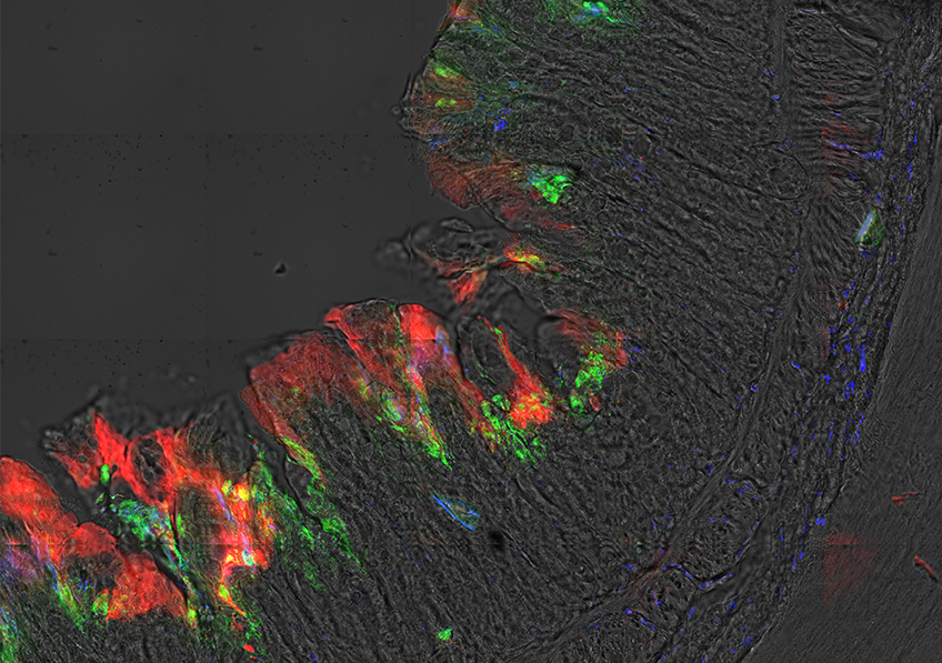
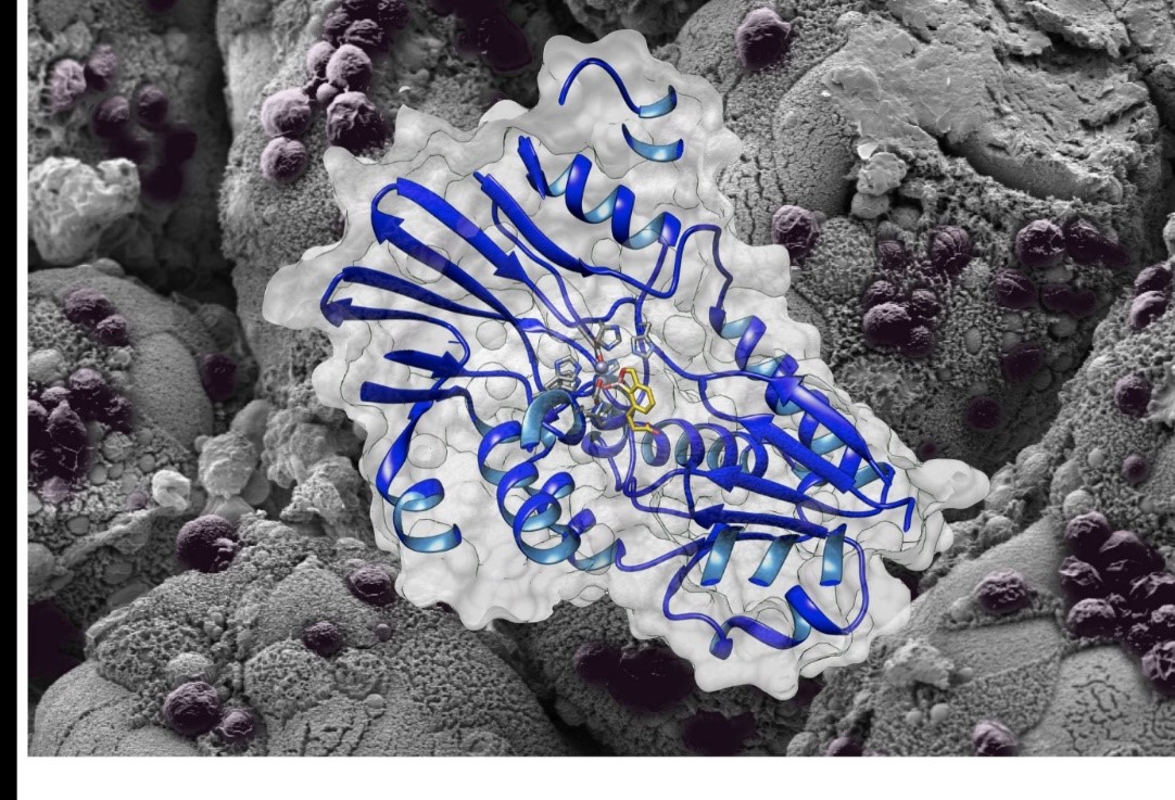
3D structure of the enzyme with the molecule AN3661 shown against the background of the intestine of an immunocompromised mouse infected with Cryptosporidium. ©Fabrice Laurent and Christopher Swale.

Researchers from Inserm and INRA working in the teams of Mohamed-Ali Hakimi (Institute for Advanced Biosciences – Inserm U 1209 / CNRS JRU 5309 / UGA) and Fabrice Laurent (INRA) have recently discovered a new candidate drug to control cryptosporidiosis, a severe intestinal disease in children, immunocompromised patients, and young ruminants. Beyond this disease, their research represents an opportunity to discover new therapeutic avenues for related infections, such as toxoplasmosis and malaria. Their findings have been published in Science Translational Medicine.

Cryptosporidiosis is a diarrheal disease caused by Cryptosporidium, a microscopic parasite that develops in the intestine of numerous mammals – notably humans. This intestinal parasite is mainly spread through contaminated drinking or pool water, where it can survive for several days in the presence of chlorine, or through contact with infected animals. Over the past 20 years, infection with Cryptosporidium has been recognized as a common cause of waterborne disease in humans. According to a recent study by the US Centers for Disease Control and Prevention (CDC), the number of Cryptosporidium epidemics is even on the increase. In humans, it causes acute and sometimes fatal diarrhea in the most vulnerable populations, including young malnourished children and immunocompromised patients (for example, those infected with HIV). The therapeutic arsenal is currently very limited and in some cases ineffective in eliminating this parasite.

This study conducted by the researchers from Inserm and INRA reveals the discovery of a candidate drug called AN3661, which drastically reduces not just Cryptosporidium infection but also that of Toxoplasma, the parasite responsible for toxoplasmosis.

The study teams have revealed the mechanism of action of this molecule by elucidating the three-dimensional structure of its target, called CPSF3, in Cryptosporidium. AN3661 binds to the heart of the enzyme CPSF3, thereby preventing the maturation of the messenger RNA, a process essential to the parasite’s survival. Preclinical tests using an animal model show remarkable efficacy in vivo with single-dose treatments of the infection in immunocompromised or baby mice.

This major discovery paves the way for new therapeutic strategies and innovations to fight not just cryptosporidiosis but also other related infections, such as toxoplasmosis and malaria.

A Vaccine to Overcome Immunotherapy Resistance

©AdobeStock

For patients with metastatic cancers such as those of the lung or bladder, or melanoma, immunotherapy represents a genuine therapeutic revolution. Unfortunately, it is only effective in 10 to 25% of those eligible to receive it. Researchers from the Cancer Research Center of Lyon (CRCL – Inserm / CNRS / Université Claude Bernard Lyon 1 / Léon Bérard cancer center) and the Léon Bérard and Gustave Roussy cancer centers have shown that a commercially-available vaccine can overcome resistance to immunotherapy. Their study, published in Science Translational Medicine, shows that not only can gastroenteritis vaccines induce the immunogenic death of cancer cells in vitro, but also that combining them with immunotherapy triggers a potent anti-tumor immune response in vivo – where immunotherapy alone had failed.

How can we overcome resistance to immunotherapies, so that as many patients as possible can benefit from these innovations? A team of researchers led by Aurélien Marabelle (Gustave Roussy and Léon Bérard cancer centers), Christophe Caux (Inserm U1052) and Sandrine Valsesia-Wittmann (Léon Bérard cancer center – Inserm UA8) has studied this question. Their idea was to use vaccines to render immunotherapy effective in those cancers in which it has been unsuccessful so far. Then, the aim was to increase the number of patients who could benefit from such therapy in cancers where it has already been shown to be effective.

In this study, our research team looked at pediatric tumors such as neuroblastomas, which are aggressive cancers that do not respond to existing immunotherapies such as anti-PD(L)1 and anti-CTLA4. In our aim to transform how they respond to immunotherapy, we used various vaccines as sources of pro-inflammatory elements because pathogens such as viruses are able to directly stimulate innate immune receptors” explains Marabelle.

Gastroenteritis vaccines

To start with, the researchers tested 14 commercially available vaccines (BCG, Cervarix, TicoVac, etc.) in vitro for their ability to stimulate these innate immune receptors.

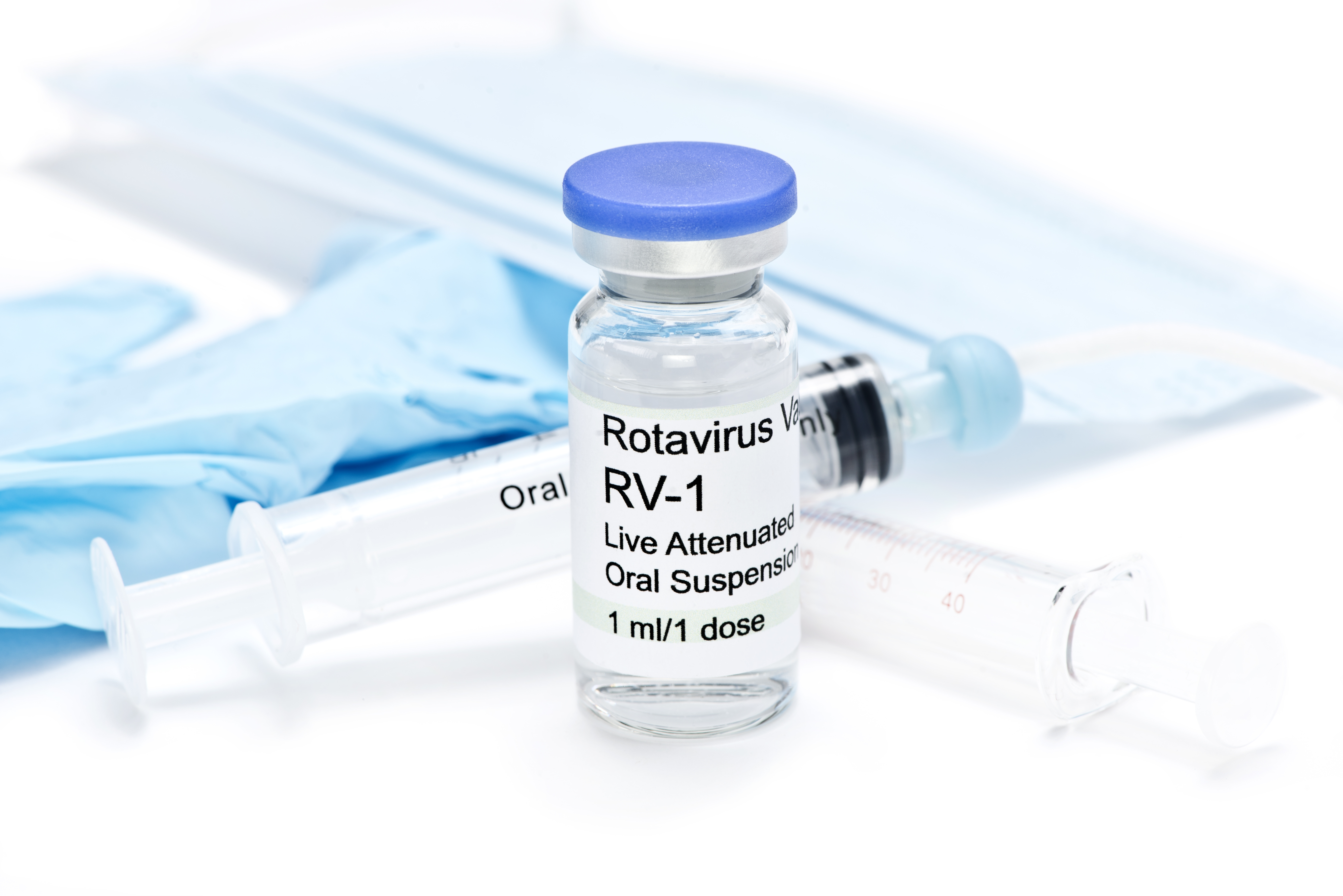
Out of the vaccines tested, those used against rotavirus (Rotarix, Rotateq), the virus responsible for gastroenteritis, were identified as having strong pro-inflammatory properties. Unexpectedly, they observed that these vaccines have an oncolytic action – namely the ability to preferentially infect and kill cancer cells in relation to normal cells and induce what is known as immunogenic death.

Vaccine plus immunotherapy – a potent combination

The researchers also conducted in vivo testing of the most pro-inflammatory vaccines using models of neuroblastoma in which the anti-PD(L)1 and anti-CTLA4 immunotherapies are ineffective in humans. This involved injecting these vaccines either systemically or intratumorally (directly into the tumors).

They observed that when the rotavirus vaccines were injected intratumorally, some of the tumors disappeared. When they then administered the vaccine in combination with anti-PD(L)1 or CTLA4 immunotherapies, all of the tumors disappeared.

While the tumors usually do not respond well to either of these treatments used alone, combining them generates a strong systemic antitumor immune response capable of eradicating both injected and non-injected tumors. “Our findings show that the rotavirus strains contained in gastroenteritis vaccines can make usually naturally-resistant tumors sensitive to immunotherapy“, highlights Caux.

The researchers also sought to explain how the rotaviruses exert a stimulant effect on the immune system. They showed that the activation of an innate immune receptor known as RIG-I (retinoic acid induced gene I) was essential for the synergistic effect of the intratumoral rotaviruses with the immunotherapies.

The findings of this study provide a strong scientific rationale in favor of the development of intratumor immunization strategies for immunotherapy-refractory cancers, particularly in pediatric oncology but also in adults,” concludes Valsesia-Wittmann.

This research was supported by the Pediatric Hematology and Oncology Institute (iHOPe), the French League Against Cancer, Inca, the French National Research Agency (ANR) and the patient associations Les Torocinelles, ALBEC and 111 des Arts.

Is physical activity always good for the heart?

International guidelines emphasize the need to be active in order to prevent cardiovascular mortality. Credits : Adobe Stock

Physical activity is thought to be our greatest ally in the fight against cardiovascular disease. But there may be significant variations in its protective effects across a range of different situations, such as regularly playing a sport, carrying heavy loads at work, or going for a walk with friends. These are the findings of a new study led by Inserm researcher Jean-Philippe Empana (U970 PARCC, Inserm/Université de Paris) in collaboration with Australian researchers. The results have been published in Hypertension.

Cardiovascular diseases are the leading cause of mortality around the world, and there is no sign that this trend is declining. However, a large number of premature deaths could be prevented by taking appropriate preventive measures. Among these measures, physical activity is often presented as having multiple benefits, and international guidelines emphasize the need to be active in order to avoid cardiovascular mortality.[1]

But physical activity is a broad concept, and few scientific studies have looked into the differences between various types of exercise may have. This was the focus of the new study published in Hypertension, which was conducted by the research teams led by Jean-Philippe Empana, Xavier Jouven, and Pierre Boutouyrie (Inserm/Université de Paris), in collaboration with Rachel Climie at the Baker Heart and Diabetes Institute, Melbourne, Australia.

“Our idea was to look at whether all types of physical activity are beneficial, or whether under some circumstances physical activity can be harmful. We wanted in particular to explore the consequences of physical activity at work, especially strenuous physical activity such as routinely carrying heavy loads, which could have a negative impact”, explains Empana.

Sport, work, or leisure

The research by Empana and his colleagues was based on data from participants in the Paris Prospective Study III. For ten years, this extensive French study has been monitoring the health status of over 10,000 volunteers, aged 50 to 75 years old and recruited during a health check-up at the Paris Health Clinic (Paris Preclinical Investigations, IPC).

Participants were asked to fill out a questionnaire about the frequency, duration, and intensity of their physical activity in three different contexts: physical activity through sport, physical activity at work (for example carrying heavy loads), and physical activity in their leisure time (such as gardening).

The cardiovascular health of participants was then assessed based on the health of their arteries using cutting-edge ultrasound imaging of the carotid artery (a superficial artery in the neck). This method, known as “echo tracking”, can be used to measure baroreflex sensitivity, a mechanism of automatic adaptation to sudden changes in blood pressure. When this system is impaired, this can lead to major health problems, and a higher risk of cardiac arrest.

Studying the arduous nature of work

In their analyses, the researchers distinguished between two components of the baroreflex: mechanical baroreflex, assessed through the measurement of arterial stiffness, and neural baroreflex, assessed through the measurement of nerve impulses sent by the receptors on the walls of the artery, in response to a distension of the vessel. Abnormalities in the mechanical component tend to be associated with aging-related cardiovascular diseases, while abnormalities in the neural component tend to be linked to heart rhythm disorders that can lead to a cardiac arrest.

The study shows that high-intensity sporting physical activity is associated with a better neural baroreflex. Conversely, physical activity at work (such as routinely carrying heavy loads) appears to be more strongly associated with an abnormal neural baroreflex and greater arterial stiffness. Such activity could therefore be harmful for cardiovascular health, and in particular may be associated with heart rhythm disorders. 

“Our findings represent a valuable avenue of research for improving our understanding of the associations between physical activity and cardiovascular disease. They do not suggest that movement at work is harmful for health, instead they suggest that chronic, strenuous activity (such as lifting heavy loads) at work may be”, highlights Empana

The researchers will attempt to replicate these results in other populations, and explore in greater detail the interactions between physical activity and health. “This study has major public health implications for physical activity at work. We now want to expand our analysis to further explore the interactions between physical activity and the health status of people in the workplace”, concludes Empana.

[1] See the Inserm collective expert review: “Physical activity: Prevention and treatment of chronic diseases” https://www.inserm.fr/information-en-sante/expertises-collectives/activite-physique-prevention-et-traitement-maladies-chroniques
Aging with HIV Linked to Increased Risk of Cognitive Impairment

Adobe Stock 

Thanks to antiretroviral therapies, it is possible to grow old with HIV under control. However, this chronic infection may not leave cognitive function unscathed. That is why Alain Makinson (Translational Research on HIV and Infectious Diseases unit, Montpellier University Hospital, Université de Montpellier, Inserm, IRD) and his team were interested in exploring the development of neurocognitive impairment (NCI), such as diminished attention, memory and motor capacity, in patients living with HIV in the ANRS EP58 HAND 55-70 study. In their latest research, published in Clinical Infectious diseases, the scientists describe the results of their observations of 200 people living with HIV enrolled in six French centers. The data collected from these patients were then compared, using the same neurocognitive evaluation methods, with those of a control population of 1,000 people of the same age, sex and education taken from Constances – a cohort recruited from the general population, with over 200,000 volunteers. The researchers reveal that among those living with HIV between the ages of 55 and 70, the risk of developing mild (and in some cases, symptomless) neurocognitive impairment increases by 50%.

Thanks to antiretroviral therapies, people living with the human immunodeficiency virus (PLHIV) can bring it under control. However, while HIV is no longer their major cause of death – having decreased substantially since the advent of these therapies – other risks must be considered. Several recent studies have highlighted the increased prevalence of neurocognitive impairment (NCI) in PLHIV compared with uninfected individuals – an increase that is all the more pronounced in those whose virus is not controlled. However, this NCI could also be caused by cardiovascular factors or the onset of depression, which is more common in this population.

In order to try to determine the nature of the link between living with HIV and developing NCI (such as diminished attention, memory and motor capacity), Alain Makinson and his coworkers studied the data of 200 PLHIV, between 55 and 70 years of age, whose HIV was under control and who were enrolled between January 2016 and October 2017 in the ANRS EP58 HAND (HIV-Associated Neurocognitive Disorder) study. Each patient was compared with five HIV-uninfected individuals of the same age, sex and education, from Constances – a cohort recruited from the general population. The same methods were used for both populations when performing the cognitive tests and collecting the study data. All in all, 1,200 people took part in this study.

Although the impairments observed in the study were either mild or without apparent symptoms (i.e. with zero to low impact on daily activities despite abnormal test results), those living with HIV were more affected by NCI: 35% versus 24% for the control group.

The risk of developing NCI is therefore 50% higher for HIV-infected individuals compared with HIV-uninfected individuals, all other criteria being equal (age, sex and education).

The conclusion remained the same after the authors took into account various confounding factors (alcohol consumption, smoking, physical activity, diabetes, hypertension, etc.), and despite the use of several methods to evaluate the cognitive tests.

Despite these highly robust results, a causal link between living with HIV and developing NCI cannot be established – for which there are several possible hypotheses. One is that HIV infection and its treatments cause recurrent brain inflammation. Another is that the complications associated with the immune deficiency can affect cognition before antiretroviral treatment is initiated, but without the subsequent deterioration occurring more rapidly in comparison with the general population. Finally, seropositivity could be associated with other risk factors (particularly drug use), which are difficult to measure fully in the two populations of this study.

The authors wish to continue to follow the same population over a longer period in order to better define the causes of NCI in this aging population and test the hypothesis of accelerated cognitive aging in PLHIV – particularly given that very few studies with a control group are available. Testing the brain inflammation hypothesis by collecting certain specific blood biomarkers represents another avenue for the team to explore in its quest to elucidate the mechanisms behind NCI.

Dysentery: Shigella, bacteria with adaptation to respiration

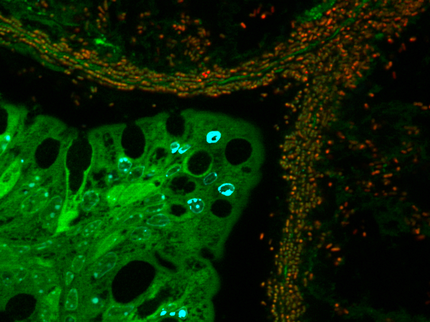
Imagerie montrant la déplétion de l'oxygène au sein de la muqueuse intestinale par Shigella (vert), induisant une hypoxie (rouge) au sein des foyers infectieux inflammatoires (neutrophiles: marqués à l'aide du Myelotracker, bleu).

Déplétion de l’oxygène au sein de la muqueuse intestinale par Shigella (vert), induisant une hypoxie (rouge) au sein des foyers infectieux inflammatoires (neutrophiles: marqués à l’aide du Myelotracker, bleu). ©Benoit Marteyn/ Inserm/ Institut Pasteur

Bacillary dysentery caused by the intestinal bacteria Shigella is a major health problem in tropical regions and developing countries. Complications from this infection lead to several hundred thousand deaths a year, primarily among infants. Researchers from Inserm and the Institut Pasteur have studied the mechanisms of Shigella virulence. They found that these bacteria are not only able to consume the oxygen in colonic tissue in order to grow and create foci of infection, but can also adapt their mode of respiration so that they can continue to grow once the oxygen in these foci has been used up. These findings, published in Nature Microbiology, open up new prospects for the development of antibiotics and vaccines to combat this group of bacteria, which is on the WHO list of 12 priority pathogens.

Shigella is a group of pathogenic enterobacteria (bacteria found in the digestive tract) that cause bacillary dysentery, which is also known as shigellosis. They are transmitted via the fecal-oral route, for example through food or water contaminated with fecal matter, and are thus primarily endemic in tropical regions, particularly in developing countries where a lack of hygiene and healthcare infrastructure favor outbreaks of disease. After ingestion, Shigella bacteria invade the cells of the intestinal wall and then the colonic mucosa, causing major inflammation combined with severe tissue damage. This causes symptoms such as abdominal pain, vomiting, diarrhea containing blood or mucus, and fever.

With no commercialized vaccine (the infection is currently treated with antibiotics), shigellosis remains a major public health problem, and results in 700,000 deaths per year around the world—primarily among children under the age of 5—from acute complications.

The emergence of new multi-drug resistant strains of Shigella has prompted inclusion of the bacteria on the WHO list of 12 “priority pathogens” for which new treatments (vaccines or antibiotics) are urgently needed.

With this in mind, a team led by Inserm researcher Benoit Marteyn within Unit 1202, “Molecular Microbial Pathogenesis” (Institut Pasteur/Inserm), sought to better understand the mechanism used by Shigella to infect tissue by modulating the levels of oxygen present. To do this, the researchers used innovative image analysis methods developed by the Imagopole at the Institut Pasteur, which allowed them to study each cell individually (single-cell analysis) and monitor variation in the levels of oxygen O2 in intestinal tissues around isolated bacteria and in foci of infection, where bacteria are numerous.

The research group also found that foci of Shigella infection had abnormally low levels of oxygen (hypoxia). The denser the population of bacteria, the greater the consumption of O2. Hypoxia was not, however, seen around isolated bacteria away from the foci of infection.

Shigella bacteria are “facultative anaerobes,” which means that while they favor aerobic respiration (which uses O2 as fuel), if oxygen is lacking they can also switch to “anaerobic” respiration, which does not require O2. This characteristic enables them to continue to grow in hypoxic, or even anoxic (O2-depleted) foci after they have consumed all the oxygen in the tissues.

The researchers have thus shown that aerobic respiration of Shigella and their capacity to modulate the oxygenation of infected tissues enables the formation of hypoxic foci of infection within the intestinal mucosa, which constitutes the first stage in their colonization strategy, with over 99% of the bacterial population growing in these areas. When these foci are depleted of oxygen, the adaptability of the bacteria to O2-poor environments gives them a crucial advantage that explains their virulence and that of other facultative anaerobic enterobacteria.

“These findings are very important in relation to the search for new antibiotics and candidate vaccines to combat Shigella infection. Their mechanisms of action will need to be confirmed in hypoxic or even anoxic conditions, to reflect the pathophysiological conditions in which Shigella primarily grow within the colonic mucosa,” concludes Benoit Marteyn.

Identified: A Protein Essential for Chikungunya Virus Replication

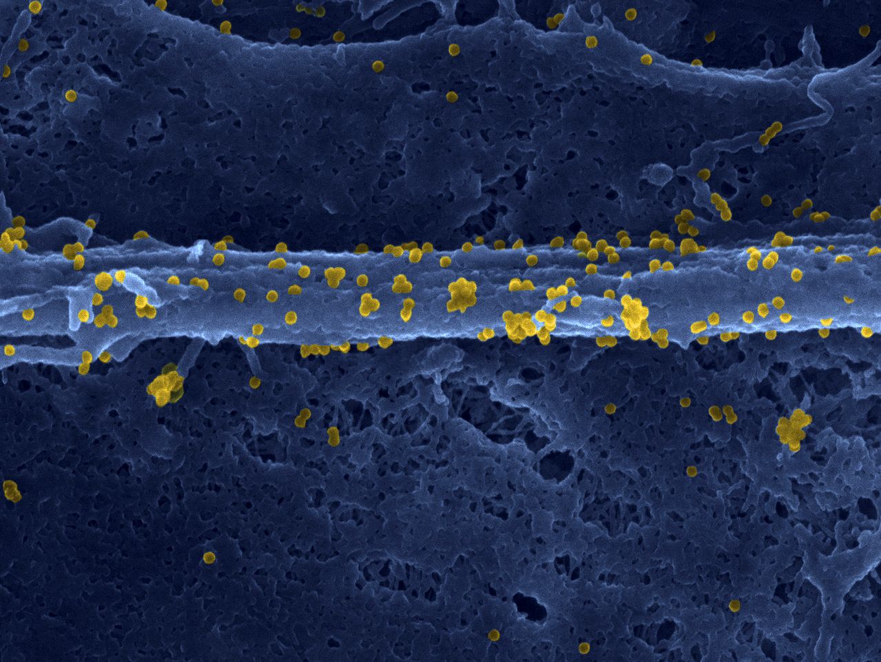
Fibroblast infected with the chikungunya virus. © Inserm/Thérèse Couderc/Marie-Christine Prévost/Marc Lecuit

Chikungunya is an infectious disease caused by a mosquito-borne virus transmitted to humans. First detected in Africa, the virus has been responsible for recent epidemics in the Americas, Asia and the Indian Ocean – particularly the Reunion Island. Chikungunya is characterized by high fever and intense joint and muscle pain that can last for several months. The mechanisms of infection of human cells with the virus remain very poorly understood. Led by Ali Amara in collaboration with Marc Lecuit researchers from Inserm, Institut Pasteur, CNRS and Université de Paris have identified a protein that is crucial in order for the virus to replicate within its target cells. This research, published in the journal Nature, opens up therapeutic avenues in the fight against chikungunya.

Originally from Africa, chikungunya is aptly named. It derives from a word in the Kimakonde language, meaning “to become contorted”, because the severe muscle and joint pains endured by the patients prevent them from moving normally or performing their daily activities.

While the clinical manifestations of the disease are well understood, the mechanisms by which the virus infects human cells and multiplies remain poorly elucidated. Several studies had already identified certain host cell factors implicated in the replication of the virus. However, none had succeeded in explaining why the virus preferentially targets the muscle and joint cells, causing these clinical signs.

Researchers from Inserm, CNRS and Université de Paris led by Dr. Ali Amara at the AP-HP Saint-Louis Hospital Research Institute in Paris, in collaboration with Marc Lecuit’s team from Institut Pasteur, Inserm and Université de Paris, have identified that the FHL1 protein is a key cellular factor for the replication and pathogenesis of chikungunya. FHL1 is a molecule present mainly in the muscle cells and fibroblasts, the preferred targets of the virus. Usually, FHL1 contributes to healthy muscle physiology and it is now thought to be diverted from that function by the virus to ensure its replication in the target cells.

To conduct this study, Amara’s team used the CRISPR-Cas9 technology to systematically screen the genome of human cells in order to identify the host factors necessary for viral replication. In doing so, it isolated the gene coding for the FHL1 protein. The team then conducted a series of experiments showing the incapacity of the virus to infect cells whose FHL1 expression had been abolished.

In addition, the researchers have shown that the virus was incapable of multiplying within cells derived from patients suffering Emery-Dreifuss muscular dystrophy – a rare genetic disease. This muscle disease is the result of mutations of the FHL1 gene responsible for the breakdown of the FHL1 protein. The researchers have shown that the cells of these patients are resistant to the virus.

Finally, the researchers performed in vivo experiments in mice whose Fhl1 gene was invalidated. They have shown that these animals are totally resistant to infection and do not develop the disease, whereas the virus multiplies and causes major muscle lesions in mice expressing a functional FHL1 protein. These observations demonstrate that the FHL1 protein plays a key role in chikungunya virus replication and pathogenesis.

The precise role played by FHL1 in the viral infection is not fully understood. The researchers have discovered that FHL1 interacts with a viral protein known as nsP3. It is when binding to this that FHL1 participates in the replication of the virus.

“We now want to understand this interaction in molecular detail. The next step is to define why FHL1 is so specific to the chikungunya virus, and to decipher its mechanism of action at the molecular level. Elucidating the molecular structure of the FHL1-nsP3 complex could represent a major step forward in the development of antivirals that block the replication of the virus“, emphasize Ali Amara and Laurent Meertens, the Inserm researchers in charge of the study.

At present, only symptomatic treatments are available for patients infected with chikungunya.

Disarming a probiotic to improve its benefits

Escherichia coli © Inserm/Cloup, Emilie/Nougayrede, Jean-Philippe

For more than a century, the Nissle 1917 strain of the bacterium Escherichia coli has been used as a probiotic to treat gastrointestinal disorders. However, this bacterium also produces a toxin, colibactin, which has deleterious effects on host DNA and might cause colon cancer. It is therefore crucial to understand the mechanisms at play in the strain if we wish to limit undesirable side effects. In a recent study, researchers at INRA, INSERM, the University of Toulouse III – Paul Sabatier, and ENVT managed to decipher certain pathways by which Escherichia coli Nissle 1917 produces both beneficial and toxic compounds. They then successfully created a modified strain with the same probiotic properties but an unactivated version of the toxin. This work was published on September 23, 2019, in PLOS Pathogens.

A German doctor discovered the bacterial strain that became known as Escherichia coli Nissle 1917 during World War 1. There had been an outbreak of dysentery in a group of soldiers; only one remained unafflicted. The doctor, Alfred Nissle, isolated the strain from this soldier and used it to treat other soldiers. Since then, E. coli Nissle 1917 has been used as a probiotic to treat various gastrointestinal disorders.

In 2006, researchers at INRA, INSERM, the University of Toulouse III – Paul Sabatier, and ENVT discovered that the genome of E. coli Nissle 1917 contained a group of genes that jointly encode a toxin: colibactin. In certain E. coli strains, colibactin acts as a virulence factor and can promote colon cancer. Until now, researchers had been unable to decouple the strain’s probiotic properties from its toxic side effects. It was therefore necessary to delve into the mechanisms underpinning both the strain’s synthesis of beneficial compounds, which make it a probiotic, and its synthesis of harmful compounds, notably colibactin, which can have negative impacts.

In a new study, this same team of researchers uncovered the important role played by the protein ClbP. It is an enzyme that has a key function in the strain’s probiotic action but that also activates colibactin. 

Using this discovery, the scientists were able to create a strain in which a tiny relevant portion of the enzyme was modified. This strain had the same probiotic properties as the original but could not activate colibactin, negating its toxicity. In vivo assays in mice confirmed the functionality of the new strain. Mice infected with Salmonella typhimurium (one of the bacteria that can cause dysentery) developed less severe symptoms of salmonellosis when treated with the modified E. coli Nissle 1917 strain.

Comparable results were obtained with the original E. coli Nissle 1917 strain.

This research underscores the importance of taking a closer look at the bacteria that we use as probiotics. Indeed, like traditional medications, they can have side effects.

This study may have further implications because genomic analyses have revealed that other E. coli strains naturally produce modified versions of this enzyme. Future work should assess whether such bacteria could also serve as probiotics.


Illustration of the experimental approach. © INRA, INRA, Eric Oswald


Illustration of the experimental approach © INRA, INRA, Eric Oswald

A Single Dose of Yellow Fever Vaccine Does Not Offer Lasting Protection to all Children

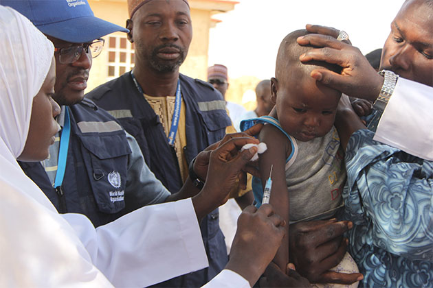
WHO©C. Onuekwe

The World Health Organization (WHO) recommends a single dose of the yellow fever vaccine for individuals aged 9 months or older living in or traveling to areas at risk of disease transmission, but there is a lack of data on its long-term efficacy when administered to infants. José Enrique Mejía, Inserm researcher at Unit 1043 Center for Pathophysiology of Toulouse Purpan and Cristina Domingo from the Robert Koch Institute in Berlin have recently shown that around half of children initially protected by the vaccination at 9 months of age lose that protection within the next 2 to 5 years, due to disappearance of the neutralizing antibodies. This research has been published in The Lancet Infectious Diseases.

Yellow fever is a viral infection spread by various species of mosquito and is rife in 34 countries in Africa and 13 in Latin America. Infection may be asymptomatic and go unnoticed or, on the contrary, it may progress rapidly to severe illness with fever, headache, muscle pain, nausea, vomiting and fatigue. The virus attacks the liver cells, often causing jaundice from which the disease gets its name. Severe bleeding occurs in 25 to 50 % of cases, with high levels of mortality observed 7 to 10 days after the onset of symptoms.

Since 2013, WHO recommends a single dose of the vaccine for life-long protection. This recommendation is based on proof of long-term efficacy, in vitro and in vivo, established in adults and children over 2 years of age.

But data on the long-term efficacy of primary vaccination in infants are absent, despite 9-12-month-olds being the main targets of routine vaccination in countries in which yellow fever is endemic. In this respect, WHO recommended research into the long-term persistence of the immunity conferred by vaccination in this age group. This research was performed by José Enrique Mejía from Unit 1043 Center for Pathophysiology of Toulouse Purpan in partnership with Cristina Domingo from Robert Koch Institute in Berlin, and researchers from the USA, Ghana and Mali, with support from the Wellcome Trust.

Their study verified whether children to whom the vaccine was administered at around 9 months of age were still protected several years later. The team studied two cohorts, one from Mali (587 children) and the other from Ghana (436 children), in whom the levels of specific antibodies to the yellow fever virus had been measured 4 weeks after vaccination. They then repeated the measurement several years later, with findings from previous studies enabling them to estimate that levels above 0.5 IU/ml should protect children from infection.

In the Malian cohort, 4.5 years after vaccination, only half of the children continued to present levels of antibodies above 0.5 IU/ml. And 19.3 % presented detectable antibodies but at levels below this recommended threshold (<0.5 IU/ml). The proportion of children seropositive for these antibodies was therefore 69.7 % as opposed to 96.7 % just after vaccination.

In the Ghanaian cohort, 2.5 years after vaccination, only around 30 % of children continued to be protected against infection and 11.7 % continued to present specific antibodies but in low concentrations (<0.5 IU/ml). All in all, 39.4 % of the children were considered seropositive as opposed to 72.7 % just after vaccination.

Irrespective of the differences in vaccine efficacy between these two groups, which could be explained by ethnic and environmental factors (urban/rural population, seasonality of vaccination, diet, exposure to other infectious agents, etc.), the results in both cases show a substantial fall – practically by half – in the levels of protective antibodies in the years following vaccination, and which predict the absence of protection against infection for large numbers of children.

“Our data suggest that a booster may be necessary when the 1st vaccination is performed in 9-12-month-olds, but we will need more precise knowledge of the decrease in antibodies over time. Maintaining immunity to the virus during childhood and in adulthood is fundamental for obtaining vaccine coverage beyond the threshold of 80 % of the population in order to prevent the risk of epidemic”, concludes Mejía.

New prebiotics: benefits without the downsides?

©Photo by Gesina Kunkel on Unsplash

Prebiotics are currently a preferred treatment for certain metabolic disorders, as they can restore the balance of dysfunctional gut microbiota, and improve the body’s metabolism. However, these substances have to be used at high doses, which can result in patients experiencing bloating and flatulence. A research group led by Matteo Serino, Inserm researcher at the Digestive Health Research Institute (Inserm/Inra/ENVT/UT3 Paul Sabatier), has recently shown three plant extracts to have prebiotic effects in obese/diabetic mice, with a shorter duration of treatment and at lower doses to the prebiotics currently in use. For Matteo Serino, this raises the question: do we need to reconsider the protocol for prebiotic administration? These results have been published in Molecular Nutrition & Food Research.

Dysbiosis of the gut microbiota corresponds to an imbalance in the populations of gut microorganisms and/or a change in their activity. This imbalance is often associated with metabolic disorders such as obesity and type 2 diabetes.

It has been shown that such dysbiosis can be targeted with prebiotics, long molecules that are not digested by the body but which increase the growth and activity of certain bacteria and thus improve patients’ carbohydrate and lipid metabolism. However, the duration of treatment is often greater than 4 weeks, and the effective dose at which an improvement in health starts to be observed can result in adverse effects such as bloating and flatulence.

A team of researchers from the Digestive Health Research Institute sought to identify other substances that might have a prebiotic effect. This led them to study extracts from three plants that had previously shown some beneficial effects: a red alga called Porphyra umbilicalis, the leaves of the lemon balm plant, Melissa officinalis L., and fruit extracts from an Indian plant, Emblica officinalis Gaertn.

The researchers administered these extracts to obese and diabetic mice for just two weeks, and at doses proportionally lower than the effective doses of traditional prebiotics.

The team considered a range of criteria in evaluating the individual prebiotic effects of the plant extracts.

The researchers first measured the impact of these substances on the diversity (the number of bacteria from different populations) and activity of the gut microbiota before and after treatment. “Our results were broadly positive for microbial diversity, with all the extracts increasing the abundance of at least one group of bacteria,” explains Matteo Serino, the Inserm researcher who led the study.

Beyond the impact on the microbiota, improved metabolism was one of the key criteria for evaluating the prebiotic efficacy of an extract. The research team observed that each of the three substances produced an increase in the gut of the quantity of the various short-chain fatty acids that are produced by bacterial metabolism of prebiotics and contribute to improving host metabolism.

The researchers also measured the presence of various markers in the blood that would enable them to demonstrate any metabolic improvements. The best result was obtained with the extract from the red alga P.umbilicalis. The researchers measured a 30% decrease in the level of blood triglycerides, large molecules with long-chain fatty acids that are present in excess levels in the blood of patients with metabolic disorders. This finding thus suggests an improvement in lipid metabolism among the treated animals.

“What’s interesting to note is that all of these effects were observed after just two weeks of treatment, and at low doses. This suggests the need for further studies to find out if this type of treatment could be effective in humans, and to test whether the extracts currently in use remain effective when given at low doses, thus limiting their side effects. That would mean reconsidering the existing protocol for prebiotic administration,” concludes Matteo Serino.

High-risk pregnancy: the interferon effect

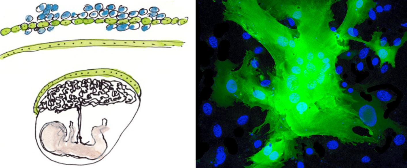
Artist’s representation of cells and placenta © Fabrice Hyber – Organoïde-Institut Pasteur/Fluorescence microscopy of fused cells (in green) and nucleus (in blue). © Institut Pasteur

Discovery of a cellular mechanism involved in abnormal placental development during some high-risk pregnancies.

High-risk pregnancies occur frequently and may be caused by various factors. It is estimated that 10 to 20% of pregnant women miscarry during their first trimester of pregnancy. Slow fetal growth may also arise as a result of maternal infection with certain microbes, parasites or viruses (such as toxoplasmosis or infection with rubella virus, cytomegalovirus, herpes or Zika) or because of genetic or autoimmune diseases. Teams from the Institut Pasteur, the CNRS, Inserm, Necker-Enfants Malades Hospital (AP-HP) and Université de Paris have identified a new cellular mechanism that alters placental development, potentially causing serious complications during pregnancy. The mechanism is linked with the production of interferon, a molecule produced in response to infection, especially viral infection. The findings are published in Science on July 11, 2019.

The placenta is both a surface for exchange and a barrier between mother and fetus – it delivers nutrients needed for fetal growth, produces hormones and protects the fetus from microbes and the maternal immune system. The external layer of the placenta, known as the syncytiotrophoblast, is composed of cells which fuse together, forming giant cells that are optimized for the placenta’s barrier and exchange functions. Cell fusion is mediated by a protein known as syncytin. If the syncytiotrophoblast fails to form correctly, it can cause placental insufficiency and hinder fetal development. An abnormal syncytiotrophoblast can be observed in conditions such as slow intrauterine growth, the lupus and in women whose fetus has Down syndrome.

Interferon is a substance produced by immune cells during infection to combat viruses and other intracellular microbes. High levels of interferon are observed in autoimmune or inflammatory diseases such as lupus, and also in some infections. In this study, the scientists demonstrated that interferon is responsible for placental abnormality and that it acts by preventing syncytiotrophoblast formation. Specifically, interferon induces the production of a family of cellular proteins known as IFITMs (interferon-induced transmembrane proteins), which block the fusion activity of syncytin.

IFITM proteins are beneficial since they prevent viral fusion with cellular membrane, thereby stopping viruses from entering and multiplying within cells. The scientists used experimental models and human cells to demonstrate that this beneficial effect can nevertheless be harmful if IFITM proteins are produced in an important level in the placenta.

“Identifying the role of IFITMs gives us a better understanding of the mechanisms involved in placental development and how it may be disrupted during infections and other diseases,” comments Olivier Schwartz, Head of the Virus and Immunity Unit at the Institut Pasteur and joint last author of the paper. The scientists want to investigate whether placental pathologies of unknown etiology, such as some early spontaneous abortions and occurrences of preeclampsia, also involve IFITM proteins. In the longer term, blocking the effects of IFITMs could represent a new therapeutic strategy to prevent interferon-related placental abnormality.

In addition to the institutions mentioned above, this research was funded by the ANRS, Sidaction, the French Vaccine Research Institute (VRI), LabEx IBEID and the European Research Council (ERC).

fermer