Menu

COVID-19: A “Programmed Cell Death” Phenomenon in Hospitalized Patients

Coronavirus SARS-CoV-2

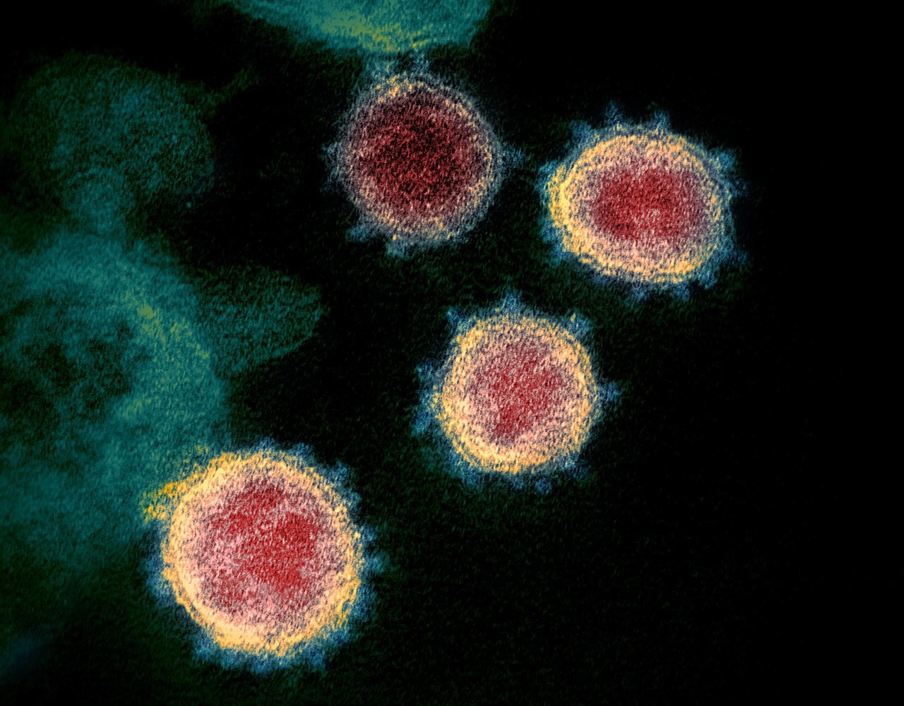
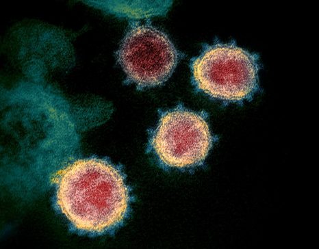
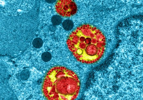
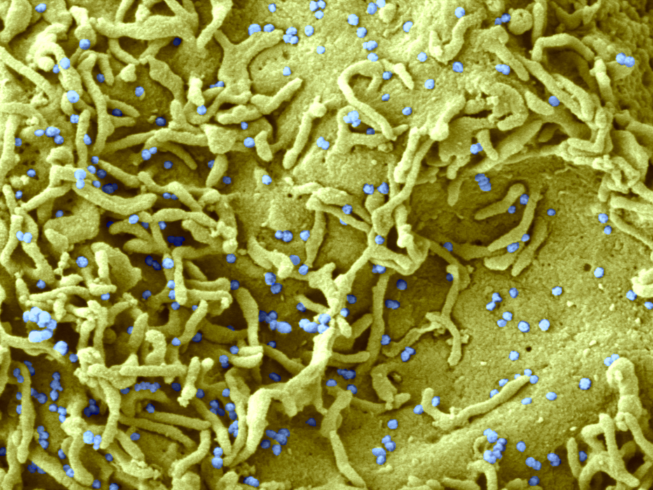
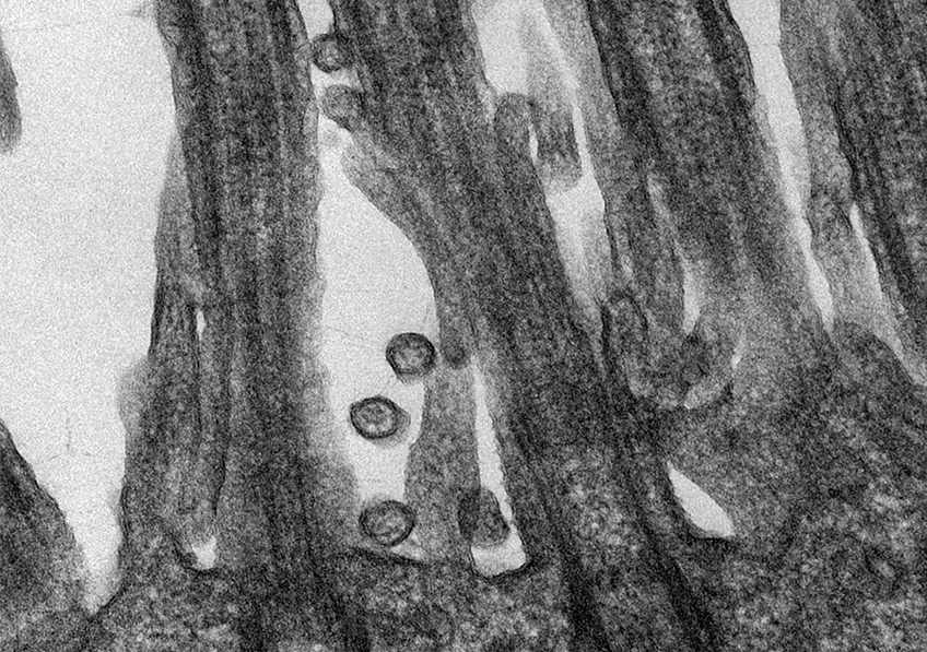
SARS-CoV-2 coronavirus responsible for COVID-19 disease attached to human respiratory epithelial cells. ©M.Rosa-Calatraval/O.Terrier/A.Pizzorno/E.Errazuriz-Cerda

Almost 60% of patients hospitalized for COVID-19 have lymphopenia – a lower than normal number of lymphocytes[1] in the blood circulation. The mechanisms underlying this condition have long been poorly understood. In a new study, researchers from Inserm and Université de Paris[2] in collaboration with Canadian and Portuguese teams (Université Laval and Life and Health Sciences Research Institute [ICVS], respectively)[3] have revealed a phenomenon of programmed cell death known as “apoptosis”[4] that would explain the loss of lymphocytes in these patients. They have also shown in vitro that this process can be reversed by using caspase inhibitors – molecules that block the action of the enzymes responsible for apoptosis. These findings, published in the January 22, 2022 issue of Cell Death & Differentiation, make it possible to envisage new therapeutic avenues for patients with severe forms of COVID-19.

COVID-19 is a disease that varies broadly from one patient to another. While most infected people are asymptomatic or have mild symptoms, others develop severe forms of the disease.

Research on hospitalized patients had until now focused mainly on the inflammatory state that characterizes serious forms, and less so on other biomarkers. Yet among these patients, 60% have lymphopenia. The number of lymphocytes in their blood is lower than normal, especially the number of CD4 T cells.

lymphocytes apoptotiques

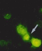
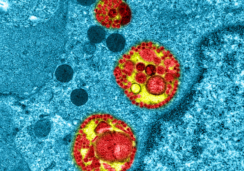
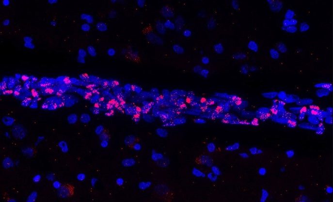
Detection of apoptotic lymphocytes with nuclear condensation and fragmentation visualizable by electron microscopy and not immunofluorescence. © Jérôme Estaquier

The team of Inserm researcher Jérôme Estaquier, at Unit 1124 (Inserm/Université de Paris) and Université Laval in Quebec, has studied this phenomenon. Thanks to their long history of researching AIDS – a disease for which a low blood CD4 level is a marker of poor prognosis – the scientists were able to apply their knowledge of these processes to COVID-19.

As part of their research, the scientists studied blood samples taken from patients hospitalized from April to June 2020 for COVID-19 (some of them in intensive care) and compared them with those of healthy donors. They showed that lymphopenia was correlated with the presence of several severity biomarkers, including the death ligand, FasL.

Programmed cell death  

They also showed that a process of programmed cell death – apoptosis – is responsible for the disappearance of CD4 T cells in hospitalized patients with COVID-19.

In an attempt to block this process, the researchers then drew on their previous HIV research in animal models, in which they had shown that administering molecules called caspase inhibitors can stop apoptosis, restore CD4 T cells, and prevent the onset of AIDS. Here they show that, with these molecules, the process of lymphocyte apoptosis is also reversible in the case of COVID-19.

These findings[5] open up new therapeutic avenues for the early treatment of hospitalized patients with lymphopenia. “The idea is now to set up phase 1 clinical trials to test the safety of caspase inhibitors in humans. CD4 cells are the cornerstone of the immune system. As such, these molecules could ultimately be useful for patients presenting with lymphopenia on admission to hospital, ” emphasizes Estaquier.


[1] Lymphocytes are white blood cells that play a key role in the immune system. They defend the body in the face of attacks.

[2] At Unit 1124 “Environmental Toxicity, Therapeutic Targets, Cellular Signaling and Biomarkers”

[3] The study was also conducted in collaboration with and the French National Center for Scientific Research (CNRS) (Institute of Human Genetics, IGH).

[4] Apoptosis is the programmed death of cells. This is a process by which cells trigger their self-destruction in response to a stress signal or death ligand.

[5] This study was funded by FRM and AbbVie. 

COVID-19: New Avenues to Explain Why Children Are at Less Risk of Severe Forms

This colorized electron microscope image shows SARS-CoV-2 isolated from a patient in the USA. Viral particles emerge from the surface of the laboratory-cultivated cells. © NIAID-RML Creative Commons.


Why are children less susceptible than adults to critical forms of COVID-19? This question has been studied by many scientists since the pandemic began. A number of interesting avenues are emerging, notably suggesting differences in immune response following SARS-CoV-2 infection. In a new study, researchers from Inserm, Université d’Angers and Angers University Hospital, members or partners of the Regional Center for Research in Cancerology and Immunology Nantes-Angers (CRCINA) have shown that the interferon response, which is part of the innate immune response, differs according to the age of the patient. Their findings have been published in Frontiers in Immunology in November 2021.

The symptoms of COVID-19 vary widely from one person to another. While some are asymptomatic following SARS-CoV-2 infection, others develop severe and possibly fatal forms of the disease. Since the start of the pandemic, age has been identified as a major risk factor for developing a severe form of COVID-19. Unlike adults, and especially the elderly who are very vulnerable to infection, children usually have no clinical signs of the disease (or only mild symptoms).

Numerous research teams are trying to identify the immune response parameters that could explain this difference in susceptibility between young and elderly people.

In this collaborative research, scientists from Inserm and Université d’Angers at the Regional Center for Research in Cancerology and Immunology Nantes-Angers, as well as the Virology and Immunology laboratories at Angers University Hospital, have hypothesized that children are protected due to a stronger local innate immune response, in the nasopharyngeal mucosa. So far, there has been less research into innate immunity to COVID-19 than into adaptive immune response[1].

A closer look at immune response

Innate immunity is the immediate response that occurs locally, at the point of entry of a pathogenic microorganism, in any individual – even in the absence of prior contact with that microorganism. It is the first barrier of defense against pathogens. In the event of a viral infection, it primarily deploys Natural Killer cells

that kill the cells infected with a virus. It also induces the production of interferons by the infected cells, and it is these interferons that protect the adjacent cells from infection.

Adaptive immunity is a response that takes 5 to 7 days to become protective when the pathogen is encountered for the first time (primo-infection), but is more rapidly effective once the pathogen has already been encountered (this is known as a memory response). In the event of a viral infection, it deploys two types of protective immune cells: antibody-producing B cells that bind to the virus and “neutralize” it, namely by preventing it from entering the cells and by promoting its elimination, and cytotoxic CD8+ T cells that kill the infected cells. The B and T cells recognize protein structures (of the virus) known as antigens.

Following infection with a virus or vaccination, the level of antibodies and lymphocytes recognizing the virus decreases over time. Nevertheless, the so-called “memory” B and T cells remain in the body and keep watch, acting faster and more efficiently if they encounter the same virus in the future.

Different interferon responses
 

In their research, the scientists analyzed nasopharyngeal samples from 226 people who had come for a PCR test at a drive-through screening center at Angers University Hospital between March 2020 and March 2021. Of these individuals, 147 were infected with SARS-CoV-2. “Our research was original in that we had not preselected the participants, so as not to bias the results, and also that we were interested in innate immunity – and more specifically the interferon response,” emphasizes Yves Delneste, an Inserm researcher who took part in this study.

When cells are infected with any given virus, they rapidly produce type I (IFN-α/β) and type III (IFN-l) interferons, which are powerful natural antiviral molecules. They are called interferons because they “interfere” with the replication of the virus and protect the adjacent cells from infection.

While these interferons all have antiviral activity, their modes of action are not redundant. Each induces an antiviral response of a different intensity and duration and has a different but complementary action on immune response[2].

An inadequate or inappropriate interferon response will not make it possible to contain the replication of the virus or it may promote a pathological immune response (for example, an exacerbation of the immune system as seen with severe forms of COVID-19).

Analysis of the samples studied by the research team revealed that in subjects infected with SARS-CoV-2, the expression profiles of type I (IFN-α/β) and type III (IFN-l) interferons differ with age. Thus, children under 15 years of age have an increased expression of type III interferons, locally-acting molecules with limited inflammatory properties, which control the virus locally at its entry point in the nasopharyngeal mucosa. Conversely, adults, especially elderly adults, preferentially express type I interferons, which are inflammatory and have a more systemic action (in the whole body).

“These findings help to explain why children are less susceptible to critical forms of COVID-19 than adults. Type III interferons, which primarily act by protecting the epithelium at local level, could control infection at the point of entry, without inducing excessive widespread inflammation, thereby preventing the slide towards the inflammatory storm with mass cell destruction that is seen in severe forms,” emphasize Pascale Jeannin (university professor and hospital practitioner) and Dominique Couez (university professor) in Angers, who led this research.

Based on these findings, the scientists will now conduct a prospective study to evaluate whether, in children with clinical signs of the disease, the characteristics of the interferon response associated with severe forms in adults are present and whether they can predict the course of infection.


1 see text box on innate and adaptive immunity

2 see text box

Covid-19 booster doses: start of inclusions in the COVIBOOST trial

Vaccin Anti Covid

French health authorities recommend that all adults over 18 years of age perform a booster injection with a MRNA to ensure maximum and prolonged vaccine protection.© AdobeStock


In a context of winter circulation of the virus, a constant increase in the number of confirmed cases and the appearance of new variants, the French health authorities recommend that all adults over 18 years of age perform a booster injection with a MRNA to ensure maximum and prolonged vaccine protection.

Administration of a 3 rd dose of a different vaccine could nevertheless have advantages in terms of efficacy and safety, but also in terms of cost and acceptability.

The COVIBOOST trial is designed to study the immune response of the two candidates based recombinant protein vaccine associated with an adjuvant developed by Sanofi Pasteur and GSK and that of a 3 rd dose of the Pfizer-Biontech vaccine.

This randomized double-blind trial, promoted by Assistance Publique – Hôpitaux de Paris, will be carried out in 11 hospitals in the COVIREIVAC network coordinated by Inserm. It starts on December 8.

300 participants who had previously received two doses of the Pfizer-BioNTech vaccine ( 2nd dose received within 5 to 7 months) and without a history of Covid-19 will be included, half of them aged 65 and over.

They will randomly receive a booster dose:

– the mRNA vaccine from Pfizer-BioNTech (Comirnaty®)

– the adjuvanted recombinant protein vaccine from Sanofi-Pasteur / GSK based on the original strain of the virus (Wuhan strain)

– the adjuvanted recombinant protein vaccine from Sanofi-Pasteur / GSK based on the beta variant (South African variant)

The data from the trial will make it possible to measure the immune response induced by the three vaccines studied as a booster, and its effectiveness on the different variants but also its persistence at 3 and 12 months, depending on age.

This is the eighth study launched by COVIREIVAC.

The booster vaccination as part of the trial will validate the health pass for the 3rd dose according to national requirements.

Today the booster dose with an mRNA vaccine has become essential. But we hope to broaden the range of possibilities with other vaccine technologies. » Explains Marie Lachâtre, infectious disease doctor (Hôpital Cochin and Hôtel Dieu / APHP) and member of COVIREIVAC. “The Sanofi-Pasteur / GSK vaccine candidates have for several months been the subject of phase 3 clinical trials as a primary vaccination or as a booster. Today we want to assess the immune response they induce by booster compared to that of the Pfizer-BioNTech vaccine ”.

This clinical study has been labeled a “national research priority” on Covid-19 by the Ministry of Health and Solidarity.

Launched in October 2020, the COVIREIVAC platform coordinated by Inserm and F CRIN in conjunction with 32 university hospitals and a network of 11 immunology laboratories aims to conduct and promote excellent clinical vaccine research in France. Since October 1, 2020, 50,000 volunteers have registered to participate in research efforts and improve knowledge about these new vaccines. This is an unprecedented initiative in our country. The platform is managed by Inserm, and the clinical operational component is coordinated by the Assistance Publique-Hôpitaux de Paris of the various CHUs. New research projects are regularly launched within the framework of COVIREIVAC.

Even if several vaccines against Covid-19 are available, it is imperative to continue research in order to deepen scientific knowledge, in particular the duration of protection and the quality of the immune response.

The objective of the clinical studies coordinated by COVIREIVAC is to provide answers to these research questions.  

Lancement du Groupement d’intérêt scientifique Obépine sur les eaux usées

Covid-19 : Le lourd impact de l’épidémie sur la santé mentale des étudiants, notamment pendant les périodes de confinements

COVID-19: Artificial Intelligence Identifies Gene Signature Specific to Patients Suffering from Critical Forms

SARS-CoV-2

Covid-19: Intracellular observation of reconstituted human respiratory epithelium MucilAir™ infected with SARS-CoV-2. © Manuel Rosa-Calatrava, Inserm ; Olivier Terrier, CNRS ; Andrés Pizzorno, Signia Therapeutics ; Elisabeth Errazuriz-Cerda  UCBL1 CIQLE. VirPath (Centre International de Recherche en Infectiologie U1111 Inserm – UMR 5308 CNRS – ENS Lyon – UCBL1). Colorisé par Noa Rosa C.


What are the molecular and genetic characteristics that distinguish patients with critical forms of COVID-19 – and particularly acute respiratory distress syndrome (ARDS)? To answer this question, researchers from Inserm and Université de Strasbourg at Unit U1109 Molecular Immunology and Rheumatology, in collaboration with clinician-researchers at the Strasbourg University Hospitals, have investigated the biological and genomic data of a targeted cohort of young patients. Patients hospitalized in intensive care with ARDS were compared with COVID-19 patients hospitalized in a non-critical care ward.

As part of a Franco-US collaboration with researchers from the company Genuity Science in Boston and the University of Southern California in Los Angeles and using the most advanced artificial intelligence techniques to interpret these data, the scientists have succeeded in identifying a gene signature that differentiates these critical patients from their non-critical counterparts. Some of the genes included in this signature could ultimately become therapeutic targets for severe forms of COVID-19 or ARDS. The findings from this research have been published in Science Translational Medicine.

COVID-19 varies broadly from one patient to another. While some are asymptomatic, others develop flu-like symptoms. Then there are others who progress towards severe forms of the disease, in some cases developing acute respiratory distress syndrome (ARDS) that requires mechanical ventilation in an intensive care unit. Although this group of patients accounts for only a small proportion of those infected with the virus, its mortality rate is high – reaching around 25%.

While age and comorbidities such as diabetes and cardiovascular diseases are the main risk factors for developing these severe, potentially fatal, forms of COVID-19, scientists do not yet know why some younger and previously healthy patients also develop these forms. In molecular and genetic terms, what is it that distinguishes these patients with severe respiratory symptoms from the others?

There has been research into the subject since the start of the pandemic and some avenues have been identified, but so far each study addressed the question from a single methodological angle, generally focusing only on one aspect – genetic and metabolic factors, immune response parameters, etc.

Young patients with no comorbidities

The scientists from Inserm and Université de Strasbourg, in conjunction with the Strasbourg University Hospitals, were in this case interested in a patient cohort with restrictive and strict inclusion criteria. The patients had been hospitalized during the first wave of the pandemic, were under 50 years of age and had no major comorbidities. A total of 72 patients were recruited into two groups, one consisting of intensive care patients with ARDS and the other of less severe COVID-19 patients hospitalized in a non-critical ward. A “control” group of 22 healthy individuals was also studied.

“We chose to focus on a restricted but very well-defined patient cohort, excluding confounding factors such as age and certain diseases so that we could really study the molecular and genetic mechanisms directly associated with the severe forms, which are exclusively linked to viral infection and not to other pre-existing risk factors,” emphasizes Seiamak Bahram [1], last author of the study.

The scientists collected various samples in order to perform a multi-omics analysis –that is say retrieve and analyze the various genomic, proteomic, transcriptomic (investigation of all messenger RNA) data and other virological, immunological, and serological data from these patients. This allowed them to confirm that ARDS is associated with a major inflammatory state and an immune system surge (the so-called “cytokine storm”).

Using artificial intelligence

However, given the considerable mass of data generated as part of this multi-omics analysis, it was impossible to take the interpretation further without the help of artificial intelligence (AI). Thus, in collaboration with the AI Research Institute of Genuity Science [2] , a biotech in Boston (USA), the team was able to identify a network of 600 genes involved in the progression towards the critical forms of COVID-19, thanks to the cross-application of several AI algorithms (including one having run on the quantum computer made available by the University of Southern California in Los Angeles).

As part of this transatlantic collaboration, these large quantities of data were modelled and analyzed with the help of AI, making it possible to more accurately identify five genes that are overexpressed in these patients.

One of them, ADAM9, is a particularly interesting “driver gene,” with previous studies having shown that it interacts with SARS-CoV-2 proteins. The findings obtained here are consistent with that, suggesting that ADAM9 overexpression would “drive” some patients towards severe forms of COVID-19 and ARDS.

The researchers then conducted in vitro experiments which showed that blocking ADAM9 in cell lines is associated with a reduction in the quantities of SARS-CoV-2 in these cells, as well as reduced replication of the virus, thereby confirming not just its importance in critical disease but also its potential as a therapeutic target.

Of course, further studies will have to be carried out to confirm this last point, but the scientists believe that these findings have opened up an interesting therapeutic avenue, especially given the current clinical trials in oncology that are testing monoclonal antibodies which inhibit ADAM9. Therapeutic repurposing strategies could therefore be considered in the longer term.


[1] Professor Seiamak Bahram, a university professor and hospital practitioner, is the director of Inserm Unit 1109, head of the Strasbourg Precision Medicine Interdisciplinary Thematic Institute, and head of the Biological Immunology Department at Strasbourg University Hospitals.

[2] It has since become the company HiberCell

Chatbot for addressing COVID-19 vaccine hesitancy

ordinateurResearchers from the CNRS, INSERM, and ENS-PSL show that such an interface is indeed capable of swaying the vaccine-hesitant. © seth schwiet on Unsplash


  • A considerable fraction of the population is reluctant to get vaccinated against COVID-19.
  • French scientists have designed a chatbot that offers personalised responses to questions posed by the curious or hesitant—and have demonstrated its effectiveness.

What if a few minutes of interaction with a chatbot could effectively address vaccine concerns? In an article published in the Journal of Experimental Psychology: Applied (28 October 2021), researchers from the CNRS, INSERM, and ENS-PSL show that such an interface is indeed capable of swaying the vaccine-hesitant.

Vaccine hesitancy is one of the major challenges in containing the COVID-19 pandemic. Previous studies have revealed that mass communication—through short messages relayed by television or radio—is not a very effective means of persuading the hesitant. In contrast, discussing your particular concerns with an expert whom you trust can be more persuasive, but having a face-to-face talk with every vaccine-hesitant individual is impractical.

To overcome this problem, a team of cognitive scientists from the Institut Jean-Nicod (CNRS / ENS-PSL) and the Laboratoire de Neurosciences Cognitives et Computationnelles (INSERM / ENS-PSL) created a chatbot that provides users with answers to 51 common questions about COVID-19 vaccines.1

Chatbots have the advantage of offering quick, personalized Q and A sessions while reaching a large number of people.

The team tested their chatbot with 338 individuals and compared their reactions to those of a control group of 305 participants who only read a brief paragraph that gave information about COVID-19 vaccines. After a few minutes of interaction with the chatbot, the number of participants with positive views of vaccination increased by 37%. People were also more open to getting vaccinated after using the chatbot: declarations of vaccine refusal fell 20%. Such changes in attitude were negligible in the control group.

It remains to be shown whether the effects of chatbot interaction are lasting, and whether they are the same across age groups, and among those most resistant to vaccination.2

Nevertheless, this study has demonstrated that a chatbot can indirectly reach a very large audience: half of the experimental group later tried to persuade others to get vaccinated, with three-quarters of them stating they drew on information provided by the chatbot to do so.

These findings suggest that a chatbot regularly updated to reflect the latest vaccine science could be an effective tool to help reduce vaccine hesitancy.


Notes

1The questions were selected on the basis of surveys on reasons for vaccine hesitancy as well as articles about vaccine-related preconceptions. Their answers were prepared from scientific sources and approved by COVID-19 vaccine specialists.

2On average, the group of participants was younger and more educated than the overall population.

COVID-19: How Does SARS-CoV-2 Infection Affect Vascular Irrigation of the Brain?

tissu cérébral humain post-mortem

Fluorescent image of post-mortem human brain tissue showing cell nuclei (blue) that reveal a blood vessel in which the vascular endothelial cells express the genetic material of SARS-CoV-2 (red). © Vincent Prévot/Inserm


A large number of researchers are currently mobilized to increase knowledge of SARS-CoV-2 in order to improve the treatment of infected patients and try to predict the future health impacts of infection with the virus. As part of an international collaboration, researchers from Inserm, Université de Lille, Lille University Hospital, and Pasteur Institute Lille within the Lille Neuroscience & Cognition laboratory, along with their colleagues from the CNRS1, have been the first to identify a direct effect of SARS-CoV-2 on the brain’s blood vessels. Certain cells, namely the cerebral vascular endothelial cells – essential components of the blood-brain barrier that protects the brain – are affected by a phenomenon of cell death. These findings, published in the journal Nature Neuroscience, particularly question the long-term impacts of the disease.

The blood vessels are comprised of endothelial cells. These include the vascular endothelial cells in the brain that make up the blood-brain barrier (BBB). The primary function of the BBB is to isolate the central nervous system from the bloodstream, preventing foreign substances or potentially toxic molecules from entering the brain and spinal cord while allowing the transfer of nutrients essential to their activity. As part of this effort, the vascular endothelial cells in the brain therefore play a key role in the proper irrigation of the organ, with their survival being essential for it to function correctly.

Within the framework of an international collaboration funded by the European Research Council3, the authors of the study looked at the vascular endothelial cells of the brain and the consequences of SARS-CoV-2 infection on their functioning.

Using preclinical research models and also by studying the cortex of patients who died as a result of SARS-CoV-2 infection, the researchers have shown that infection leads to the death of endothelial cells in the brain, resulting in the appearance of “ghost vessels” in the brain (empty tubes with no endothelial cells).

As a result, these essential cells can no longer perform their function in the BBB.

How does this endothelial cell death occur? What are the mechanisms involved? Thanks to state-of-the-art techniques2, the team has discovered that SARS-CoV-2 generates the manufacturing, from its own genetic material, of molecular scissors by the endothelial cells it infects. These scissors cleave a protein called NEMO which, being necessary for the endothelial cells to survive, therefore leads to their death.


The impacts of endothelial cell death on brain function

According to the scientists, the death of vascular endothelial cells in the brain can have two major consequences:

  • A temporary rupture of the BBB causing microbleeds in regions where the blood is not meant to have free access.
  • Hypoperfusion of some brain regions (due to the presence of non-functional ghost vessels), which is a decrease in blood flow that in the most serious cases can be fatal.

However, the study shows that the situation is reversible.

Furthermore, the scientists are interested in the long-term impacts of this phase of vulnerability during which brain irrigation is decreased. According to them, even if this hypothesis remains to be verified, this window of time could predispose certain people with the disease to develop cognitive or neurodegenerative disorders, or even dementia.

“This awareness of the severity of SARS-CoV-2 infection and its impacts on proper brain function is vital to enable the best possible management of infected patients in the years to come,” concludes Vincent Prévot, Inserm Research Director.


1 At the Center for Infection and Immunity of Lille (CNRS/Inserm/Institut Pasteur Lille/Université de Lille/Lille University Hospital)

2 Such as transgenesis, single-cell RNA sequencing, mass spectrometry and super-resolution microscopy.

3 Program funded by the European Research Council (ERC Synergy), with the participation of Drs. Prévot (Inserm, France), Nogueiras (University of Santiago de Compostela, Spain), and Schwaninger (University of Lübeck, Germany).

Study on the role of “ceramides” in infection by SARS-CoV-2 which could constitute a biomarker of severity and a therapeutic target in the management of Covid-19


SARS-CoV-2

Electron microscopy of a cell infected with SARS-CoV-2 © Philippe Roingeard, Anne Bull-Maurer, Sonia Georgeault, unité Inserm U1259 MAVIVH & Université de Tours, France.


Teams from the psychiatry and addictology department of the Corentin-Celton AP-HP hospital, the University of Paris and Inserm in collaboration with the University of Erlangen-Nuremberg and the University of Duisburg-Essen , coordinated by Dr Nicolas Hoertel, Prof Jo hannes Kornhuber and Prof Erich Gulbins, published on October 4, 2021 in the journal Molecular Psychiatry (Nature Publishing Group) a summary of the results of the international literature on the central role that “ ceramides ”, a class of lipids, may play a role in SARS-CoV-2 infection.

Results from in vitro data [1, 2] indicate that an enzyme found in cell lysosomes, acid sphingomyelinase (ASM), is activated by the virus upon binding to its ACE-2 cell receptor, inducing synthesis of a specific class of lipids, the “ceramides”, in the membrane of cells. These studies [1, 2] demonstrate that these ceramides serve as a gateway for the virus to infect cells.

Indeed, the reduction in the quantity of ceramides by functional inhibitors of ASM (called FIASMA, comprising in particular certain antidepressants such as fluoxetine or else fluvoxamine) or the use of anti-ceramide antibodies make it possible to greatly reduce infection in vitro according to these same studies [1-2] .

In addition, clinical data [3, 4] indicate that elevated plasma levels of ceramides are significantly and strongly associated with the clinical severity of infection and the severity of inflammation in patients with Covid-19.

Finally, preclinical [1, 2, 5, 6] , observational [7-9] and three clinical trials [10-12] , including two randomized placebo-controlled trials [10, 12] , conclude on potentially strong efficacy of fluvoxamine and fluoxetine against Covid-19. Several clinical trials using fluvoxamine or fluoxetine, necessary to confirm these very encouraging results, are underway in several countries (United States, Canada, South Africa, Brazil and Croatia).

This publication concludes that the activity of the enzyme ASM and the plasma levels of ceramides could allow a better understanding of this infection and its risk factors for poor prognosis, as well as the antiviral, anti-inflammatory and clinical effects observed with drugs that are functional ASM inhibitors, including fluoxetine and fluvoxamine.


References

  1. Carpinteiro, A. et al. Pharmacological inhibition of acid sphingomyelinase prevents uptake of SARS-CoV-2 by epithelial cells. Cell Rep. Med. 100142 (2020).
  2. Carpinteiro, A. et al. Inhibition of acid sphingomyelinase by ambroxol prevents SARS-CoV-2 entry into epithelial cells. J. Biol. Chem. 100701 (2021). doi: 10.1016 / j.jbc.2021.100701
  3. Marín-Corral J, Rodríguez-Morató J, Gomez-Gomez A, Pascual-Guardia S, Muñoz-Bermúdez R, Salazar-Degracia A, et al. Metabolic signatures associated with severity in hospitalized COVID-19 patients. Int J Mol Sci. 2021; 22: 4794.
  4. Khodadoust, M. Ceramide levels and COVID-19 respiratory distress, a causal relationship. Research Square. 2021. https://doi.org/10.21203/rs.3.rs-443020/v3 .
  5. Schloer, S. et al. Targeting the endolysosomal host-SARS-CoV-2 interface by clinically licensed functional inhibitors of acid sphingomyelinase (FIASMA) including the antidepressant fluoxetine. Emerg. Microbes Infect. 9, 2245–2255 (2020).
  6. Zimniak M. et al. The serotonin reuptake inhibitor Fluoxetine inhibits SARS-CoV-2 in human lung tissue. Sci Rep. 2021 Mar 15; 11 (1): 5890.
  7. Hoertel, N. et al. Association between antidepressant use and reduced risk of intubation or death in hospitalized patients with COVID-19: results from an observational study. Mol. Psychiatry (2021) .doi: 10.1038 / s41380-021-01021-4
  8. Diez-Quevedo C et al. Mental disorders, psychopharmacological treatments, and mortality in 2150 COVID-19 Spanish inpatients. Acta Psychiatr Scand. 2021 Jun; 143 (6): 526-534.
  9. Hoertel, N. et al. Association between FIASMAs and reduced risk of intubation or death in individuals hospitalized for severe COVID-19: an observational multicenter study. Clin Pharmacol Ther. 2021. https://doi.org/10.1002/cpt.2317 .
  10. Lenze, EJ et al. Fluvoxamine vs Placebo and Clinical Deterioration in Outpatients With Symptomatic COVID-19: A Randomized Clinical Trial. JAMA 324, 2292–2300 (2020).
  11. Seftel, D. & Boulware, DR Prospective Cohort of Fluvoxamine for Early Treatment of Coronavirus Disease 19. Open Forum Infect. Say. 8, ofab050 (2021).
  12. Reis, G., et al. Effect of Early Treatment with Fluvoxamine on Risk of Emergency Care and Hospitalization Among Patients with COVID-19: The TOGETHER Randomized Platform Clinical Trial. medRxiv . 2021; https://doi.org/10.1101/2021.08.19.21262323 .

Good tolerance of the BNT162b2 vaccine (Pfizer / BioNTech) and good efficacy of the immune response against SARS-CoV-2 variants in patients with systemic lupus

vaccin anti covid

Covid vaccine © Adobe Stock


Teams from the immunology department and internal medicine department 2 of the Pitié-Salpêtrière AP-HP hospital, Sorbonne University and Inserm, measured the antibody and cellular responses generated by a messenger RNA vaccine in patients with systemic lupus (LS). This work, coordinated by Professors Guy Gorochov (CIMI-Paris) and Zahir Amoura (National Lupus Reference Center) shows that vaccination is both well tolerated and effective, even against worrying variants. For the first time, the factors associated with a poor vaccine response in some of these patients have been identified in order to better anticipate who should benefit from reinforced protective measures and / or adapted vaccine protocols. This work was the subjectfrom an October 4 publication in the journal Annals of Rheumatic Diseases.

Systemic lupus (LS) is a chronic autoimmune disease preferentially affecting young women. The attacks are mainly articular, cutaneous, renal, cardio-respiratory, neurological and hematological. The disease is characterized by the production of autoantibodies directed against nuclear antigens. It typically evolves in spurts. For these patients, two hypotheses were considered concerning the effects of the messenger RNA vaccination: the occurrence of more frequent adverse effects, or even the exacerbation of the disease in patients with LS, or an ineffective response to vaccination for patients with LS. patients on immunosuppressants.

The teams compared the effects of vaccination in 136 patients with systemic lupus (LS). 126 of them, who received 2 doses of the BNT162b2 vaccine (Pfizer / BioNTech) and followed the entire clinical-biological surveillance course, were included in the final analysis. The two vaccine doses were separated by 21 to 28 days with a clinical-biological evaluation from the day of the first injection and up to 42 days after it (D42). The clinical activity of the disease was measured at each visit (D0, D7-14, D21-28, D42) using standardized indices. Some patients were vaccinated while their lupus disease was active.

During the 40-day follow-up, no significant change in disease activity was observed, either in patients active at the time of vaccination, or in those who showed no signs of the disease. The only notable side effects related to the vaccination were mild or moderate pain at the injection site.

A pseudo virus expressing the envelope (Spike) of the reference SARS-CoV-2 (D614G) or that of the variants B.1.1.7 (Alpha), B.1.617.1 (Kappa), B.1.617.2 (Delta ), B.1.617.3, B.1.1.28 (Gamma) and B.1.351 (Beta) was used to measure the neutralizing activity of the serum collected on D42. 82% of the patients tested were able to effectively neutralize the reference strain and the Alpha variant. As expected, a slight decrease in the neutralization efficiency of the other variants was noted, mainly for the Beta variant, neutralized by the serum of 60% of the patients tested, while the Delta variant was neutralized in 76% of cases.

At present, the vaccine response criteria are still poorly understood. Among the various treatments received against lupus, this work was able to combine methotrexate and mycophenolate mofetil, two immunosuppressive treatments, with lower rates of IgG responses to the human receptor binding domain (RBD) of SARS-CoV-2. (independently of immunosuppressive treatments). Long-term corticosteroid intake (median prednisone dose: 19 mg / day) was not, however, associated with a poor vaccine response.

This work evaluated certain therapeutic and biological criteria of vaccine response.

A decreased vaccine response, i.e. a lower level of protective antibodies, has been shown when the patient with LS is treated with methotrexate or mycophenolate mofetil (two immunosuppressive treatments). Conversely, taking corticosteroids, regularly prescribed in LS, was not associated with a poor vaccine response.

The study also looked at the immune status of patients at the start of the vaccination protocol. The level of circulating naive B lymphocytes (CD19 + CD27-IgD +) and the overall concentration of IgG antibodies on D0 were associated with a more intense vaccine response.

In conclusion, the vaccination of lupus patients with BNT162b2 is very well tolerated and its efficacy is reduced only in patients treated with methotrexate or mycophenolate mofetil. A pre-existing alteration in the adaptive humoral response is also associated with the poor vaccine response.

fermer