Menu

Blood Stem Cell Immune Memory: A New Research Avenue in COVID-19

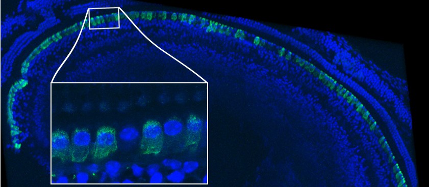
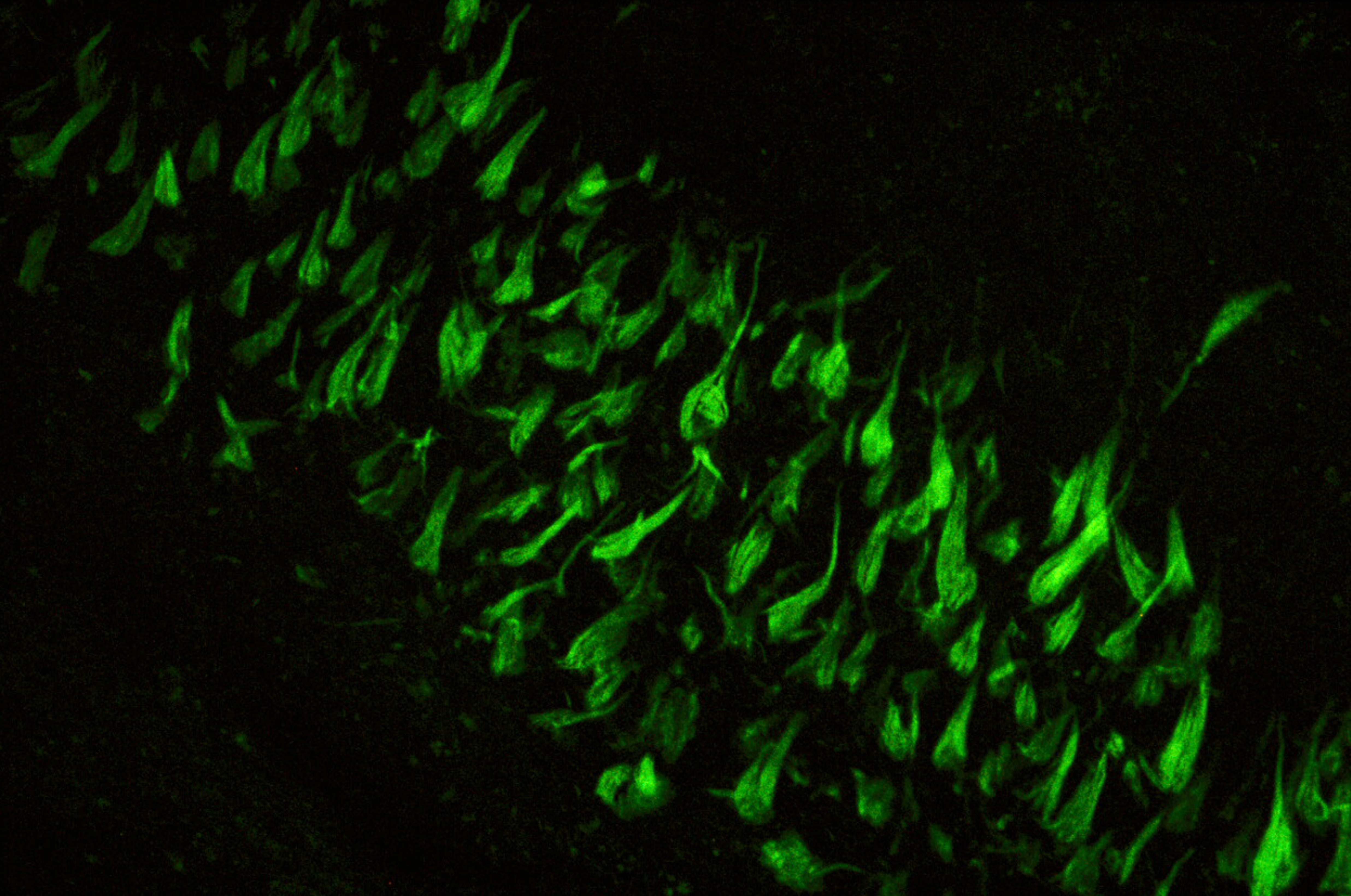
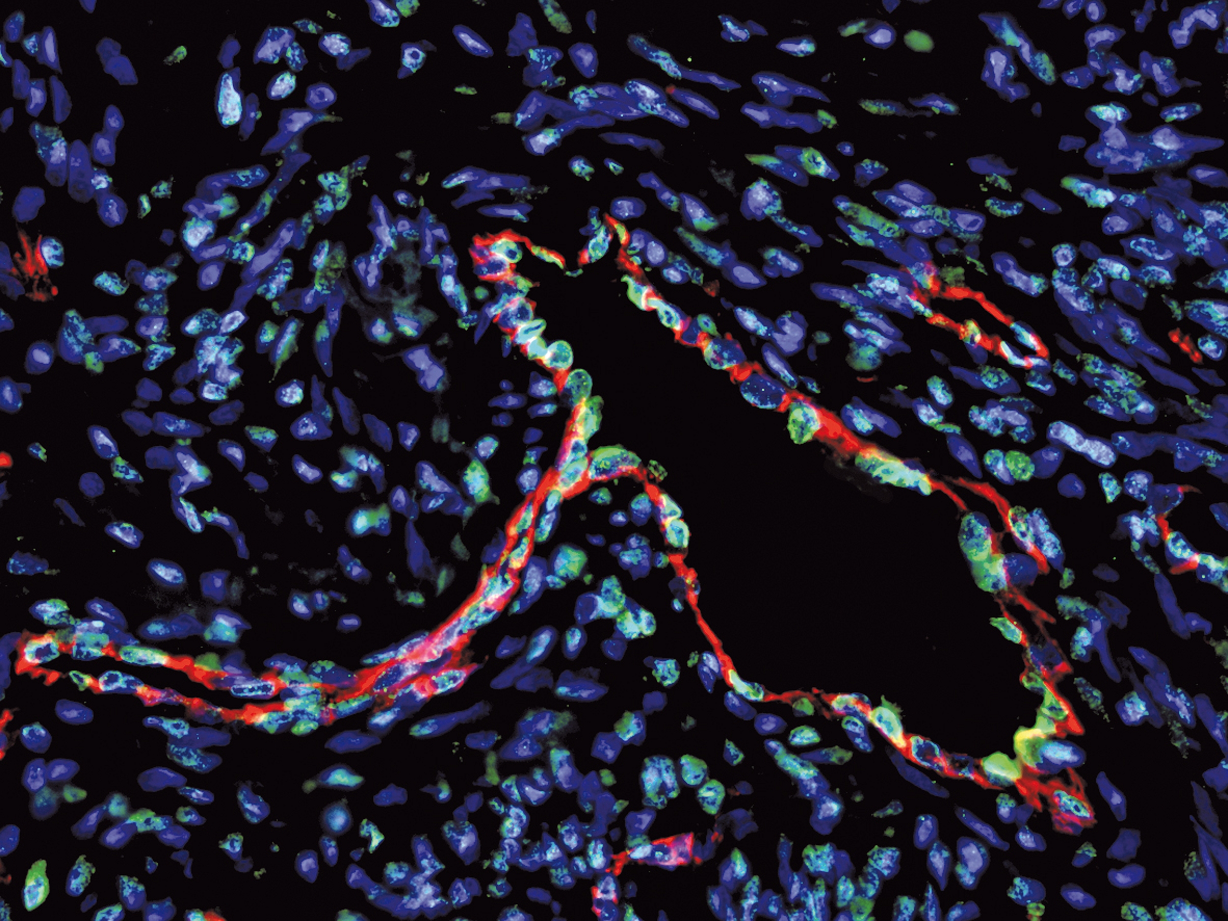
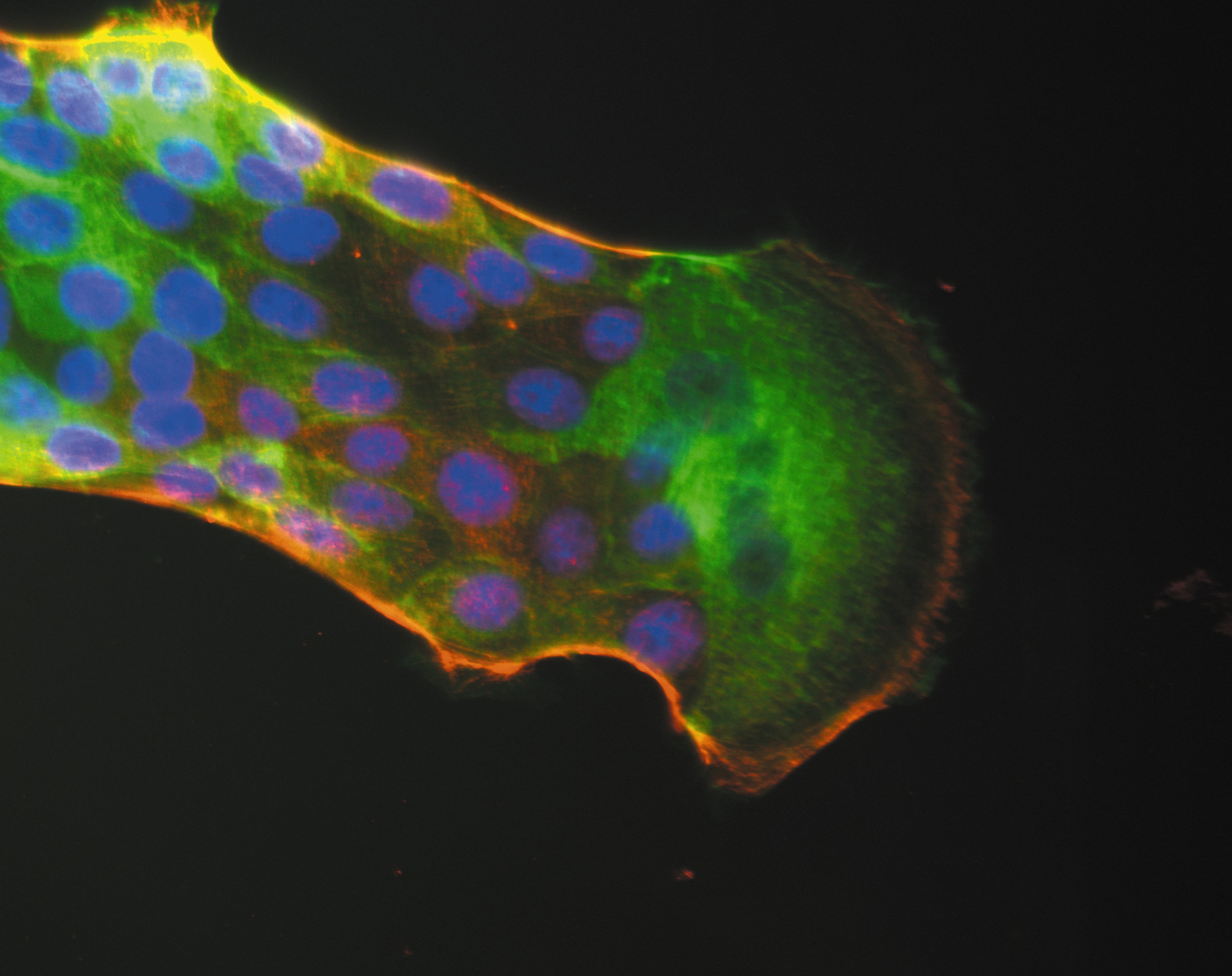
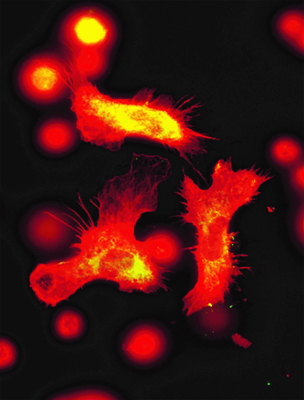
Immune cells seen by fluorescence microscopy. Blood immune cells store information from past infections and then produce more immune cells like the macrophages captured in this image.© Sieweke lab/CIML.

Blood stem cells have a surprising ability. In addition to ensuring the continuous renewal of blood cells, they keep track of past infections so that faster and more effective immune responses can be triggered in the future. This is according to a new study co-led by Inserm researcher Sandrine Sarrazin and CNRS researcher Michael Sieweke at the Center of Immunology Marseille-Luminy (CNRS/Inserm/Aix-Marseille Université, France) and the Center for Regenerative Therapies Dresden (Germany). This discovery could have a significant impact on future vaccination strategies, particularly those being explored for COVID-19, and also further research into new treatments that modulate the immune system. These findings have been published in Cell Stem Cell.

It has long been known that the adaptive immune system has a memory. Following exposure to an infectious pathogen, lymphocytes in the blood become specific to it, with some of them remaining in the body long-term. The principles of vaccination are based on the knowledge of these immune mechanisms.

More recent studies suggest that the innate immune system, which enables immediate defense of the body in response to an infection, also has a form of memory. For example, researchers have shown that the innate immune system continues to be more efficient in the event of reinfection despite the very short lifespan of the immune cells, such as monocytes or granulocytes. They went on to suspect that this innate immune system memory is in fact inscribed in the blood stem cells, which have a very long lifespan and are at the origin of various mature immune cells.

To verify this hypothesis, scientists at the Center of Immunology Marseille-Luminy (CNRS/Inserm/Aix-Marseille Université) and the Center for Regenerative Therapies Dresden (Germany) carried out research whose findings have been

published in Cell Stem Cell. The researchers began by exposing mice to a molecule found on the surface of the E. coli bacterium (lipopolysaccharide or LPS), a pathogen which is commonly used in laboratories to mimic infections.

They then transferred blood stem cells taken from these animals to non-infected mice whose immune systems had previously been destroyed. The aim was to fully reconstitute their immune systems based on these stem cells.

The researchers then infected mice from this group with a live bacterium of the species P. aeruginosa, observing a mortality rate of just 25%. However, in the control mice whose stem cells had never been exposed to a pathogen, this rate was 75%. 

“This research strongly demonstrates that the blood stem cells have a memory function that we did not know existed. Initial exposure to a pathogen makes them better equipped to face subsequent infections”, explains Sandrine Sarrazin.

This mechanism is not specific to pathogens because, in another experiment, an initial exposure of the blood stem cells to a viral antigen protected the mice from secondary exposure to P. aeruginosa. The scientists made the surprising discovery that the protection afforded by this immune system memory extends beyond the infectious agent used for the first infection.

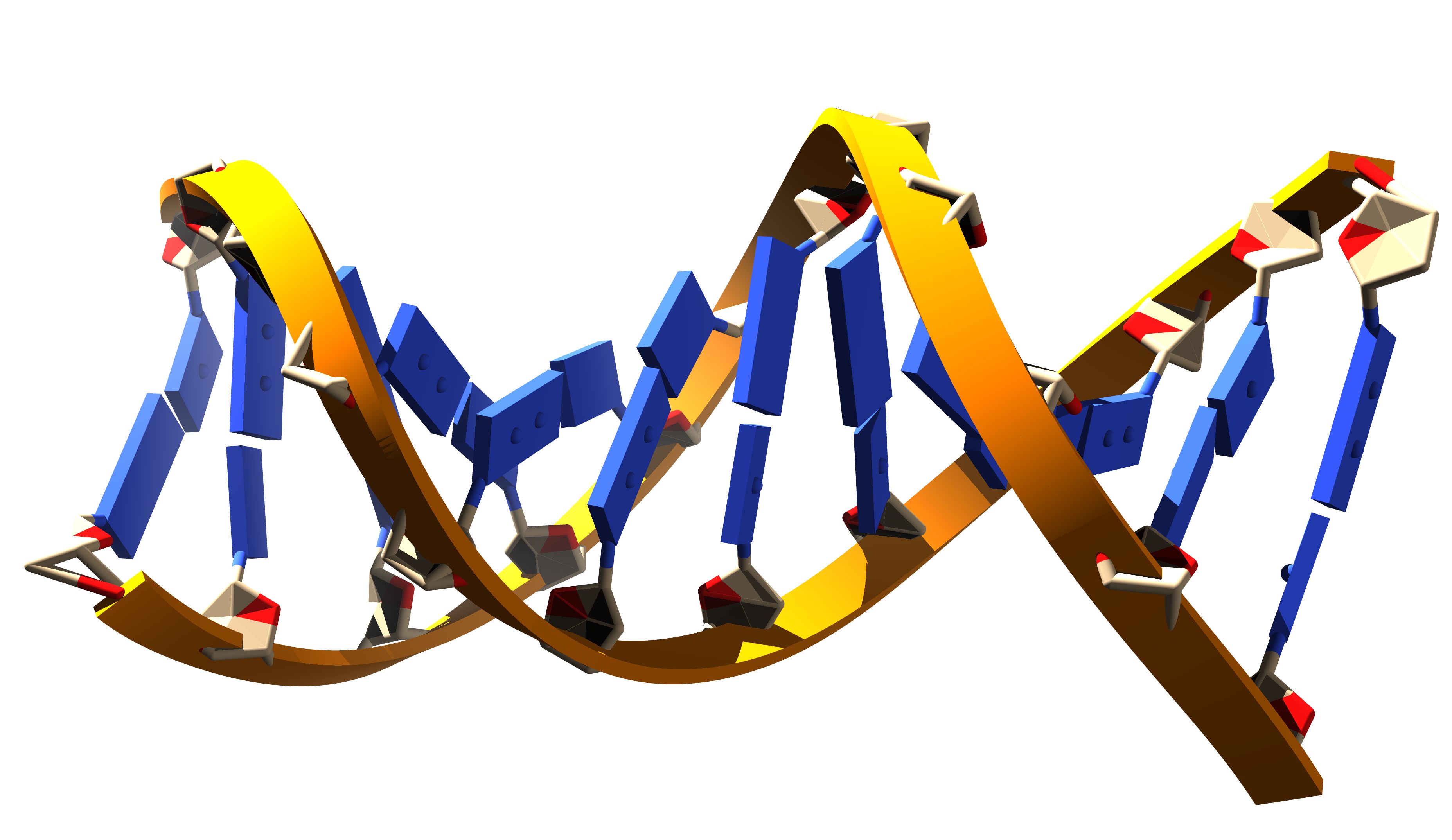
The researchers then looked at how this memory is coded. When studying the genome of the blood stem cells of the infected mice, they observed lasting modifications in its spatial organization. Changes that are likely to modify the expression of some genes implicated in the innate immune response. “At the time of first contact with the pathogen, genes required for the immune response are in fact put forward long-term so as to rapidly activate the immune system in the event of a second infection”, explains Bérengère de Laval, lead author of the study. Finally, the team looked for molecules implicated in this change of genome structure and discovered that the protein C/EBP beta played a major role.

Research relevant in fighting COVID-19?

These results are particularly relevant during this period of SARS-Cov-2 coronavirus pandemic.

Recent findings suggest that the BCG vaccine – it too known for inducing innate immune memory – also acts at blood stem cell level and offers a certain degree of protection from respiratory infections. Studies are ongoing in order to test its utility against COVID-19.

The team’s discoveries could elucidate the molecular mechanisms at play in this protection and open up new avenues for vaccines – particularly against COVID-19.

“Our discoveries represent a major contribution to understanding immune system memory and blood stem cell functions. They also point towards new strategies for stimulating or limiting immune response in various disease states and could make it possible to refine current vaccination strategies for better protection from various pathogens, including SARS-CoV-2″, hopes Michael Sieweke.

Rapid Genome Analysis Aids Diagnosis of Neonatal ICU Patients

Adobe/Stock

Teams from Dijon-Bourgogne University Hospital, Inserm and CEA have recently established the results of the whole-genome analysis of severely ill neonates, hospitalized in neonatal ICUs – the time of which was slashed from the current 18-month average to just 38 days. Thanks to this rapid analysis, the resulting diagnosis of two-thirds of the infants enrolled in the project enabled one third of them to receive faster and more appropriate treatment. The deployment of this process over the coming years will make it possible to optimize the management of these patients.

Although a number of countries use whole-genome sequencing for diagnostic purposes and while France has recently launched its 2025 Genomic Medicine Plan (PFMG2025)1, its urgent use in a neonatal setting is not very widespread at present. Yet rapid genetic examination is a crucial factor when a diagnosis is required urgently – a common situation when it comes to rare diseases with early pediatric onset or rapid progression. Teams from Dijon-Bourgogne University Hospital, Inserm and CEA conducted a feasibility study of fast high-throughput genome sequencing before envisaging such a process for the future, in the framework of the PFMG2025.

In this pilot study, called Fastgenomics2, some thirty children hospitalized in neonatal ICUs across eight university hospitals belonging to the AnDDI-rares network3 underwent fast genome analysis in the previous nine months. High-throughput sequencing of the genomes of the children and their parents and a primary bioinformatics analysis were carried out on the sequencing platform of the French National Research Center for Human Genomics (CEA-CNRGH), in collaboration with the Very Large Computing Center (TGCC) of the CEA and the Computing Center of Université de Bourgogne (CCuB). The genomics data were interpreted by the TRANSLAD University Hospital Federation (FHU TRANSLAD), in close collaboration with the Inserm U1231 GAD (Genetics of Developmental Disorders) research team.

Mobilizing the teams meant that it was possible to obtain the analysis results within 49 days, with the most rapid turnaround being 38 days. This is particularly fast for a genetic analysis, given that despite the considerable advances made, the average time to obtain a genetic diagnosis in France continues to remain long: 1.5 years on average, and up to 5 years for 25% of patients. Rapid analysis of the genomes of these neonates made it possible to diagnose two thirds of them, with one third able to receive quicker and more appropriate treatment.

Such analysis has been made possible thanks to major advances in the high-throughput sequencing of the gene set. The new-generation high-throughput DNA sequencing technologies – which analyze a person’s entire genome – have emerged in recent years as a tool of choice in the study of rare diseases. These cutting-edge technologies are used at CNRGH and have already implicated numerous genes in numerous diseases. The FHU TRANSLAD team from Dijon-Bourgogne University Hospital was one of the first in France to demonstrate the value of exome sequencing (with the exome representing 1% of the total size of the genome) in the diagnosis of severe diseases with early pediatric onset, as well as developmental abnormalities and intellectual disability.

Diagnosing rare diseases in the neonatal period

Rare diseases (affecting fewer than 1 in 2,000 people) are a major public health problem because they represent around 8,000 conditions and affect more than 3 million people in France. Given that the majority of these diseases are of pediatric onset, they are responsible for 10% of deaths before 5 years of age. Up to 80% of these diseases are considered to have a genetic origin. Establishing a diagnosis brings multiple benefits to the patients and their families: it clarifies the cause and the prognosis, enables access to treatment or clinical trials, determines risk of recurrence, cuts out needless diagnostic tests, enables the management of known complications, facilitates the acquisition of specific financial and practical support, and gives them the possibility to forge links with other families dealing with the same challenges.

Obtaining a diagnosis is a major challenge in diseases with early pediatric onset and rapid progression, and whose genetic causes are highly heterogenous, such as epilepsy, metabolic diseases, cardiac diseases, musculoskeletal diseases and other polymalformative syndromes. The 3rd French National Plan for Rare Diseases (PNMR3) envisages reducing diagnostic delay to one year, given that it is responsible for “the potential worsening of the condition of patients, the delayed possibility for genetic counseling and the wastage of medical resources (due to multiple diagnostic consultations)“.

When it comes to severe neonatal diseases, obtaining rapid diagnosis is particularly important. Diagnosis, when accurate, makes it possible to modify how the patient is managed, whether in terms of treatment adaptation (such as in metabolic diseases or epilepsy), referral to specialists, dietary adjustments, additional examinations, reassessment of any need for surgery, or the consideration of these results when discussing the continuation of care.


1 In 2016, France launched its 2025 Plan for Genomic Medicine (PFMG2025). It aims to implement mass whole-genome sequencing for the diagnosis of rare diseases through the establishment of very high-throughput platforms, as well as pilot studies to define the prescribing conditions for such investigations.

2 Fastgenomics: French national pilot study prepared by the AnDDI-rares national healthcare network, FHU TRANSLAD and CEA-CNRGH, and supported by a financial donation from SANOFI-GENZYME.

3 AnDDI-rares: National rare diseases network dedicated to diseases with somatic and cognitive developmental abnormalities. http://anddi-rares.org

Rôle des consultations génétiques dans le diagnostic des enfants et des adolescents atteints de troubles du spectre de l’autisme


A Gene Therapy Tested in the Treatment of Myotubular Myopathy


Inserm and CNRS researchers from the Institute of Genetics and Molecular and Cellular Biology (Inserm/CNRS/Université de Strasbourg) have discovered how myotubularin – a protein deficient in myotubular myopathy – interacts with amphiphysin 2 and suggest targeting the latter in order to treat patients. This research was published on March 20, 2019 in Science Translational Medicine.

Myotubular myopathy is a rare genetic disease affecting around one in 50,000 children. Linked to a mutation in the MTM1 gene located on the X-chromosome, it manifests as reduced muscle-cell adhesion and an alteration of the muscle fibers. This phenomenon causes major muscle weakness – including at the respiratory level – and leads to premature death with two thirds of patients not surviving beyond two years of age. At present, there is no treatment.

When exploring the interactions of myotubularin (coded by the MTM1 gene) with another protein, amphiphysin 2 (coded by the BIN1 gene), which is also expressed in the muscles and involved in similar myopathies, Inserm’s “Pathophysiology of neuromuscular diseases” team, in conjunction with the CNRS at the Institute of Genetics and Molecular and Cellular Biology (CNRS/Inserm/Université de Strasbourg), discovered how these proteins work together and suggests a new therapeutic target. Previous research had shown that myotubularin and amphiphysin 2 can physically interact by binding to each other.

To explore this functional link between the two, the researchers developed a model of MTM1-deficient transgenic mice and crossed these animals with other mice – some of which do not express BIN1 and some of which, on the contrary, overexpress it. They were unable to obtain any animals deficient in both MTM1 and BIN1, proving that at least one of the two proteins is necessary for muscle-fiber development and fetal survival. Conversely – and this came as a pleasant surprise – the overexpression of BIN1 made it possible to correct the myopathy linked to the MTM1 deficiency and obtain life expectancy equivalent to that of wild animals. Upon closer analysis of the muscles, the researchers observed satisfactory muscle-fiber organization and size with good cell adhesion, thereby leading to the hypothesis that MTM1 is an in vivo activator of the bin1 protein and that large quantities of the latter could make it possible to “do without” MTM1.

To verify whether BIN1 is a good therapeutic target, the researchers went on to conduct a gene therapy experiment in MTM1-deficient mice. They administered the human BIN1 gene using an AAV viral vector by systemic (intraperitoneal) injection following the birth of the rodents. A procedure that markedly reduced the symptoms of the condition and increased the survival of the diseased mice to that of healthy mice.

“There we have the proof of concept that the human BIN1 gene offers major potential in the treatment of myotubular myopathy linked to myotubularin deficiency, with spectacular results in mice. We would now like to continue this development in the form of preclinical trials and hope in the long-term to be able to propose a treatment for patients currently facing a therapeutic desert”, concludes Jocelyn Laporte, head of the Inserm team having performed this research.

Gene therapy durably reverses congenital deafness in mice

Immunofluorescence imaging of the cochlear sensory epithelium in a mouse treated with gene therapy © Institut Pasteur

In collaboration with the universities of Miami, Columbia and San Francisco, scientists from the Institut Pasteur, Inserm, CNRS, Collège de France, Sorbonne University and the University of Clermont Auvergne have managed to restore hearing in an adult mouse model of DFNB9 deafness – a hearing disorder that represents one of the most frequent cases of congenital genetic deafness. Individuals with DFNB9 deafness are profoundly deaf as they are deficient in the gene coding for otoferlin, a protein which is essential for transmitting sound information at the auditory sensory cell synapses. By carrying out an intracochlear injection of this gene in an adult DFNB9 mouse model, the scientists successfully restored auditory synapse function and hearing thresholds to a near-normal level. These findings, published in the journal PNAS, open up new avenues for future gene therapy trials in patients with DFNB9.

Over half of nonsyndromic profound congenital deafness cases have a genetic cause, and most (~80%) of these cases are due to autosomal recessive forms of deafness (DFNB). Cochlear implants are currently the only option for recovering hearing in these patients.

Adeno-associated viruses (AAVs) are among the most promising vectors for therapeutic gene transfer to treat human diseases. AAV-based gene therapy is a promising therapeutic option for treating deafness but its application is limited by a potentially narrow therapeutic window. In humans, inner ear development is completed in utero and hearing becomes possible at approximately 20 weeks of gestation. In addition, genetic forms of congenital deafness are generally diagnosed during the neonatal period. Gene therapy approaches in animal models must therefore take this into account, and gene therapy efficacy must be demonstrated following a gene injection when the auditory system is already in place. In other words, therapy must reverse existing deafness. The team led by Saaïd Safieddine, a CNRS researcher in the Genetics and Physiology of Hearing Unit (Institut Pasteur/ Inserm) and coordinator of the project, used a mouse model of DFNB9, a form of human deafness that represents 2 to 8% of all cases of congenital genetic deafness.

DFNB9 deafness is caused by mutations in the gene coding for otoferlin, a protein that plays a key role in transmitting sound information at the inner hair cell synapses. Mutant mice deficient in otoferlin are profoundly deaf as these synapses fail to release neurotransmitters in response to sound stimulation, despite the absence of detectable sensory epithelial defects. DFNB9 mice therefore constitute an appropriate model for testing the efficacy of viral gene therapy when it is administered at a late stage. However, as AAVs have limited DNA packaging capacity (approximately 4.7 kilobase (kb)), it is difficult to use this technique for genes whose coding region (cDNA) exceeds 5 kb, such as the gene coding for otoferlin, which has a 6 kb coding region. The scientists have overcome this limitation by adapting an AAV approach known as dual AAV strategy because it uses two different recombinant vectors, one containing the 5’-end and the other the 3’-end of the otoferlin cDNA

A single intracochlear injection of the vector pair in adult mutant mice was used to reconstruct the otoferlin coding region by recombining 5′ and 3′-end DNA segments, leading to long-term restoration of otoferlin expression in the inner hair cells, and then restored hearing.

The scientists have therefore obtained initial proof of the concept of viral transfer of fragmented cDNA in the cochlea using two vectors, showing that this approach can be used to produce otoferlin and durably correct the profound deafness phenotype in mice.

The outcomes achieved by the scientists suggest that the therapeutic window for local gene transfer in patients with DFNB9 congenital deafness could be wider than thought, and offers hope of extending these findings to other forms of deafness. These results are the subject of a patent application filed

In addition to the institutions mentioned in the first paragraph, this research was funded by the French Foundation for Medical Research, the European Union (TREAT RUSH) and the French National Research Agency (EargenCure and Lifesenses LabEx).


Oreille humaine anglais

The left panel is a schematic representation of the human ear. Sound waves are collected by the outer ear made up of the pinna and ear canal. The middle ear, composed of the eardrum and ossicles, transmits sound waves to the inner ear, which features the cochlea – the hearing organ responsible for transmitting auditory messages to the central nervous system. The right panel shows an immunofluorescence image of the auditory sensory epithelium within an injected cochlea. The inner hair cells have been stained for otoferlin in green. Otoferlin is detected in almost all of these cells. The inset is a high magnification area showing an inner hair cell that has not been transduced. © Institut Pasteur

[1] Research published by the Institut Pasteur and Inserm Genetics and Physiology of Hearing Unit (UMRS1120): “Otoferlin, defective in DFNB9 deafness, is essential for synaptic vesicle exocytosis at the auditory ribbon synapse” Cell, October 20, 2006

Alzheimer’s: identification of potential target protein aggregates for treating the disease

Aggregates of Tau protein in Alzheimer’s disease. Inserm/U837, 2008

The propagation of tau protein aggregates in the brain contributes to the progression of Alzheimer’s disease. Researchers at the Neurodegenerative Diseases Laboratory: mechanisms, therapies, imaging (CNRS/CEA/Université Paris-Sud, MIRCen), working in collaboration with the Ecole Normale Supérieure, Sorbonne University and Inserm, have just identified the targets of these aggregates. Published in the EMBO Journal on 10 January 2019, this work will enable the development of tools capable of blocking these key elements of aggregate propagation and thus combating their pathological effect.

The aggregation of alpha-synuclein proteins in Parkinson’s disease and tau proteins in Alzheimer’s disease is intimately linked to the progression of these neurodegenerative diseases. These aggregates propagate from one neuronal cell to another, attaching themselves to the cells.

They multiply[1] during this propagation. It has already been shown that the propagation and amplification of these protein aggregates are harmful and contribute to the progression of these diseases.

Understanding the formation of these aggregates, their propagation and their multiplication in the cells of the central nervous system offers potential for treatments: it would make it possible to target these processes and to act on their consequences.

Protein propagation

The key step in the propagation of the pathogenic aggregates is the attachment of aggregates released from affected neuronal cells to the membranes of unaffected cells. Having already identified the targets of pathogenic aggregates of the alpha-synuclein protein (Shrivastava et al., 2015 EMBO-J), the team at the Neurodegenerative Diseases Laboratory (CNRS/CEA/Université Paris-Sud , MIRCen, Fontenay-aux-Roses), in collaboration with the Ecole normale supérieure, Sorbonne University and Inserm, has just identified the targets of tau protein aggregates. The targets are the sodium / potassium pump and glutamate receptors, two essential proteins for the survival of neurons The experiment was carried out on mouse neurons in culture.

Neuron membrane modification

The researchers also showed that the pathogenic aggregates modify the neuron membranes by redistributing the membrane proteins. The integrity of the membrane—and particularly of the synapses, the essential nodes for communication between neurons—is affected. These changes have a deleterious effect on the neurons because they cause abnormal communication between the neurons, as well as their degeneration.

This work therefore explains the early malfunctioning of the synapses and the degradation of normal communication observed in the neuronal networks as the disease progresses.

Towards new treatments

It also paves the way for the development of new treatment strategies based on protecting the integrity of the synapses, restoring the activity of the tau protein membrane receptors through the use of decoys to prevent harmful interaction between the pathogenic tau protein aggregates and their neuron membrane targets. These therapeutic approaches could be developed using human neurons, since researchers at the laboratory have just developed cultures of this type in collaboration with the I-Stem (Institute for Stem Cell Therapy & Exploration of Monogenic Diseases, AFM-Téléthon/Insem/Génopole/University of Evry-Val-d’Essonne) laboratory and Sorbonne University. This latter study is also published on 10 January 2019, in Stem Cell Reports[2].


[2] Propagation of α-Synuclein strains within human reconstructed neuronal network. Simona Gribaudo, Philippe Tixador, Luc Bousset, Alexis Fenyi, Patricia Lino, Ronald Melki, Jean-Michel Peyrin, Anselme Louis Perrier, Stem Cell Reports, 10 January 2019.

[1] They multiply by recruiting the endogenous alpha-synuclein and tau proteins from the affected cells during this propagation


About the Neurodegenerative Diseases Laboratory: mechanisms, therapies, imaging (LMN), a joint research unit of the CEA, CNRS and Université Paris-Sud.

The laboratory brings together nearly 60 scientists with research interests in neurosciences covering the mechanisms of degeneration, animal models, brain imaging and the study of gene-, cell- and drug-based strategies for treating neurodegenerative diseases such as Alzheimer’s disease, Parkinson’s disease and Huntington’s disease.

The LMN is located at MIRCen (Molecular Imaging Research Centre), a preclinical research facility developed by the CEA and Inserm. MIRCen is one of the departments of the CEA’s François Jacob Institute of Biology, on the Fontenay-aux-Roses site at CEA Paris-Saclay. 


Discovery of novel mechanisms that cause migraines

©Photo by Anh Nguyen on Unsplash

Researchers at CNRS, Université Côte d’Azur and Inserm have demonstrated a new mechanism related to the onset of migraine. In fact, they found how a mutation, causes dysfunction in a protein which inhibits neuronal electrical activity, induces migraines. These results, published in Neuron on December 17, 2018, open a new path for the development of anti-migraine medicines.

Even though 15% of the adult population worldwide suffers from migraines, no long-term, effective, curative treatment has been marketed to date. Migraine episodes are related, among other factors, to electric hyperexcitability in sensory neurons. Their electrical activity is controlled by proteins that generate current called ion channels, specifically by the TRESK channel, which inhibits electrical activity. The researchers have shown that a mutation in the gene encoding for this protein causes a split between two dysfunctional proteins: one is inactive and the other targets other ion channels (K2P2.1) inducing a great stimulation of the neuronal electrical activity causing migraines.

Though researchers had already shown the hereditary nature of migraines, they did not know the mechanism underlying migraine. By demonstrating that the TRESK split induces hyperexcitability in sensory neurons leading to migraine, this work, carried out at the Institut de Biologie Valrose (CNRS/Inserm/Université Côte d’Azur), opens new research path for the development of anti-migraine medicines. A patent application has been filed1: the scope is targeting K2P2.1 channels to reduce the electrical activity of neurons and prevent migraines from being triggered.

What is more, the researchers propose that this new genetical mechanism, causing the formation of two proteins instead of just one, has now to be considered for the study of other genetic diseases and for diagnosing them.


1 Patent PCT/EP2018/067581 “Methods and compositions for treating migraine”

Cancer under pressure: visualizing the activity of the immune system on tumor development

Cancérogenèse : Surexpression de TRF2, marqué en vert, dans les vaisseaux tumoraux, marquage rouge, dans un cancer ovarien. ©Inserm/Wagner, Nicole, 2014

As tumors develop, they evolve genetically. How does the immune system act when faced with tumor cells? How does it exert pressure on the genetic diversity of cancer cells? Scientists from the Institut Pasteur and Inserm used in vivo video techniques and cell-specific staining to visualize the action of immune cells in response to the proliferation of cancer cells. The findings have been published in the journal Science Immunology on November 23, 2018.

Over time, the uncontrolled proliferation of tumor cells results in the accumulation of new mutations and changes to their genome. This gradual process creates significant genetic diversity among the cancer cells in any given patient. And although the cells in the immune system, especially T cells, are potentially able to eliminate these abnormal cells, tumor diversity can have a harmful effect, complicating the action of the immune system and rendering some therapies ineffective. Understanding this frantic race between tumor development and the immune response is key to the success of future immunotherapy techniques.

Scientists in the Dynamics of Immune Responses Unit (Institut Pasteur/Inserm), directed by Philippe Bousso, in collaboration with Ludovic Deriano, Head of the Genome Integrity, Immunity and Cancer Unit (Institut Pasteur), investigated how spontaneous immune responses to tumors influence this tumor heterogeneity. They demonstrated that the immune system can employ mechanisms to significantly reduce tumor diversity, favoring the emergence of more genetically homogeneous tumor cells.

In their study, the scientists marked each cancer cell subclone with a separate color in a mouse model. By monitoring these different colors they were therefore able to characterize the evolution of tumor heterogeneity in time and space. They were also able to observe the contacts between T cells and cancer cells and determine how some tumor cells are destroyed. Their research highlights the drastic impact the immune system can have on tumors by reducing their heterogeneity.


Visualizing the action of stained immune cells.
In this video, the tumor cells are shown in gray. The tumor-specific T-cells, in purple, come into contact with the cancer cells and destroy them. The killed cells are shown in blue. In green, the control cells circulate but do not kill the tumor cells. © Institut Pasteur / Philippe Bousso


Visualizing different clusters of cancer cell clones.
This video illustrates how tumor subclones, each marked by a different color (blue, orange and green), develop in the bone marrow. The vessels are shown in white. © Institut Pasteur / Philippe Bousso

The same impact on the heterogeneity of tumor cells has also been observed in response to immunotherapies that release the brakes on the immune system, an approach which was awarded the Nobel Prize in Physiology or Medicine this year.

This research shows that taking into account the interaction between immunotherapies and tumor heterogeneity could contribute to the development of optimum therapeutic combinations and sequences.

In addition to the organizations mentioned above, this research was funded by the Fondation de France, the French National Cancer Institute (INCa) and the European Research Council (ERC).

The origins of asymmetry: A protein that makes you do the twist

©Inserm/Cochet-Escartin, Olivier, 2014

Asymmetry plays a major role in biology at every scale: think of DNA spirals, the fact that the human heart is positioned on the left, our preference to use our left or right hand … A team from the Institute of biology Valrose (CNRS/Inserm/Université Côte d’Azur), in collaboration with colleagues from the University of Pennsylvania, has shown how a single protein induces a spiral motion in another molecule. Through a domino effect, this causes cells, organs, and indeed the entire body to twist, triggering lateralized behaviour. This research is published in the journal Science on November 23, 2018.

Our world is fundamentally asymmetrical: think of the double helix of DNA, the asymmetrical division of stem cells, or the fact that the human heart is positioned on the left … But how do these asymmetries emerge, and are they linked to one another?

At the Institute of biology Valrose, the team led by the CNRS researcher Stéphane Noselli, which also includes Inserm and Université Cote d’Azur researchers, has been studying right–left asymmetry for several years in order to solve these enigmas. The biologists had identified the first gene controlling asymmetry in the common fruit fly (Drosophila), one of the biologists’ favoured model organisms. More recently, the team showed that this gene plays the same role in vertebrates: the protein that it produces, Myosin 1D,[1] controls the coiling or rotation of organs in the same direction.

In this new study, the researchers induced the production of Myosin 1D in the normally symmetrical organs of Drosophila, such as the respiratory trachea. Quite spectacularly, this was enough to induce asymmetry at all levels: deformed cells, trachea coiling around themselves, the twisting of the whole body, and helicoidal locomotive behavior among fly larvae. Remarkably, these new asymmetries always develop in the same direction.

In order to identify the origin of these cascading effects, biochemists from the University of Pennsylvania contributed to the project too: on a glass coverslip, they brought Myosin 1D into contact with a component of cytoskeleton (the cell’s “backbone”), namely actin. They were able to observe that the interaction between the two proteins caused the actin to spiral.

Besides its role in right–left asymmetry among Drosophila and vertebrates, Myosin 1D appears to be a unique protein that is capable of inducing asymmetry in and of itself at all scales, first at the molecular level, then, through a domino effect, at the cell, tissue, and behavioral level.

These results suggest a possible mechanism for the sudden appearance of new morphological characteristics over the course of evolution, such as, for example, the twisting of snails’ bodies. Myosin 1D thus appears to have all the necessary characteristics for the emergence of this innovation, since its expression alone suffices to induce twisting at all scales.


[1] Myosins are a class of proteins that interact with actin (a constituent of cell skeletons or cytoskeletons). The most well-known of them, muscular myosin, makes muscles contract.

Identifying a genetic factor causing lung fibrosis complicating rheumatoid arthritis

©Inserm/Cantagrel, Alain, 1993

Teams of rheumatology, respiratory medicine, genetics and the university hospital department FIRE Hospital Bichat Claude Bernard AP-HP, in collaboration with INSERM, Université Paris Diderot, the Broad Institute of MIT and Harvard, Cambridge, Massachusetts, USA, have discovered that a rare allele of rs35705950 variant gene  MUC5B  multiply by six the risk of occurrence of pulmonary fibrosis in patients with rheumatoid arthritis (RA). This broad study of genetic association demonstrates the existence of a common genetic basis between pulmonary fibrosis associated with rheumatoid arthritis and idiopathic pulmonary fibrosis (IPF). These results, obtained with the participation of national and international network of pulmonologists and rheumatologists, are published in the journal  The New England Journal of Medicine  October 20, 2018.  They are a first step in understanding the diffuse interstitial lung disease (PID) of rheumatoid arthritis, a serious complication whose therapeutic management is not currently codified.

The teams tested the influence of the major genetic risk factor for idiopathic pulmonary fibrosis (IPF): rs35705950 variant gene MUC5B . 

This genetic association study case-control, coordinated by Professor Philippe Dieudé in collaboration with Prof. Bruno Crestani, compared 620 patients with rheumatoid arthritis with diffuse interstitial lung disease (PID), 614 patients with rheumatoid arthritis and without PID 5448 control individuals, from 7 different countries (France, Greece, Netherlands, Japan, China, Mexico and USA).

The results of this international collaboration show a varying contribution MUC5Brs35705950 in the determination of the PID in rheumatoid arthritis. 

The presence of  the risk allele multiply by 3 the risk of occurrence of interstitial lung disease  diffuse (PID)  and  by 6 that of usual interstitial pneumonia (PIC) (the most severe form of PID). Finally, an exploratory phase exploring 12 more REIT susceptibility markers suggests the existence of a common genetic architecture between IPF and pulmonary fibrosis in rheumatoid arthritis.

This work is the first to show that there are  common pathways between the two diseases – pulmonary fibrosis in rheumatoid arthritis and idiopathic pulmonary fibrosis – and provides a strong argument for promoting therapeutic intervention studies in pulmonary fibrosis associated with rheumatoid arthritis, using anti-fibrotic drugs already validated in the idiopathic pulmonary fibrosis.


To know more

The diffuse interstitial lung disease (PID)  is a common extra-articular manifestation and severe  rheumatoid arthritis (RA) , which affects nearly 30% of RA patients, and gradually progresses to irreversible pulmonary fibrosis in about 40 to 50 % of cases. Thus, a PID occurrence is responsible for about 7 to 10% of deaths in patients with rheumatoid arthritis and median survival varies between 2 and 5 years after the onset of Respiratory symptoms. The risk factors and pathophysiological mechanisms underlying the onset of pulmonary fibrosis in RA remain largely unknown. 

Idiopathic pulmonary fibrosis (IPF)  is a lung disease characterized by progressive fibrosis, in which there is no extra-respiratory disorder for which the genetic origins are known. 

Pulmonary fibrosis in rheumatoid arthritis has similarities with idiopathic pulmonary fibrosis, including a  high prevalence of interstitial pneumonia (PIC) , shared environmental risk factors (such as smoking) and a very poor prognosis.

fermer