Menu

Pandoravirus: giant viruses invent their own genes

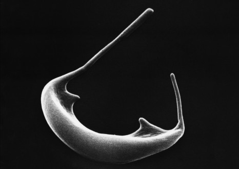
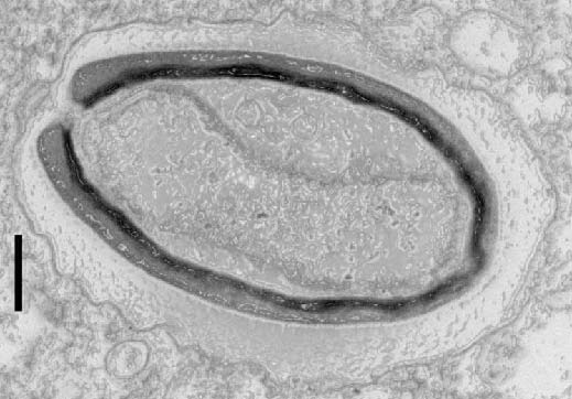
Pandoravirus quercus, found in Marseille, France. Thin section, viewed via electron microscopy. Scale bar: 100 nm.  ©IGS- CNRS/AMU.

Three new members have been isolated and added to the Pandoravirus family by researchers at the Structural and Genomic Information Laboratory (CNRS/AixMarseille Université), working with partners at the Large Scale Biology Laboratory (CEA/Inserm/Université GrenobleAlpes) and at CEA-Genoscope. This strange family of viruses, with their giant genomes and many genes with no known equivalents, surprised the scientists when they were discovered a few years ago. In the 11 June 2018 edition of Nature Communications, researchers offer an explanation: pandoviruses appear to be factories for new genes – and therefore new functions. From freaks of nature to evolutionary innovators, giant viruses continue to shake branches on the tree of life!

In 2013, the discovery of two giant viruses unlike anything seen before blurred the line between the viral and cellular world. Pandoraviruses are as big as bacteria, and contain genomes that are more complex than those found in some eukaryotic organisms[1]. Their strange amphora shape and enormous, atypical genome[2] led scientists to wonder where they came from. 


The same team has since isolated three new members of the family in Marseille, continental France, Nouméa, New Caledonia, and Melbourne, Australia. With another virus found in Germany, the team compared those six known cases using different approaches. Analyses showed that despite having very similar shapes and functions, these viruses only share half of their genes coding for proteins. Usually, however, members of the same family have more genes in common.

Furthermore, these new members contain a large number of orphan genes, i.e. genes which encode proteins that have no equivalent in other living organisms (this was already the case for the two previously discovered pandoraviruses). This unexplained characteristic is at the heart of many a debate over the origin of viruses. What most surprised researchers was that the orphan genes differed from one pandoravirus to another, making it less and less likely that they were inherited from a common ancestor!

Bioinformatic analysis showed that these orphan genes exhibit features very similar to those of non-coding (or intergenic) regions in the pandoravirus genome. Findings indicate the only possible explanation for the gigantic size of pandoravirus genomes, their diversity and the large proportion of orphan genes they contain: most of these viruses’ genes may originate spontaneously and randomly in intergenic regions. In this scenario, genes “appear” in different locations from one strain to another, thus explaining their unique nature.  

If confirmed, this groundbreaking hypothesis would make these giant viruses craftsmen of genetic creativity – a central, but still poorly explained component of any understanding of the origin of life and its evolution.


[1] Organisms whose cells contain nuclei, unlike the two other kingdoms of living organisms, bacteria and archaea.

[2] Up to 2.7 million base pairs.


This research received funding from the Bettencourt Schueller Foundation, through the “Coup d’Elan Prize for French Research” awarded to Chantal Abergel in 2014.


Credit: IGS- CNRS/AMU

Discovery of a first genetic cause of Whipple’s disease

A Franco-American team involving researchers from Inserm, Université Paris-Descartes and doctors grouped in the Institute Imagine the hospital Necker-Enfants Malades AP-HP and Rockefeller University New York discovered a genetic cause of Whipple’s disease, chronic intestinal pathology. By studying families with 4 members developed symptoms, the team found that the mutation of the gene causes IRF4 an impaired immune response to the bacteriaTropheryma whippleiAt the origin of the disease.This bacterium, common and experienced by many individuals, then causes in carriers of the mutation potentially fatal chronic infection without treatment. With this discovery, the first step towards a genetic explanation of the disease was done.

Chronic bacterial infection, Whipple’s disease occurs around the age of 50 and may result in clinical signs such as diarrhea, malabsorption, fever, weight loss, joint diseases, cardiovascular or central nervous system. In the absence or failure of antibiotic treatment, it can progress to death.

The disease is caused by Tropheryma whipplei , a bacterium that many of us encounter in our life (up to 50% of the members of some populations are carriers), but which affects a very small portion of individuals: only a subject on a million developing the symptoms of the disease.

The team led by Professor Jean-Laurent Casanova, laboratory director of Human Genetics of Infectious Diseases at the Institute. Imagine – Inserm, Paris Descartes University, AP-HP-member of Immunology, Hematology and Pediatric Rheumatology Hospital Necker Children AP-HP, Dr. Jacinta Bustamante, a research professor in the same laboratory and within the diagnostic center immune deficiencies at the Hospital Necker Children AP-HP, and Dr. Laurent Abel, co-director of the laboratory of human genetics of infectious diseases at Imagine , found that many families have multiple members affected by the disease, suggesting a genetic origin.

The researcher Antoine Guerin, first author of the scientific paper on this discovery, studied a French family with 5 healthy carriers and 4 members affected by the disease. These four patients are carriers of a mutation of the IRF4 gene, which codes for the production of a protein having a key role in immunity, and rendered non-functional by mutation. Dysfunction of this gene makes these patients vulnerable to infection with T. whipplei . The study also highlights the mode of transmission of the disease, hereditary, autosomal dominant: inherit a single mutated allele is enough to be reached.

With this study and the discovery of this mutation, the research team laid the foundation stone for a genetic understanding of the disease. Sequencing of a cohort of patients has not found other mutations in the same gene showing genetic heterogeneity of the disease. It remains to find and understand the mechanism immune deficiency caused by the mutation of the gene and to find other genetic mutations that may explain the vulnerability to disease.

This breakthrough helps explain why some patients exposed to the bacteria are sick or not, improve diagnosis, genetic counseling to families and the care of patients with the signs of the disease.

These works are the subject of a publication on www.elifesciences.org ”  IRF4 haploinsufficiency in a family with Whipple’s disease “, published March 14, 2018.

Discovery of a future therapy for hemoglobinopathies

©Inserm/Féo, Claude

With the genome editing CRISPR-case.9 technique, researchers at INSERM, the hospital Necker Children-AP-HP, and Université Paris Descartes in the Institute Imagine managed to reactivate a gene that may improve the appearance of red blood cells of patients with hemoglobinopathies such as sickle cell anemia and beta-thalassemia. The teams Annarita Miccio, Inserm researcher, Prof. Marina Cavazzana AP-HP and Isabelle André-Schmutz, Inserm researcher show this new avenue of research and treatment of β -hémoglobinopathies in the journal Blood .

Millions of people are affected by severe forms of these diseases worldwide. They are characterized by an alteration in the expression of the gene encoding the β-globin: an essential component of hemoglobin . These changes can lead to a failure of expression of the β-globin gene, as in the case of β-thalassemia, or by an aggregation of hemoglobin into fibrils leading to the deformation of the red blood cells, in the case of sickle cell disease.

The laboratory Annarita Miccio, Inserm researcher, was particularly interested in the reactivation mechanisms of fetal hemoglobin . This hemoglobin is unusual act, instead of β-globin, other globin, γ-globin called, which is only expressed during fetal development.

Most patients suffering from β-hemoglobinopathies have a non-altered form of the gene encoding this protein. Its reactivation in patients with thalassemia and sickle cell would replace β-globin mutated γ-globin. This change would result in a significant improvement in the observed state of red blood cells for these diseases and thus associated symptoms (pain associated with vaso-occlusive crises in sickle-cell anemia or correction of anemia in both diseases).

The results show that certain genetic sequences responsible for blocking the expression of globine- γcan be modified, including a DNA sequence which inhibits the production of globin γ after fetal development. Its removal, using “genetic scissors” CRISPR / case.9, reactive synthesis globine- γ at levels sufficient to be considered in the future treatment protocol.

This study also improves the state of knowledge on the expression regulation mechanism to β globin γ during our development.

It also contributes to developing curative therapeutic protocols for these diseases with the majority of current treatments remain symptomatic and very heavy for patients.

In figures  :

Beta-thalassemia and sickle cell disease affect alone nearly 100 million healthy carriers or sick people in the world. 60,000 new cases of β-thalassemia and sickle cell 300,000 are diagnosed annually worldwide.

Besides the high mortality observed, the less severe forms greatly affect the quality of life of these patients and their management represents a very significant cost to health systems.

In developing countries where these diseases have the highest incidence, β-hemoglobinopathies are a major public health issue.

Beta-thalassemia :

– 90 million people affected worldwide, about 288 000 patients

– 60,000 new cases diagnosed each year.

Vichinsky EP: Changing patterns of thalassemia worldwide. Ann NY Acad Sci 2005, “Gene Therapy in Patients with Transfusion-Dependent β-Thalassemia”

Alexis A. Thompson, et al. https://www.nejm.org/doi/full/10.1056/NEJMoa1705342

Sickle:

– 43 million people in the world carrying the S allele moderately affected. Global Burden of Disease Study 2013, Collaborators (22 August 2015), The Lancet.

– 4.4 million homozygous patients worldwide, so very sick . GBD 2015 Disease and Injury Incidence and Prevalence, Collaborators. (8 October 2016), The Lancet.

– newborns diagnosed sickle 300,000 worldwide each year.

– 114,000 deaths worldwide in 2015. GBD 2015 Mortality and Causes of Death, Collaborators. (October 8, 2016), The Lancet.

New gene therapy success in beta thalassemia: 22 patients treated in France, United States, Thailand and Australia

©AdobeStock

In an article in the New England Journal of Medicine published on April 19, the intermediate results of a clinical trial (HGB-205) led by Pr. Marina Cavazzana and her teams at the Necker-Enfants malades hospital AP-HP in collaboration with the Imagine Institute (AP-HP/Inserm/Paris Descartes University) as well as those of an international multicenter trial (HGB-204) conducted in the United States, Thailand and Australia, show that gene therapy is effective in improving the state of health or curing patients with beta thalassemia. These two clinical trials have used the same therapeutic vector “LentiGlobin”, developed at Harvard University in Boston and at the CEA in Fontenay-aux-Roses by Pr. Philippe Leboulch, in collaboration with the American company bluebird bio, for which he is the founder.

Pr. Marina Cavazzana, director of the biotherapy department at Necker-Enfants malades hospital AP-HP, laboratory co-director of the Inserm human lymphohematopoiesis laboratory at the Imagine Institute, and her team have treated patients who now produce a sufficient amount of therapeutic hemoglobin to stop the need for monthly blood transfusions.

Eight years after the first gene therapy in this disease, conducted by Pr. Cavazzana and Leboulch (Cavazzana et al. 2010), the lentiviral vector “LentiGlobin” of this therapy has been produced under the leadership of Pr. Leboulch of the Paris-Sud and Harvard universities and his colleagues, such as Dr. Emmanuel Payen, at the French Alternative Energies and Atomic Energy Commission (CEA) where Pr. Leboulch is Senior Advisor for medical innovation of the CEA Fundamental Research Division and Honorary Scientific Director of the François Jacob research institute. These trials are promoted by the American company bluebird bio, which was founded by Pr. Leboulch in Boston. Cumulating 15 to 42 months of monitoring, the patients of the two trials have no adverse effect and have resumed their professional or academic activities.

A young woman testifies:

I am nearly 24 years old and I benefited from an autograft 4 years ago. Now, thanks to that, I no longer have transfusions but above all I no longer have Desferal, which was my treatment in the form of a subcutaneous injection that I had to do every day to lower my ferritin. It was quite complicated, mentally more than anything because I was young and I did not feel like the others… Psychologically, I feel better now. I only take an oral treatment, which is an antibiotic, because they had removed the spleen damaged by iron deposits related to the transfusions, editor’s note] and a hormone treatment. I am monitored at Archet hospital […] as well as Necker hospital by Dr. Cavazzana and Dr. Semeraro. I go to Paris roughly every 6 months for my health to be assessed (a test) but everything is fine. I am delighted, I was lucky enough to benefit from this autograft and I wish the same for any sick person.

Beta thalassemia is one of the most common monogenic genetic diseases. It is caused by more than 200 mutations of the beta-globin gene (HBB) and affects almost 288,000 people worldwide with 60,000 new cases per year. Passed on as an autosomal recessive trait, it disrupts the production of the hemoglobin beta chain, resulting in a more or less severe anemia. In its major form, beta-thalassemia requires monthly transfusions and a treatment against the harmful effect of iron deposits caused by these transfusions. These only have a palliative effect. The curative treatment offered to these patients is generally an allogeneic transplant of bone marrow cells, when their clinical condition is not too fragile and one of their siblings is a compatible donor, which is only possible in about 25% of cases. In addition, the success rates are uneven and patients remain vulnerable to infections in the months following the transplant and to the “graft versus host disease”.

In the phase 1-2 trials HGB-204 and HGB-205, started in 2013, the researchers took blood stem cells from patients. They modified them using the vector LentiGlobin BB305 to give them a healthy replacement gene, before transplanting them into patients previously conditioned by a myeloablative treatment.

This way, these therapeutic stem cells have produced a sufficient amount of red blood cells with healthy hemoglobin levels. According to patient genotypes, gene therapy has freed them from all transfusions (12 out of 13 patients with a non-beta0/beta0 genotype), or has reduced their volume of 73% and reduced the frequency of transfusions (3 of the 9 patients with a beta0/beta0 genotype or two copies of the IVS1-110 mutation).

“After the therapeutic proof of principle that we had obtained in a thalassemic patient and a sickle cell patient, these international multicenter trials confirm the consistency and magnitude of the therapeutic efficacy of our vector in many patients. The phase 3 clinical trials are now underway on several continents before applying to market this biological medicinal product” said Pr. Leboulch.

“Gene therapy has again shown its therapeutic potential, provided that the expertise from different fields is combined. As such, I thank all medical teams at the Necker Hospital and AP-HP for providing us with this indispensable expertise, key to the success of this treatment. Our effort must now be focused on extending this approach to a large number of patients” emphasized Pr. Cavazzana.

The life of these patients has already changed dramatically. In the framework of trials HGB-204 and HGB-205, they will continue to be monitored for 13 years.

The article “Gene Therapy in Patients with Transfusion-Dependent β-Thalassemia” is published on April 19 in the New England Journal of Medicine.

Flunarizine: a New Drug Candidate in the Treatment of Spinal Muscular Atrophy

©Adobestock

A team of researchers from Inserm (“Toxicology, pharmacology and cell signaling” JRU 1124) and the universities of Paris Descartes and Paris Diderot have recently discovered that flunarizine – a drug already used to treat migraine and epilepsy – enables the repair of a molecular defect related to spinal muscular atrophy, a severe and incurable disease. This discovery is the culmination of research efforts ongoing since 1995, when the Inserm team – comprising Suzie Lefebvre, leader of the current research projects – identified the gene responsible for infantile spinal muscular atrophy. The results of the initial animal tests, published in Scientific Reports, demonstrate a marked improvement in health. These extremely promising findings must now be confirmed in humans.


 Spinal muscular atrophy is a rare genetic disease, affecting between 1 and 9 out of every 100,000 people. It is caused by degeneration of the motor neurons in the spinal cord, resulting in progressive muscle loss. In the majority of cases, symptoms appear either following birth – with the infant unable to hold up his or her head, or a little later in early childhood – with the inability to walk. More rarely, symptoms can begin in adolescence, in which case the muscular disorders are substantial but compatible with a more-or-less normal life.

The disease is caused by a mutation of the SMN1 gene, leading to a deficiency in the SMN protein. The SMN2 gene, which is virtually identical, then takes over. However, the SMN protein that it produces is for the most part truncated and not highly functional.


An SMN protein targeting problem

In healthy individuals, the SMN protein is drawn into cell nucleus structures known as Cajal bodies. There, small non-coding RNA is formed, which is implicated in a maturation step of the messenger RNA (known as splicing), a precursor of the proteins. In spinal muscular atrophy, the truncated SMN proteins are unable to reach the Cajal bodies. The Cajal bodies then function poorly and the production of the small non-coding RNA is altered. As such, many messenger RNA present maturation problems and result in abnormal or deficient proteins – a phenomenon occurring in all tissues.

In an attempt to restore this mechanism, the researchers tested therapeutic molecules in vitro, on cells taken from patients with a severe form of the disease. The objective was to find one or more cells able to retransport the SMN proteins to the Cajal bodies so that they regain their function.


Flunarizine effective on cells from a variety of patients

Just one molecule has demonstrated an effect on a large number of cells from various patients: flunarizine, which is already used in the treatment of migraine and epilepsy. In a second step, it was used to treat mice with spinal muscular atrophy, at a rate of one spinal cord injection per day from birth. Their life expectancy increased by 40% on average, from 11 to 16 days and even up to 36 days in one case. Analysis of the motor neurons and muscles show that they are preserved for longer in the treated animals. “The molecule presents a major neuroprotective effect even if we currently don’t know why that is,” declares Lefebvre, research leader and a member of the team having discovered the gene responsible for infantile spinal muscular atrophy in 1995. In addition, her team observed that flunarizine makes it possible to restore the functioning of the small non-coding RNA produced in the Cajal bodies for the maturation of the messenger RNA.


Findings to be confirmed in humans

Flunarizine remains to be tested in humans, a stage which will face the challenge of enrolling patients in the context of a rare disease. In addition, most of these patients are already enrolled in a clinical trial to evaluate a new-generation drug that was granted marketing authorization in 2016, meaning that they cannot be mobilized to participate in a second trial. Ultimately, the two therapeutic approaches – each of which targeting a different mechanism – could very well complement each other to promote patient survival and quality of life.

A new gene implicated in hypertension

A team of researchers led by Maria-Christina Zennaro, Inserm research Director at the Paris Cardiovascular Research Center (Inserm/ Paris-Descartes University), in collaboration with German colleagues[1], has identified a new gene implicated in hypertension. This study has been published in Nature Genetics.

These new findings highlight the role of genetic predisposition in the onset of common diseases and the importance of the French Plan for Genomic Medicine 2025. A plan in which one objective is to enable access to genetic screening, even for common diseases, in order to provide personalized medical care.

Hypertension is a major cardiovascular risk factor which affects up to 25% of the population. In around 10% of cases, it is due to dysfunction of the adrenal gland, which produces too much aldosterone – a hormone that regulates blood pressure. This is known as primary aldosteronism. Patients with this condition have hypertension that is often severe and resistant to standard treatments. They are also at greater risk of experiencing cardiovascular events, notably myocardial infarction or stroke.

To elucidate the causes of this disease, Maria-Christina Zennaro and Fabio Fernandes-Rosa, Inserm researchers in Paris, analyzed the exomes (the part of the genome that codes for proteins) of patients affected by primary aldosteronism before the age of 20. By doing so, they identified a mutation in a previously unknown gene – CLCN2 – which codes for a chloride channel, whose presence and effects in the adrenal gland were at that point unknown. 


Autonomous aldosterone production

Thanks to their partnership with a German team, the researchers studied the mechanisms by which this mutation could induce autonomous aldosterone production and trigger hypertension. They discovered that the mutation led to a permanent opening in the chloride channel.

In an animal model, the researchers showed that this channel was indeed expressed in the area of the adrenal glands that produces aldosterone.

Through electrophysiology and cellular biology experiments, they showed that the influx of chloride through the mutated channel led to increased chloride flow and depolarization of the cell membrane. The cells of the adrenal cortex then produced more aldosterone in the presence of the mutated channel and expressed to a greater degree the enzymes implicated in its biosynthesis.

This study reveals a previously unknown role of a chloride channel in the production of aldosterone. It opens up entirely new prospects for the pathogenesis and management of hypertension.


[1] From the Leibniz Institute for Molecular Pharmacology (FMP) and the Max Delbrück Center for Molecular Medicine (MDC) in Berlin.

UK-France Summit. United Kingdom of Great Britain and Northern Ireland. Genomic Medicine, the Focus of the Agreement Supported by Aviesan

©Adobe stock

To become the most advanced and competitive genomics research and healthcare system in the world: such was the ambition declared by Inserm, and its partners Aviesan and Genomics England Ltd, during the UK-France Summit on January 18, 2018. An agreement was signed by Sir John Chisholm, Executive Chairman of Genomics England Ltd, and Yves Levy, Chairman and Chief Executive Officer of Inserm, and Chairman of Aviesan, which heads up the governmental “French Plan for Genomic Medicine 2025”. They signed it in the presence of the President of the French Republic, Emmanuel Macron, and UK Prime Minister, Theresa May.

France and the United Kingdom share the same ambition to build and operate the most advanced and competitive genomics research and healthcare system in the world. This agreement is based on a partnership between two national programs: “100,000 genomes” by Genomics England and the “French Plan for Genomic Medicine 2025” supported by Aviesan.

From the discovery of the DNA double helix in 1953, which earned a Nobel Prize for Englishman Francis Crick, to the development of the use of genomics in medicine, the two nations have been undisputed international leaders in genomic medicine. This is embodied by the creation of two programs (“UK Genome” and the “French Plan for Genomic Medicine 2025” supported by Aviesan), from research to health care. Both countries have currently made the most ambitious, and most significant public commitments worldwide, to building infrastructure, mobilizing the necessary talents, and thus promoting globally renowned proposals in genomic medicine for the 21st century.

In practice, as part of their national programs, France and the United Kingdom are developing joint approaches to guarantee the harmonization and availability of the most relevant technological advances, most suited to changes in this sector.

By combining the strengths, efforts, and research and healthcare infrastructure of each nation, this agreement will thus make it possible to accelerate developments and achieve the defined objectives.

“This common, shared vision of genomics and our national strengths represents a genuine opportunity to intensify our partnerships and delve into the era of genomic medicine. Tailored therapies can only become a reality for patients through exhaustive knowledge of the human genome and by enlisting our best scientific talents,” asserts Yves Lévy, Chairman and CEO of Inserm and Chairman of Aviesan.

Gut bacteria can help to predict how the body will respond to fatty foods

Scientists have found that certain compounds, produced by microbes in the guts of mice, could be used to show which animals are at greater risk of becoming obese, or developing health conditions such as diabetes or cardiovascular disease.

The group, led by scientists at Imperial College London and INSERM UMRS 1138 in Paris, tested the urine of mice for a number of these microbial compounds, finding that certain key chemical signatures could accurately predict how the animals would respond to a high-fat diet before they received it.

High-fat diets are a major driver of obesity and related health conditions, such as diabetes and cardiovascular disease. However, evidence from previous studies suggests different people eating the same high-fat diet may have different outcomes, making it hard to define a one-size-fits-all ‘healthy diet’.

Previous research has shown that the hundreds of species of bacteria and other microbes which inhabit our gut work with our own cells to carry out a number of roles, and that this microbial garden can be shaped by what we eat or medicines we take, such as antibiotics.

In the latest study, published in Cell Reports, researchers used genetically similar mice to highlight the role that gut bacteria played in how the body responds to changes in diet and the impacts on health.

Before animals switched diets, their urine was screened for compounds produced by their gut bacteria using magnetic resonance spectroscopy, giving the mice a profile of chemical signatures, generated by metabolites from their microbiomes.

The team found that once the mice were switched to the same high-fat diet, they had a range of outcomes, with some animals gaining more weight than others, or becoming less tolerant to glucose – one of the early warning signs of diabetes.

Analysis revealed that key chemical signatures in their urine were predictive of some outcomes, such as changes in behaviour, weight gain and tolerance to glucose. One compound in particular, trimethylamine-N-oxide (TMAO), was shown to be predictive of glucose tolerance.

“We know that our environment and genetics can influence our risk of obesity and disease, but the effects of these communities of bacteria living inside us are less well understood,” said Dr Marc-Emmanuel Dumas, from the Department of Surgery & Cancer at Imperial, who led the research. “By using a group of mice with the same genetic makeup, we were able to zoom in on the variability in animals switched to a high-fat diet.”

“This study shows that value of a diet is determined not only by your genes, but also the genes of your gut microbes. This work has implications in lots of different areas, which is why it’s so exciting.” Senior investigator on the study, Dr Dominique Gauguier, from INSERM-Paris and a visiting professor at Imperial, said: “Our results illustrate the strong capacity of an organism’s gut microbiome to drive the adaptation to environmental challenges regardless of genetic variation and underline the need of deeper physiological and molecular phenotyping of individuals in large scale genetic studies.”

The findings will be explored further as part of an ongoing large clinical trial of 2,000 patients, where details of their lifestyle, diet, medication and other factors, as well as their microbiomes being characterised. Pulling together all of these data, and building on previous findings, they will be able to reveal how people react to different diets, and how their microbiomes influence the outcome.

According to the researchers, the hope is that in future, a patient’s profile could be generated from urine and blood samples and used to predict which diet they will respond to best.

“Our findings reveal that measuring metabolites in urine before the diet switch, we can predict which animals will get fat and become intolerant to glucose and which ones won’t,” added Dr Gauguier. “These findings open up really strong perspectives into designing personalised diets and harnessing our gut bacteria to promote health.”

A major advance towards a treatment for accelerated ageing

In a study published today in the journal EMBO Molecular Medicine1, the team led by Prof. Nicolas Lévy identifies the mechanism associated with the accumulation of progerin, a toxic protein produced in the course of ageing, and demonstrates the therapeutic potential of a new drug – MG132 – to treat progeria, a rare syndrome involving premature and accelerated ageing. Nicolas Lévy and his team have demonstrated the ability of this drug to considerably reduce progerin production and simultaneously degrade it. This drug, along with other compounds from the same family, is undergoing evaluation for the treatment of other rare diseases, as well as more common diseases including certain types of cancer.

This work, supported by Inserm, Aix-Marseille University, the A*Midex foundation and AFM-Téléthon, paves the way to a therapeutic trial and the development of compounds to reduce the effects of accelerated and physiological ageing.

 Hutchinson Gilford progeria syndrome (HGPS) is an extremely rare and severe genetic disease that causes precocious and accelerated ageing in children. Although it spares the brain functions, it progressively leads to ageing in the vast majority of the organs, with particularly dramatic consequences being observed in the skin, adipose tissue, cardiovascular system and bones. Constantly fatal, death usually occurs around the age of 13 years. This disease, which affects 1 birth per 10–20 million worldwide, is caused by a mutation in the LMNA gene taht leads to the production  and  accumulation  of  a toxic protein, progerin, in cells nuclei. Progerin causes serious cellular dysfunctions (defects in DNA breaks repair, failure of cell proliferation    and    differentiation, etc…). Progeria is thus a unique model for understanding major mechanisms involved in natural ageing. Since 2003, Nicolas Lévy and his team have identified the gene and mechanism inducing progeria and other premature ageing diseases, developed therapeutic approaches, and conducted the first European trial in 12 children affected with the disease.

In the study published today, Nicolas Lévy’s team – UMR_S910, Aix-Marseille University/Inserm – has identified the mechanism whereby progerin accumulates without being degraded, and has identified a family of drugs that not only allow a tremendous reduction in its initial production, but also the simultaneous elimination of the remaining produced progerin. This study, using cells from children affected with progeria as well as a mouse model developed within this same team£, paves the way for a clinical trial for  progeria and other severe diseases of accelerated ageing. It will also be exploited in order to define the potential of each drug identified in the family, with respect to rare genetic diseases, cancers and natural ageing. For Dr Karim Harhouri, first author of the study, “These 5 years of work have enabled us to discover the real mechanism whereby progerin accumulates without being degraded, and a class of drugs that had not been exploited before, with a seemingly major therapeutic potential.”

“This work is part of the main thrust of our research in the area of rare genetic diseases, continuously aimed at translating knowledge of fundamental mechanisms into the most efficient possible treatments for our patients. This could not have been achieved without the convergence of talents, human skills and expertises to reach a common ambition, that of expanding effective treatments for our patients while reducing the access time; this is the philosophy we should be adopting, that of integrated research on care-related problems, and which we are upholding with the creation of the GIPTIS Institute*,” explains Nicolas Lévy, principal investigator, senior author of the study and proponent of the GIPTIS Institute*, which should open its doore in Marseille in 2020.

This work is the subject of a joint patent application – WO2016/113357 – holded by Aix- Marseille University, Inserm, AFM-Téléthon, CNRS and the ProGeLife** biotech company.

*GIPTIS : Genetics Institute for Patients, Therapies, Innovation and Science (www.giptis.com)

** www.progelife.com

An alternative route for cholesterol

illustration

©F Alpy/IGBMC

Cholesterol plays a central role in many living processes. In a new study, a team led by Catherine-Laure Tomasetto, Inserm research director at the Institute of Genetics and Molecular and Cellular Biology (Inserm/CNRS/Université de Strasbourg) reveals the role played by the STARD3 protein in the distribution of cholesterol within cells. A little like molecular velcro, this protein has the capacity to form membrane contacts between two cell organelles, enabling it to transport cholesterol from one organelle to another.

This research has been published in the EMBO Journal

Cholesterol is a component of biological membranes and essential for human cell functioning. A cell has two ways of obtaining cholesterol: by capturing it in the blood and internalizing it using endosomes, or by producing it in the endoplasmic reticulum, a network covering the inside of the cell that synthesizes most lipids. Once captured or synthesized, cholesterol is redistributed throughout the cell’s membranes via mechanisms that have not all been elucidated.

Since cholesterol is not water-soluble, its movements within the cell are very limited. To ensure its transport, the cells have specialized transporters. Catherine-Laure Tomasetto’s team is interested in one of them, protein STARD3, the role of which had remained quite a mystery until now. In this new study, the researchers unraveled some of that mystery. STARD3 is anchored to the endosomes, cell organelles that ensure communication between the outside and the inside of cells. Within the cell, STARD3 attaches to VAP, a protein that is itself fixed to the endoplasmic reticulum. This association creates close appositions between the endosome and the endoplasmic reticulum that are known as membrane contact sites. At these sites, the membranes of the two organelles are very close (less than 30 nm), thus facilitating communication and exchange. In this study, the researchers demonstrated that these membrane contact sites between the endosomes and the endoplasmic reticulum form a type of bridge, enabling STARD3 to transfer cholesterol from the membrane of the endoplasmic reticulum to that of the endosome, thereby rerouting some of the cholesterol that was intended for the plasma membrane.

These results therefore identify a new pathway that regulates cholesterol flow within cells. Understanding how cells balance the two available cholesterol sources will probably help us better understand the mechanisms of certain neurodegenerative or cardiovascular disorders presenting alterations in cholesterol distribution.

This study was funded by the French National Cancer Institute (INCA), the French foundation for medical research (FRM), the French League against cancer, the Ara Parseghian Medical Research Foundation, and the Vaincre les Maladies Lysosomales (overcoming lysosomal diseases) association.

figurev2

The formation of a membrane contact site for the transport of cholesterol

Image credit: F Alpy/IGBMC

Communication between neurons implicated in autism spectrum disorders and intellectual disabilities

vignettecp-web

© Fotolia

An international collaborative study coordinated by Frédéric Laumonnier (Unit 930 “Imaging and Brain” Inserm/University of Tours) and Yann Hérault of the Institute of Genetics and Molecular and Cellular Biology (Inserm/ CNRS/ University of Strasbourg) provides new and original findings on the pathophysiological role of the contact areas between neurons in certain brain disorders. The study reveals that mutation of one of the genes involved in intellectual disability and autism spectrum disorder leads to dysfunction of the synapses, which are essential for neuronal communication. The research was published on April 18, 2017, in Molecular Psychiatry.

Autism spectrum disorder (ASD) and intellectual disability (ID) are neurodevelopmental disorders that generally emerge when a child’s brain is developing and often persist into adulthood. Behavioral disorders and inabilities to communicate and establish social interactions are observed in people with ASD. In addition, those with ID present difficulties with comprehension, memory, and learning. While the origins of these disorders remain poorly understood, we now know that a significant proportion are associated with genetic mutations.

During the brain development process, synapse formation is essential for brain functions such as memory and learning. Synapses are the points of contact between neurons which enable neurons to connect with each other and propagate information.  Some synapses are inhibitory and others excitatory, to enable the establishment of functional neuronal networks. However, mutations of the so-called PTCHD1 (Patched Domain containing 1) gene, which is located on the X chromosome and enables the expression of a protein potentially involved in synaptic functioning, have recently been identified in boys with the aforementioned disorders. These mutations stop the gene from expressing itself.

In order to validate the involvement of PTCHD1 gene mutations in ASD and ID, Hérault and his co-workers created a mouse model that was deficient for the PTCHD1 gene. In these animals, they observed major memory deficits and significant symptoms of hyperactivity, thus confirming the gene’s involvement in ASD and ID. Parallel studies by Laumonnier’s team showed a presence of the PTCHD1 protein in the excitatory synapses and also detected changes in the same mice’s synapses.

These changes to synaptic structure and activity in the excitatory neuronal networks were found to be particularly significant in a central brain region known as the hippocampus. This region plays a major role in cognitive processes, particularly those involving memory and the formation of new memories.

Genetic abnormalities impacting the structure or functioning of these synapses constitute a pathophysiological target in ASD and ID. In this context, this research defines a new “synaptic disease” caused by a PTCHD1 gene mutation. This dysfunction emerges during the development of the central nervous system and is associated with ID and ASD. Understanding of the pathophysiological mechanisms that underlie these neurodevelopmental disorders, particularly through the study of model organisms, is essential to improve therapeutic strategies.

fermer