Menu

Human “Jumping Genes” Caught in the Act!

©Photo AdobeStock

Over the course of evolution, the genomes of most living organisms have grown more complex thanks to transposable elements, a.k.a. “jumping genes,” or DNA fragments that can move and copy themselves from one chromosome location to another. Researchers from Inserm, the CNRS, Université Côte d’Azur, and Université de Montpellier were able to capture these “jumping genes” just after they moved. The researchers compared their observations with existing databases. Their work, to be published in Molecular Cell, shows that the integration of “jumping genes” in humans is not random. Instead, it is thought to be influenced by specific genome properties. These results open up new perspectives for interpreting whole genome sequencing data.

Transposable elements, also known as “jumping genes,” are small DNA fragments that can multiply and move in the chromosomes of most living organisms. They have proliferated so intensely in mammals and primates that they make up more than half of our chromosomes! Of course, they don’t jump all at once in all of our cells. Of all the copies present in our DNA, only a small fraction remain active. All the rest are molecular remnants reflecting millions of years of evolution, during which harmful insertions were eliminated and beneficial ones retained.

In humans, the most active jumping genes are L1 retrotransposons. They can alter or destroy genes when they jump, triggering the manifestation of genetic diseases like hemophilia and muscular dystrophy. L1 retrotransposons are also particularly active in some forms of cancer, and could be involved in cellular aging or in some mental illnesses.

Do L1 retrotransposons target specific chromosome regions, or do they choose their positions at random? Teams led by Inserm head researchers Gaël Cristofari and Simona Saccani working at the Nice Institute for Research on Cancer and Aging (IRCAN, Inserm, CNRS, Université Côte d’Azur), along with their colleagues at Université de Montpellier, were able to use a “high-speed” genome sequencing technique to catch actively jumping genes right after they jumped to a new position.

After comparing their observations with genomic and epigenomic databanks, the researchers were able to identify which genome characteristics influenced the integration of the L1 retrotransposons. The most notable characteristic was DNA replication, and natural selection phenomena after integration played a preponderant role.

“We already knew that L1 retrotransposons tend to accumulate in specific areas of our chromosomes, especially heterochromatin. But we didn’t know whether that reflected a particular attraction to those regions, or if they are simply tolerated in those regions and eliminated elsewhere through natural selection. When we know where they jump to and which copies are retained over the course of evolution, we can discover – by deduction – the regions where they can do damage,” explains Cristofari.

Their results make it easier to understand how jumping genes can trigger mutations in humans, and how they contribute to the evolution of our genetic heritage. In the future, this research could be used to interpret whole genome sequencing data, particularly in personalized medicine and vast sequencing programs.

The research was made possible with financial support from the Fondation pour la Recherche Médicale, Cancéropôle PACA, the European Research Council, the French National Research Agency, the Labex Signalife, the Groupement de Recherche sur les Eléments Transposables (CNRS, GDR 3546), the FHU OncoAge, and the European Erasmus Mundus Mobility with Asia program.

The origins of asymmetry: A protein that makes you do the twist

©Inserm/Cochet-Escartin, Olivier, 2014

Asymmetry plays a major role in biology at every scale: think of DNA spirals, the fact that the human heart is positioned on the left, our preference to use our left or right hand … A team from the Institute of biology Valrose (CNRS/Inserm/Université Côte d’Azur), in collaboration with colleagues from the University of Pennsylvania, has shown how a single protein induces a spiral motion in another molecule. Through a domino effect, this causes cells, organs, and indeed the entire body to twist, triggering lateralized behaviour. This research is published in the journal Science on November 23, 2018.

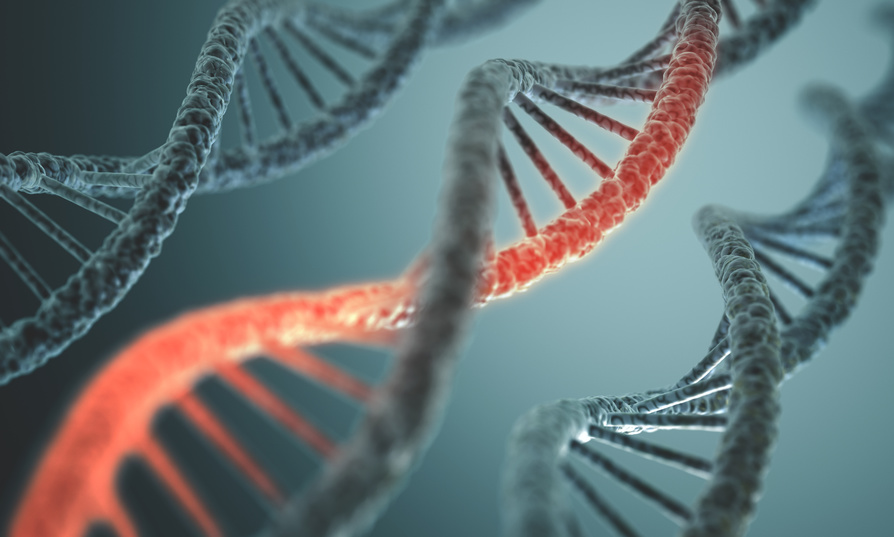
Our world is fundamentally asymmetrical: think of the double helix of DNA, the asymmetrical division of stem cells, or the fact that the human heart is positioned on the left … But how do these asymmetries emerge, and are they linked to one another?

At the Institute of biology Valrose, the team led by the CNRS researcher Stéphane Noselli, which also includes Inserm and Université Cote d’Azur researchers, has been studying right–left asymmetry for several years in order to solve these enigmas. The biologists had identified the first gene controlling asymmetry in the common fruit fly (Drosophila), one of the biologists’ favoured model organisms. More recently, the team showed that this gene plays the same role in vertebrates: the protein that it produces, Myosin 1D,[1] controls the coiling or rotation of organs in the same direction.

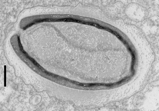
In this new study, the researchers induced the production of Myosin 1D in the normally symmetrical organs of Drosophila, such as the respiratory trachea. Quite spectacularly, this was enough to induce asymmetry at all levels: deformed cells, trachea coiling around themselves, the twisting of the whole body, and helicoidal locomotive behavior among fly larvae. Remarkably, these new asymmetries always develop in the same direction.

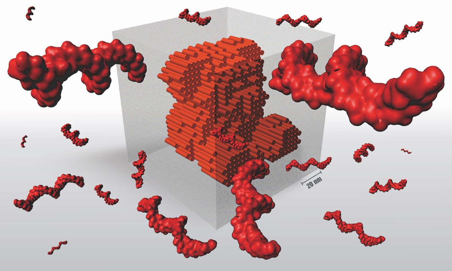
In order to identify the origin of these cascading effects, biochemists from the University of Pennsylvania contributed to the project too: on a glass coverslip, they brought Myosin 1D into contact with a component of cytoskeleton (the cell’s “backbone”), namely actin. They were able to observe that the interaction between the two proteins caused the actin to spiral.

Besides its role in right–left asymmetry among Drosophila and vertebrates, Myosin 1D appears to be a unique protein that is capable of inducing asymmetry in and of itself at all scales, first at the molecular level, then, through a domino effect, at the cell, tissue, and behavioral level.

These results suggest a possible mechanism for the sudden appearance of new morphological characteristics over the course of evolution, such as, for example, the twisting of snails’ bodies. Myosin 1D thus appears to have all the necessary characteristics for the emergence of this innovation, since its expression alone suffices to induce twisting at all scales.


[1] Myosins are a class of proteins that interact with actin (a constituent of cell skeletons or cytoskeletons). The most well-known of them, muscular myosin, makes muscles contract.

Pandoravirus: giant viruses invent their own genes

Pandoravirus quercus, found in Marseille, France. Thin section, viewed via electron microscopy. Scale bar: 100 nm.  ©IGS- CNRS/AMU.

Three new members have been isolated and added to the Pandoravirus family by researchers at the Structural and Genomic Information Laboratory (CNRS/AixMarseille Université), working with partners at the Large Scale Biology Laboratory (CEA/Inserm/Université GrenobleAlpes) and at CEA-Genoscope. This strange family of viruses, with their giant genomes and many genes with no known equivalents, surprised the scientists when they were discovered a few years ago. In the 11 June 2018 edition of Nature Communications, researchers offer an explanation: pandoviruses appear to be factories for new genes – and therefore new functions. From freaks of nature to evolutionary innovators, giant viruses continue to shake branches on the tree of life!

In 2013, the discovery of two giant viruses unlike anything seen before blurred the line between the viral and cellular world. Pandoraviruses are as big as bacteria, and contain genomes that are more complex than those found in some eukaryotic organisms[1]. Their strange amphora shape and enormous, atypical genome[2] led scientists to wonder where they came from. 


The same team has since isolated three new members of the family in Marseille, continental France, Nouméa, New Caledonia, and Melbourne, Australia. With another virus found in Germany, the team compared those six known cases using different approaches. Analyses showed that despite having very similar shapes and functions, these viruses only share half of their genes coding for proteins. Usually, however, members of the same family have more genes in common.

Furthermore, these new members contain a large number of orphan genes, i.e. genes which encode proteins that have no equivalent in other living organisms (this was already the case for the two previously discovered pandoraviruses). This unexplained characteristic is at the heart of many a debate over the origin of viruses. What most surprised researchers was that the orphan genes differed from one pandoravirus to another, making it less and less likely that they were inherited from a common ancestor!

Bioinformatic analysis showed that these orphan genes exhibit features very similar to those of non-coding (or intergenic) regions in the pandoravirus genome. Findings indicate the only possible explanation for the gigantic size of pandoravirus genomes, their diversity and the large proportion of orphan genes they contain: most of these viruses’ genes may originate spontaneously and randomly in intergenic regions. In this scenario, genes “appear” in different locations from one strain to another, thus explaining their unique nature.  

If confirmed, this groundbreaking hypothesis would make these giant viruses craftsmen of genetic creativity – a central, but still poorly explained component of any understanding of the origin of life and its evolution.


[1] Organisms whose cells contain nuclei, unlike the two other kingdoms of living organisms, bacteria and archaea.

[2] Up to 2.7 million base pairs.


This research received funding from the Bettencourt Schueller Foundation, through the “Coup d’Elan Prize for French Research” awarded to Chantal Abergel in 2014.


Credit: IGS- CNRS/AMU

Reducing Protein Intake to Fight Tumors More Effectively

©Brooke Lark on Unsplash

What if immune system efficacy against cancerous cells could be reinforced by a diet in which calories are not reduced but nutrients are precisely determined? This what Inserm researchers from Université Côte d’Azur, through a study of the effects of restrictive diets on tumor growth in mice, have been exploring.  They have observed that a low-protein diet restricts tumor development by increasing immune response.  The findings, to be published in Cell metabolism, have proved promising in understanding anti-tumor immunity in mice and pave the way for new studies in humans. 

Despite the recent popularity of fasting in preventing cancer, in reinforcing chemotherapy and in extending life expectancy in patients with tumors, there is no solid scientific proof to support its efficacy at present. In reality, clinical trials are virtually non-existent in humans and the findings obtained from animal models are highly debatable.  Prolonged calorie reduction can be an aggravating factor in the undernourishment and loss of muscle mass (sarcopenia) frequently associated with chemotherapy.

An Inserm team at Université Côte d’Azur decided to focus on a hypothesis by which modulating the intake of macronutrients (carbohydrates, fats and proteins) rather than that of calories, could restrict tumor growth.  The researchers compared the effect of various diets, with varying levels of carbohydrates and proteins but the same number of calories, on tumor growth in mice.  The results show that it was a low-protein and not a low-carbohydrate diet that had a positive impact on limiting tumor growth and prolonging life expectancy in mice.

Analysis of the tumor cell content of mice on a low-protein diet showed an increased quantity and more intense activity of the specific anti-tumor cells of the immune system.  The researchers observed that the restriction of tumor growth was not due to inhibited cancer cell proliferation as could be believed, but to an increased efficacy of the immune response, also known as immunosurveillance, in destroying the cancerous cells.

When studying the molecular mechanisms linked to this phenomenon, the researchers observed that this strengthened immunosurveillance was linked to tumor cell secretion of immune system alert proteins, known as cytokines. According to the study, reducing proteins in the diet renders the available quantity of certain amino acids (constituents of proteins) insufficient – and these are substances to which cancer cells are highly sensitive.  When access to amino acids is reduced, stress is triggered in the tumor cells, which then release cytokines and thereby activate a strong immune response against the tumor.

While these findings in mice are promising in terms of understanding the anti-cancer immunosurveillance activation mechanisms, several major unknowns remain to be elucidated. These include a precise definition of the protein reduction necessary and sufficient for the diet to be effective, the identification of the amino acids implicated in tumor cell stress, and the transposability of the results to humans, whose immunosurveillance and metabolism are notably different to those of mice.  Finally, ongoing human clinical trials must take into account the difficulty of imposing such a rigorous long-term diet on patients.

Flunarizine: a New Drug Candidate in the Treatment of Spinal Muscular Atrophy

©Adobestock

A team of researchers from Inserm (“Toxicology, pharmacology and cell signaling” JRU 1124) and the universities of Paris Descartes and Paris Diderot have recently discovered that flunarizine – a drug already used to treat migraine and epilepsy – enables the repair of a molecular defect related to spinal muscular atrophy, a severe and incurable disease. This discovery is the culmination of research efforts ongoing since 1995, when the Inserm team – comprising Suzie Lefebvre, leader of the current research projects – identified the gene responsible for infantile spinal muscular atrophy. The results of the initial animal tests, published in Scientific Reports, demonstrate a marked improvement in health. These extremely promising findings must now be confirmed in humans.


 Spinal muscular atrophy is a rare genetic disease, affecting between 1 and 9 out of every 100,000 people. It is caused by degeneration of the motor neurons in the spinal cord, resulting in progressive muscle loss. In the majority of cases, symptoms appear either following birth – with the infant unable to hold up his or her head, or a little later in early childhood – with the inability to walk. More rarely, symptoms can begin in adolescence, in which case the muscular disorders are substantial but compatible with a more-or-less normal life.

The disease is caused by a mutation of the SMN1 gene, leading to a deficiency in the SMN protein. The SMN2 gene, which is virtually identical, then takes over. However, the SMN protein that it produces is for the most part truncated and not highly functional.


An SMN protein targeting problem

In healthy individuals, the SMN protein is drawn into cell nucleus structures known as Cajal bodies. There, small non-coding RNA is formed, which is implicated in a maturation step of the messenger RNA (known as splicing), a precursor of the proteins. In spinal muscular atrophy, the truncated SMN proteins are unable to reach the Cajal bodies. The Cajal bodies then function poorly and the production of the small non-coding RNA is altered. As such, many messenger RNA present maturation problems and result in abnormal or deficient proteins – a phenomenon occurring in all tissues.

In an attempt to restore this mechanism, the researchers tested therapeutic molecules in vitro, on cells taken from patients with a severe form of the disease. The objective was to find one or more cells able to retransport the SMN proteins to the Cajal bodies so that they regain their function.


Flunarizine effective on cells from a variety of patients

Just one molecule has demonstrated an effect on a large number of cells from various patients: flunarizine, which is already used in the treatment of migraine and epilepsy. In a second step, it was used to treat mice with spinal muscular atrophy, at a rate of one spinal cord injection per day from birth. Their life expectancy increased by 40% on average, from 11 to 16 days and even up to 36 days in one case. Analysis of the motor neurons and muscles show that they are preserved for longer in the treated animals. “The molecule presents a major neuroprotective effect even if we currently don’t know why that is,” declares Lefebvre, research leader and a member of the team having discovered the gene responsible for infantile spinal muscular atrophy in 1995. In addition, her team observed that flunarizine makes it possible to restore the functioning of the small non-coding RNA produced in the Cajal bodies for the maturation of the messenger RNA.


Findings to be confirmed in humans

Flunarizine remains to be tested in humans, a stage which will face the challenge of enrolling patients in the context of a rare disease. In addition, most of these patients are already enrolled in a clinical trial to evaluate a new-generation drug that was granted marketing authorization in 2016, meaning that they cannot be mobilized to participate in a second trial. Ultimately, the two therapeutic approaches – each of which targeting a different mechanism – could very well complement each other to promote patient survival and quality of life.

3D Objects of Unequaled Precision Made from DNA

A revolution in the field of nanotechnology! An Inserm researcher[1] in collaboration with Harvard University has succeeded in creating 3D shapes of unprecedented sophistication, thanks to the four DNA bases A, T, C and G. In practice, these researchers can create nanoscopic (10-9 m) objects from 30,000 DNA sequences that fold and self-assemble like LEGO® bricks. In time, this will make it possible to manufacture new tools adapted to the size of our cells. These results have been published in Nature.

Nanotechnology represents a rapidly expanding scientific field, particularly when it comes to creating materials with increasingly specific properties. This is the case of carbon nanotubes, for example, which are light but solid and possess very high thermal and electrical conductivity. However, a slightly less well-known field of research exists: that of DNA-based nanotechnology,  whose objective is to model living matter to use as a therapeutic tool on a scale compatible with that of a human cell. However, this technology, which was developed in 2012 and is called DNA LEGO® bricks, encountered challenges in programming DNA sequences sufficient to create increasingly complex objects.

In this paper published in Nature, the researchers have reached a turning point. Their objects, manufactured according to the LEGO® brick method, use one million DNA bases, a size comparable to the genome of a bacterium, whereas until then the objects comprised only one thousand bases.

So, how does it work?

The method uses bricks, like LEGOs®, each comprising 52 DNA bases. One of the properties of DNA is based on the fact that the nucleobases of a DNA strand (A, T, C and G) can interact with those of another strand by always pairing in the same way: A with T, and C with G. Like LEGOs®, these units all have the same general shape but the internal order of the 52 bases determines which bricks will be able to interlock with which and at what level.

Next comes the choice of shape, which is either designed or selected from a database of 3D forms (cube, teddy bear, rabbit, Möbius strip, etc.). Then, each “voxel” [2] of the design is translated into DNA bricks using software developed by the researchers, called Nanobricks. “Nanobricks ‘codes’ the DNA by specifying in advance the order of the 52 bases of each brick that will subsequently be used. This step determines how the 30,000 initial sequences will fit together to produce one final 3D structure,” explains Gaëtan Bellot, Inserm researcher and co-author of this paper.

Once the IT procedures are complete, the 30,000 sequences are synthesized in a laboratory and mixed in a tube. The 30,000 DNA sequences are then completely destructured by means of a denaturing step performed at 80°C. The mixture is then gradually cooled to 25°C at a rate of 0.5°C/hour, which is when the self-assembly takes place. The molecules spontaneously fold and adopt a final shape in accordance with the 3D model selected. In this paper, the researchers produced 13 different objects.

To make objects from 30,000 sequences, they had to increase the diversity of the DNA brick sequences. By exploring various brick sizes, the research teams were able to define an optimal brick size (52 bases) making it possible both to maintain a 3D geometry similar to that of a LEGO brick and increase the diversity of individual bricks to 67 million.

This means that it is possible to obtain increasingly sophisticated objects with a greater diversity of individual bricks. The researchers have succeeded in creating objects with cavities. This level of precision is necessary if we are to design useful and effective tools. “With a key, you can open a car. With a DNA tool you can, for example, build a capsule into which you can place a medicine. And if this object has cavities, you can create a biological chain reaction depending on the products present in each cavity. Taking inspiration from life, this approach will enable the reproduction, on a nanometer scale, of solutions and inventions that occured after millions of years of evolution,” explains  Bellot.

This method offers two advantages. The first is that unlike industrial assembly processes, such as car production lines, this technology compresses all the stages into one. Imagine placing a car’s various components in the presence of each other for them to spontaneously self-assemble! The second resides in its rapidity: 30,000 components self-assemble within several hours into an object duplicated a billion times inside the same tube.

Unlike carbon nanotubes, DNA nanotechnologies are biocompatible and can be rapidly eliminated from the human body and the environment.  Nevertheless, even if the DNA molecules used are synthetic and as such not biologically active, potential interaction with the DNA present in living organisms cannot be ruled out.


[1] From the Institute for Functional Genomics (Inserm/CNRS/Université de Montpellier)

[2] Voxel, a contraction of the words “volume” and “element”, is a 3D pixel.

The cause of uncombable hair syndrome identified

Surprised disheveled preschooler girl with long hair

In 1973, the rare syndrome of uncombable hair or ‘pili trianguli et canaliculi‘ was described by a Toulouse dermatologist. More than 40 years later, Michel Simon, Inserm research director his colleagues at the ‘Epidermal Differentiation and Rheumatoid Autoimmunity’ Unit [UDEAR] (Inserm/CNRS/Toulouse III – Paul Sabatier University) have identified its genetic cause. These results are published in The American Journal of Human Genetics.

Uncombable hair syndrome is a rare disease of the hair, the prevalence of which is unknown. It generally begins during childhood between 3 months and 12 years. Dry and unruly, the hair of affected children becomes gradually silver-blond or straw coloured. Hairs stand up on the scalp and grow in all directions. It is impossible to comb it or to flatten it with a comb. In detail, scanning electron microscopy reveals a longitudinal groove running their entire length, with a triangular or kidney-shaped cross-section. However, this syndrome is not disabling and undergoes spontaneous at the end of childhood.


uncombable-hair

he researchers, working with a team from the Human Genetics Institute at Bonn University and dermatologists or geneticists from 7 different countries, have discovered that the disease is due to recessive mutations of a trio of genes that contribute to forming the hair: the gene coding for one of its structural components, trichohyalin (TCHH); or two genes coding for enzymes that take it in turns as target: peptidyl-arginine deiminase 3 (Pad3) and transglutaminase 3 (TGase3).

Furthermore, the researchers have also shown, in mice, that inactivating the Pad3 gene alters the shape of the fur and whiskers of animals, as had already been reported in TGase3-deficient mice.

In conclusion, the absence of TCHH or failure of the biochemical cascade that results in stiffening the hair stem are responsible for the hair formation abnormalities characteristic of uncombable hair syndrome or ‘pili trianguli et canaliculi‘.

These results, as well as describing the molecular cause of the disease and enabling better diagnosis, provide knew knowledge about the hair and the mechanisms of its formation£ concludes Michel Simon, Inserm research director.

For further information

Although extremely rare, the syndrome has long been known. It was brought to public awareness by the famous literary figure ‘Struwwelpeter’ created by children’s author Heinrich Hoffmann in 1845. The book was subsequently translated into English by Mark Twain as ‘Slovenly Peter’. Although he never said so, one might even think that it inspired director Tim Burton to make his film Edward Scissorhands.

Jumping genes: all guilty?

Transposable elements, also known as “jumping genes” are DNA fragments that can move or copy themselves from one location to another on the chromosomes. They have invaded the genomes of most living organisms, from bacteria to humans, via the plants. When they jump, they bring about complex modifications in genes near which or in which they insert themselves, and can thereby alter or abolish their function. This phenomenon contributes to the evolution and adaptation of species.

However, in the shorter term, “jumping genes” can have harmful effects. In humans, the only currently active transposon family, the LINE-1 type retrotransposons, causes new cases of genetic diseases, such as haemophilia and muscular dystrophy. This is why their activity is normally under tight control. However, in nearly half of epithelial cancers, they manage to escape the many cellular defence mechanisms that protect our DNA, and jump actively, contributing to the emergence and progression of cancers. Moreover, in studies they are often used as tumour biomarkers for diagnostic or prognostic purposes.

DNA Structure

(c) Fotolia

One of the main problems raised by the study of “jumping genes” is related to their extremely repetitive nature. Our DNA contains thousands of copies of them, almost identical to one another, and every individual contains hundreds of copies that are not listed in the reference map of the human genome. Furthermore, until now, it had been impossible to know whether activation of “jumping genes” results from a general disruption, leading to massive mobilisation of all copies, or whether, on the contrary, only a small number of them manage to escape the protective controls. Through a new approach, published in the journal eLife, involving high-throughput sequencing, genomics, epigenomics and bioinformatics, the team led by Gaël Cristofari, Inserm Research Fellow, and his collaborators at Unit 1081, “Institute for Research on Cancer and Aging, Nice (IRCAN),” has succeeded in measuring the activity of the “jumping genes” in normal and cancer cells with unprecedented resolution.

According to their results, only a small number of copies are really guilty: those located in permissive regions of our chromosomes. And these regions vary depending on the type of cell. Furthermore, all these active copies are not present in all individuals!

“The important concept here is that the small group of LINE-1 transposons that escapes control is different from one cell type to another: in certain cancers, one group is important, in another type of cancer, it will be another group of copies. This observation suggests that behind each LINE-1 group, there is a mechanism and signals specific for a particular type of organ or tissue,” explains Gaël Cristofari.

These results provide a better understanding of how new mutations may emerge, they suggest the existence of genetic factors behind this phenomenon, and they provide new data for the rational use of LINE-1 retrotransposons as biomarkers in oncology, by focusing on the active copies in a given type of cell.


This work was made possible by financial support from ARC French Foundation for Cancer Research, Fondation pour la Recherche Médicale, Cancéropôle PACA, the European Research Council, the French National Research Agency (ANR) (Labex Signalife), and the Research Group on Mobile Genetic Elements (CNRS, GDR 3546).

Light shed on the underside of the “cocktail effect” of endocrine disruptors

Chemical substances that are safe for humans when taken in isolation can become harmful when they are combined. Three research teams bringing together researchers from Inserm and CNRS[1] in Montpellier have elucidated in vitro a molecular mechanism that could contribute to the phenomenon known as the “cocktail effect.” This study is published in the journal Nature Communications.

[1] Centre for Structural Biochemistry (CBS) (CNRS UMR5048 – Inserm U1054) at the Cancer Research Institute (Inserm U1194), and the Functional Genomics Institute (CNRS UMR5203 – Inserm U661)

Every day we are exposed to many exogenous compounds such as environmental pollutants, drugs or substances in our diet. Some of these molecules, known as endocrine disruptors, are strongly suspected of interacting inappropriately with regulatory proteins in our cells, and inducing numerous physiological or metabolic disorders (cancers, obesity, diabetes, etc.). Moreover, the combination of these molecules in complex mixtures with which we are in routine contact might exacerbate their toxicity.

In an article to be published in Nature Communications, researchers have unveiled a mechanism that might contribute to this effect of mixing, for which no rational explanation has been offered until now. They show that some oestrogens such as ethinyloestradiol (one of the active ingredients of contraceptive pills) and organochlorine pesticides such as trans-nonachlor, although very weakly active on their own, have the ability to bind simultaneously to a receptor located in the cell nucleus, and to activate it synergistically. 

Analyses at molecular level indicate that the two compounds bind cooperatively to the receptor, i.e. binding of the first molecule promotes binding of the second. This cooperativity is due to strong interactions at the level of the receptor binding site, so that the binary mixture induces a toxic effect at substantially lower concentrations than the individual molecules.


These results obtained in vitro constitute a proof of concept that opens the way to a wide field of study. There are actually about 150,000 compounds in our environment that could have unexpected effects on human health through combined action, given their recognised or assumed safety as isolated substances. If these studies are confirmed in vivo, important consequences are expected in the areas of endocrine disruption, toxicology, and the assessment of risks associated with the use of chemicals.

Figure

Separately, ethinyloestradiol (EE2) and trans-nonachlor (TNC) bind to the xenobiotic receptor (PXR) only at high concentrations, and are weak activators of this receptor. When they are used together, the two compounds mutually stabilise each other in the binding pocket of the receptor. The “supramolecular ligand” thus created has increased affinity for PXR, so that it can induce a toxic effect at doses at which each compound is inactive individually. © Vanessa Delfosse, William Bourguet 

Intelligent bacteria for detecting disease

Another step forward has just been taken in the area of synthetic biology. Research teams from Inserm and CNRS (French National Centre for Scientific Research) Montpellier, in association with Montpellier Regional University Hospital and Stanford University, have transformed bacteria into “secret agents” that can give warning of a disease based solely on the presence of characteristic molecules in the urine or blood. To perform this feat, the researchers inserted the equivalent of a computer programme into the DNA of the bacterial cells. The bacteria thus programmed detect the abnormal presence of glucose in the urine of diabetic patients. This work, published in the journal Science Translational Medicine, is the first step in the use of programmable cells for medical diagnosis.

Bacteria have a bad reputation, and are often considered to be our enemies, causing many diseases such as tuberculosis or cholera. However, they can also be allies, as witnessed by the growing numbers of research studies on our bacterial flora, or microbiota, which plays a key role in the working of the body. Since the advent of biotechnology, researchers have modified bacteria to produce therapeutic drugs or antibiotics. In this novel study, they have actually become a diagnostic tool.

Medical diagnosis is a major challenge for the early detection and subsequent monitoring of diseases.In vitro” diagnosis is based on the presence in physiological fluids (blood and urine, for example) of molecules characteristic for a particular disease. Because of its noninvasiveness and ease of use, in vitro diagnosis is of great interest. However, in vitro tests are sometimes complex, and require sophisticated technologies that are often available only in hospitals.

This is where biological systems come into play. Living cells are real nano-machines that can detect and process many signals and respond to them. They are therefore obvious candidates for the development of powerful new diagnostic tests. However, they have to be provided with the appropriate “programme” for them to successfully accomplish the required tasks.

To do this, Jérôme Bonnet’s team in Montpellier’s Centre for Structural Biochemistry (CBS) had the idea of using concepts from synthetic biology[1] derived from electronics to construct genetic systems making it possible to “programme” living cells like a computer.

The transcriptor: the cornerstone of genetic programming

The transistor is the central component of modern electronic systems. It acts both as a switch and as a signal amplifier. In informatics, by combining several transistors, it is possible to construct “logic gates,” i.e. systems that respond to different signal combinations according to a predetermined logic. For example, a dual input “AND” logic gate will produce a signal only if two input signals are present. All calculations completed by the electronic instruments we use every day, such as smartphones, rely on the use of transistors and logic gates.

During his postdoctoral fellowship at Stanford University in the United States, Jérôme Bonnet invented a genetic transistor, the transcriptor.

The insertion of one or more transcriptors into bacteria transforms them into microscopic calculators. The electrical signals used in electronics are replaced by molecular signals that control gene expression. It is thus now possible to implant simple genetic “programmes” into living cells in response to different combinations of molecules[2].

In this new work, the teams led by Jérôme Bonnet (CBS, Inserm U1054, CNRS UMR5048, Montpellier University), Franck Molina (SysDiag, CNRS FRE 3690), in association with Professor Eric Renard (Montpellier Regional University Hospital) and Drew Endy (Stanford University), applied this new technology to the detection of disease signals in clinical samples.

Clinical samples are complex environments, in which it is difficult to detect signals. The authors used the transcriptor’s amplification abilities to detect disease markers, even if present in very small amounts. They also succeeded in storing the results of the test in the bacterial DNA for several months.

The cells thus acquire the ability to perform different functions based on the presence of several markers, opening the way to more accurate diagnostic tests that rely on detection of molecular “signatures” using different markers.
bonnet

Figure 1: Principle of the use of modified bacteria for medical diagnosis. ©J. Bonnet/ Inserm.

“We have standardised our method, and confirmed the robustness of our synthetic bacterial systems in clinical samples. We have also developed a rapid technique for connecting the transcriptor to new detection systems. All this should make it easier to reuse our system,” says Alexis Courbet, a postgraduate student and first author of the article.

As a proof of concept, the authors connected the genetic transistor to a bacterial system that responds to glucose, and detected the abnormal presence of glucose in the urine of diabetic patients.

“We have deposited the genetic components used in this work in the public domain to allow their unrestricted reuse by other public or private researchers,[3] says Jérôme Bonnet.

“Our work is presently focused on the engineering of artificial genetic systems that can be modified on demand to detect different molecular disease markers,” he adds. In future, this work might also be applied to engineering the microbial flora in order to treat various diseases, especially intestinal diseases.

This work received financial support from Inserm, CNRS, the Stanford-France Center for Interdisciplinary Studies, and Stanford University. Jérôme Bonnet is a recipient of the Atip-Avenir programme award, and is supported by the Bettencourt-Schueller Foundation.

[1] aimed at the rational engineering of artificial biological systems and functions

[2] (Bonnet et al. Science, 2013).

[3] available on: https://biobricks.org/bpa/

fermer