

Press Contact
Institut curie
Catherine Goupillon-Senghor
Tél. 01 56 24 55 23
rf.eiruc@esserp.ecivres
In order to better understand this long and extremely complex process, and to discover new therapeutic possibilities, it is crucial to elucidate all stages of tumour progression, from the initial mutation until the development of metastases.
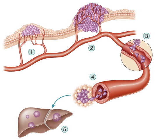
From tumour to metastases, a multitude of events
© Eléonore Lamoglia/Institut Curie
A blend of theory and experiment
“With the help of a mathematical model compiling the data from over 200 scientific publications, we first identified two vital components in the epithelial-mesenchymal transition of intestinal cells,” explains Inna Kuperstein, a researcher from Emmanuel Barillot’s team. This transition converts the epithelial cells into a form known as mesenchymal[1]. These less specialised and more plastic cells lose their capacity to adhere to one another, and acquire properties that enable them to migrate and “blend” into their immediate environment. The epithelial-mesenchymal transition may represent the first step for tumour cells towards dissemination.
“For this to happen, our model shows that two events must occur in the intestinal cells: the Notch receptor must be activated, and the p53 gene must be lost,” comments Andrei Zinovyev, coordinator of the mathematical study at Institute Curie.
The researchers subsequently developed an animal model carrying these two alterations in the intestinal tissue. “This model makes it possible to study tumour cells throughout their development and thus better understand the modifications required for metastases to form,” explains Sylvie Robine, an Inserm Research Director at Institut Curie.
First observation: these mice developed numerous metastases, in several organs. The combination of alterations in Notch and p53 provides the starting ground needed for the development of metastatic colon cancer.
“When colon cancer cells begin to disseminate, they progressively lose the features of epithelial cells, the tissue from which they originate, to acquire the specific features of mesenchymal cells,” she continues.
In addition, the hallmarks of mesenchymal tissue are uniquely present in the cells of the invasive front of the tumour, cells that are moving towards the “exit” from the intestinal tissue (the stroma). The cells that escape from the original tissue are therefore cells that have begun the epithelio-mesenchymal transition. “This result has been corroborated by analysing samples from invasive colon cancers and metastases taken from patients,” states Prof. Daniel Louvard[2], CNRS Research Director at Institut Curie. “The cells present in these specimens have the characteristic features of mesenchyme, not of epithelia.”
Thanks to research carried out jointly by bioinformaticians from Emmanuel Barillot’s team and biologists from Prof. Daniel Louvard’s team, the stages in tumour progression and the various pathways leading to colon cancer are gradually yielding up their secrets.
“The combination of alterations in p53 and Notch creates the most conducive conditions for the development of metastases in colon cancers” explains Prof. Daniel Louvard.
The mice developed constitute an excellent preclinical model, and as such they may provide the basis of a search for new therapeutic targets. By knowing the specific alterations of the tumour from an individual, better targeted and more effective personalised treatments can be created.
[1] Mesenchyme is an embryonic connective tissue that gives rise to various types of connective tissue in the adult.
[2] Prof. Daniel Louvard is the Honorary Director of the Institut Curie Research Centre, and is currently advisor to the President on international relations of Institut Curie. Maia Chanrion, a member of Daniel Louvard’s team, contributed to the experimental work, Inna Kuperstein and David Cohen, members of Emmanuel Barrillot’s team, participated in the bioinformatics study.
Institut curie
Catherine Goupillon-Senghor
Tél. 01 56 24 55 23
rf.eiruc@esserp.ecivres
Concomitant Notch activation and p53 deletion trigger epithelial-to-mesenchymal transition and metastasis in mouse gut
M.Chanrion, I. Kuperstein, C. Barrière, F. El Marjou, D. Cohen, D. Vignjevic, L. Stimmer, P. Paul-Gilloteaux, I. Bièche, S. Dos Reis Tavares, G. Fulvio Boccia, W. Cacheux, D. Meseure, S. Fre, L. Martignetti, P. Legoix-Né, E. Girard, L. Fetler, E. Barillot, D. Louvard, A. Zinovyev, S. Robine
Nature Communications, 8 octobre 2014