Menu

History of stroke and coronary heart disease – a fatal combination

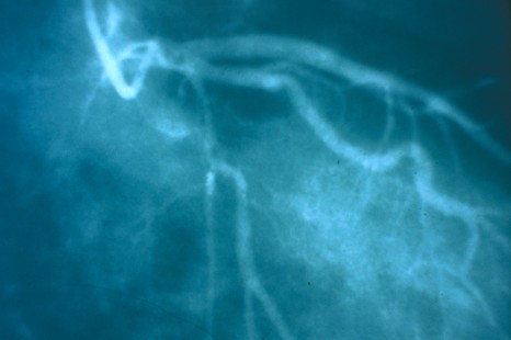
The cardiology service team at the Hôpital Bichat and the Mixed INSERM Unit 698 (AP-HP, Université Paris Diderot), in collaboration with international teams of researchers, studied a cohort of patients suffering from coronary disease. The study showed that those patients with a history of stroke or transient ischæmic attack (TIA) are not only at higher risk of cardio-vascular episodes but also of haemorrhagic events, stressing the therapeutic challenge involved in treating such patients. The research is published online in Circulation*.


© Serimedis/Inserm

Heart and cerebro-vascular disorders represent the two leading causes of death throughout the world. They are sometimes combined in a single patient and their combination represents both a considerable risk to the patient and a therapeutic challenge

Today’s anti-thromobotic, blood-thinning medication provides effective treatment for coronary heart disease. Several randomised tests to assess new anti-thrombotic treatments in coronary patients have identified the fact that a history of stroke or TIA constitutes a marker for increased risk of intracranial bleeding that could prove fatal for the patient. The study was conducted by the Cardiology Service Team of the Bichat Hospital and INSERM Unit 698, working with several international teams, and consisted in monitoring and analysing 26,389 coronary patients from REACH, the international register of athero-thrombic patients, over a four-year period, in order to very accurately assess the ischaemic and haemorrhagic risk associated with a history of stroke or TIA in coronary patients.

The results of this study highlight the frequency of the phenomenon. It showed that 4,460 patients, 17% of the cohort of coronary patients, had a history of stroke or TIA. This stroke or TIA antecedent was associated, moreover, with an approximate 50% increase (in relation to coronary patients without a history of stroke) of dying from a heart attack or stroke, with increased risk of ischæmic and hæmorrhagic stroke.

This study also aims at investigating the therapeutic challenge of treating such patients. In fact, higher doses of anti-platelet or anti-coagulant are associated with a particularly high increased risk of hæmorrhage.

The conclusions of the study are important since they emphasise the difficulty in treating this type of patient, a type that is frequent but whose prognosis is particularly serious. It also shows how important it is to provide the right anti-thrombotic treatment. Additional clinical trials are currently being conducted to test new treatment strategies for these patients”, explained Professor Steg of the Cardiology Department at the Hôpital Bichat.

Stroke is where there is a blockage or rupture of a blood vessel that transports blood inside the brain. The result is a lack of oxygen to the brain, which put one or more areas of the brain in danger.

A stroke occurs when the blood circulation to or within the brain is interrupted by a blocked blood vessel (an ischæmic stroke), the commonest form, or a blood vessel ruptures (a hæmorrhagic stroke), the latter occurring in less than 20% of cases.

Ischæmic stroke. Where the artery becomes blocked by a high-cholesterol platelet (atherosclerosis), the patient suffers an ischæmic stroke (40 to 50% of strokes), or where a blood clot blocks the artery, in which case it is a cerebral embolism (30% of AVCs).

Hæmorrhagic stroke (20% of cases). This is most frequently due to a ruptured aneurism. Tumours, hypertensive crisis and miscellaneous blood-clotting disorders can also cause hæmorrhagic strokes.

Transient Ischæmic Attack (TIA) When the obstruction in the cerebral artery reabsorbs itself and produces no sequel, the event is described as a transient ischæmic attack. The symptoms are the same as those of a stroke, but they only last a few seconds or minutes before a return to normal. An AIT may even pass unnoticed and be mistaken for mere discomfort. Yet it indicates an important risk of a more serious AVC.

The efficacy of certain anti-cancer vaccines depends on how they are administered

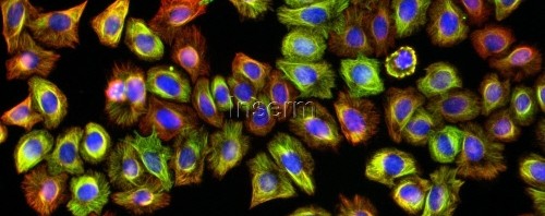
The therapeutic efficacy of certain anti-cancer vaccines depends on how they are administered. This is what the team of researchers headed by Eric Tartour at the Paris-Centre de recherche Cardiovasculaire (Université Paris Descartes, INSERM Unit 970 PARCC, AP-HP), in collaboration with researchers from the CNRS[1], have just demonstrated. A paper on the subject was published on 13 February 2013 in Science Translational Medicine. In the case of so-called mucosal cancers of the lungs or ENT area, the vaccine should be administered directly via the mucosa if it is going to be effective (intra-nasally for example). The same vaccine administered in the usual way that immunisation is performed, i.e. intramuscular or sub-cutaneous injection, will be ineffective. In this research, the investigators also identified an important method by which the vaccine acts against tumours. These results will have a major impact on the creation and effectiveness of anti-cancer vaccines targeting mucous tumours.


© Fotolia

Vaccines to treat cancer represent a new and promising avenue of therapy. The aim is to stimulate certain cells in the immune system, such as the T-CD8 lymphocytes, in order to cause the tumour to shrink. Many hopes are being pinned on these types of vaccines. Numerous trials are currently in progress, but some have suffered setbacks in the clinical phase even though they proved successful on animal models.  

So as to understand why this happens, Eric Tartour and his team (Université Paris Descartes, INSERM  Unit 970 PARCC, AP-HP) used mice to test the efficacy of two different routes of administration, the intramuscular route, on the one hand, and the intranasal route on the other. The tests were performed on a candidate vaccine against mucous, oropharyngeal cancers developed in collaboration with Ludger Johannes at the Institut Curie.

They proved that only vaccination via the mucosa will shrink a tumour in the lung or the ear, nose and throat (ENT) area. The same anti-cancer vaccine administered in the usual immunisation way, i.e., intramuscularly or subcutaneously, is ineffective.

Our results may explain the failure of anti-tumoural vaccines designed to treat tumours of the mucosa in humans, and should lead to a change in how anti-cancer vaccines are used to target mucosal tumours”, explained Eric Tartour, Professor at Paris Descartes University and hospital doctor at the Hôpital Européen Georges Pompidou (AP-HP).

To try and explain the difference in efficacy, researchers sought to identify the mechanisms that operated differently depending on the method of administration. They showed that vaccination through the mucosa caused a certain protein (the mucous integrin CD49a) to be produced that enabled the T-CD8 lymphocytes (whose production is stimulated by the administration of the vaccine) to migrate into the tumour. Once they are at the site of the tumour, the T-CD8 lymphocytes destroy the cancerous cells.

In the opposite case, if this molecule is blocked it prevents the penetration of T lymphocytes into the mucosal tumour and prevents their anti-tumoural activity. The integrin CD49a is essential in the promotion of migration of the anti-tumoural lymphocytes produced through vaccination in the case of pulmonary tumours and ENT tumours and thus the efficacy of the vaccination in combatting mucosal cancers.

To check the applicability of these results in humans, the researchers analysed samples from patients suffering from lung cancer. Here again, they demonstrated the elevated expression of mucosal integrin CD49a on T-CD8 lymphocytes present in patients with lung tumours[1].

These results are encouraging for the future extrapolation of the study to humans.


[1] Travaux menés par Laurence Zitvogel, Unité immunologie des tumeurs et immunothérapie (Université Paris-Sud/Inserm) ; Institut Gustave Roussy, Villejuif 


[1] Laboratories involved: Unité de pharmacologie chimique et génétique et d’imagerie (CNRS/Université Paris Descartes/Inserm/Chimie ParisTech); Compartimentation et dynamique cellulaires (CNRS/Institut Curie/UPMC). 

“Simplified” brain lets the iCub robot learn language

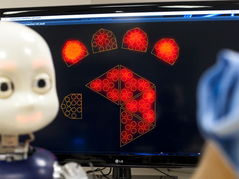
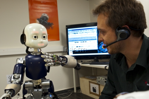
The iCub humanoid robot on which the team directed by Peter Ford Dominey, CNRS Director of Research at Inserm Unit 846 known as the “Institut pour les cellules souches et cerveau de Lyon” [Lyon Institute for Stem Cell and Brain Research] (Inserm, CNRS, Université Claude Bernard Lyon 1) has been working for many years will now be able to understand what is being said to it and even anticipate the end of a sentence. This technological prowess was made possible by the development of a “simplified artificial brain” that reproduces certain types of so-called “recurrent” connections observed in the human brain. The artificial brain system enables the robot to learn, and subsequently understand, new sentences containing a new grammatical structure. It can link two sentences together and even predict how a sentence will end before it is uttered. This research has been published in the Plos One journal. 

robot ICub apprentissage

© P Latron/inserm

INSERM and CNRS researchers and the Université Lyon 1 have succeeded in developing an “artificial neuronal network” constructed on the basis of a fundamental principle of the workings of the human brain, namely its ability to learn a new language. The model was developed after years of research in the INSERM 846 Unit of the Institut de recherche sur les cellules souches et cerveau, through studying the structure of the human brain and understanding the mechanisms used for learning.

One of the most remarkable aspects of language-processing is the speed at which it is performed. For example, the human brain processes the first words of a sentence in real time and anticipates what follows, thus improving the speed with which humans process information. Still in real time, the brain continually revises its predictions through interaction between new information and a previously created context. The region inside the brain linking the frontal cortex and the striatum plays a crucial role in this process.

Based on this research, Peter Ford Dominey and his team have developed an “artificial brain” that uses a “neuronal construction” similar to that used by the human brain.

Thanks to so-called recurrent construction (with connections that create locally recurring loops) this artificial brain system can understand new sentences having a new grammatical structure. It is capable of linking two sentences and can even predict the end of a sentence before it is provided.

To put this advance into a real-life situation, the INSERM researchers incorporated this new brain into the iCub humanoid robot.

In a video demonstration, a researcher asks the iCub robot to point to a guitar (shown in the form of blue object) then asking it to move a violin to the left (shown by a red object). Before performing the task, the robot repeats the sentence and explains that it has fully understood what it has been asked to do.

For researchers, the contribution that this makes to research into certain diseases is of major importance. This system can be used to understand better the way in which the brain processes language. “We know that when an unexpected word occurs in a sentence, the brain reacts in a particular way. These reactions could hitherto be recorded by sensors placed on the scalp”, explains Peter Ford Dominey. The model developed by Dr Xavier Hinaut and Dr Peter Ford Dominey makes it possible to identify the source of these responses in the brain. If this model, based on the organisation of the cerebral cortex, is accurate, it could contribute to possible linguistic malfunctions in Parkinson’s disease.

This research has another important implication, that of contributing to the ability of robots to learn a language one day. “At present, engineers are simply unable to program all of the knowledge required in a robot. We now know that the way in which robots acquire their knowledge of the world could be partially achieved through a learning process – in the same way as children”, explains Peter Ford Dominey.

©P Latron/Inserm

Cancer du col de l’utérus : première résolution 3D d’une oncoprotéine du virus à papillome humain

Treatrush (TreatRetUsher): combating Usher Syndrome blindness – European collaboration in the service of a rare disease

On the occasion of International Rare Disease Day: 28 February 2013

How has research into Usher Syndrome – a particularly debilitating hereditary disease that damages the two main senses, hearing and sight – overcome a major hurdle in understanding the mechanisms causing pigmentary retinopathy? How has it managed to improve clinical diagnosis and develop molecular diagnosis? How has it prepared gene therapy tests for the retinopathy aspect of the syndrome? Scientists who have joined forces in the European TREATRUSH (TreatRetUsher) project have been reporting progress. Twelve partners in seven countries came together to form this network, financed by the European Commission and coordinated by the Pierre and Marie Curie University (UPMC). In France, it brings together researchers from the Collège de France, INSERM, the Institut Pasteur, the CNRS and the UPMC, working at the Institut de la Vision and the Institut Pasteur, as well as clinicians from the XV-XX Hospital and the Armand-Trousseau Hospital.

Usher Syndrome is a hereditary disease attacking hearing and sight. It affects about one person in 10,000. It is the main cause of deafness associated with a loss of vision in young people. The three clinical types of the syndrome, USH1, USH2 and USH3, are distinguished by the severity of hearing loss, early onset of retinitis pigmentosa and the presence or otherwise of a vestibular (inner ear) attack that translates into difficulties with balance. In each of these aspects of the disease the USH1 form is the most incapacitating.

Going beyond the major advances previously achieved in identifying the genes responsible for the syndrome and understanding the pathogenesis of the damage done to hearing by the syndrome, the Treatrush project has set itself the aim of improving diagnosis, understanding the cellular and molecular mechanisms behind retinopathy and preparing gene therapy trials.

  • Responding to the imperative of early diagnosis. 

A reduction in vision only manifests years after the loss of hearing, so the absence of early diagnosis of the syndrome can cause parents to make the wrong choice, and cause their children to learn sign language as young as possible, rejecting early cochlear implantation. Consequently, the European Consortium’s ENT specialists and ophthalmologists have jointly drawn up clinical investigation protocols that should result in a diagnosis being available for children under one year old. At the same time, a robust and swift molecular diagnosis of all forms of the syndrome has been developed. An initial study[1] enabled the detection of mutations in 54 patients. It was supplemented by the introduction of a new method[2] that now makes it possible to analyse a cohort of more than 350 patients recruited throughout Europe (France, Germany, Italy, Slovenia, Spain).

  • Clarifying the physio-pathological mechanisms that cause retinopathy.

While mutant mice in which there are defective Usher-1 genes are profoundly deaf, they do not present with retinopathy. Consequently, the defective mechanisms causing retinopathy in those suffering from the syndrome were totally unknown. Three years ago, teams headed by Professor Christine Petit, co-ordinator of the Treatrush Project, and Professor José-Alain Sahel, attempted to understand the physio-pathological mechanisms causing retinitis pigmentosa in patients suffering from Usher Syndrome, by using other animal models[3]. They were successful and thus laid the foundation for discovering a new type of mechanism that causes pigmentary retinopathy, one that also involves the structures of photoreceptors, an aspect hitherto neglected, the role of which is still unknown. This data is essential for the creation of any protocol designed to improve treatment.

Project teams in Italy, France and the United States are currently working to develop gene therapy via viral vectors associated with the adenovirus (AAV) capable of effectively transferring tiny therapeutic genes into retinal photoreceptors with the aim of preventing their degeneration. These vectors have already proven their worth in other forms of retinitis. They are currently being tested on animals to identify some of the Usher genes.

The advances achieved in curing this rare illness open the way to an understanding and treatment of commoner causes of deterioration in vision and hearing. As Christine Petit and José-Alain Sahel explain, “frequent disorders are of a more complex origin and are thus harder to study than rare monogenic diseases. Yet many of the contributing factors should belong to the same elementary mechanisms as those which, when defective, lead to a particular, rarer disease. As proof, take the example of the USH1 Syndrome. The molecules encoded by these genes lie at the heart of the auditory transduction machinery, machinery that converts a sound signal into an electric signal in the auditory sensory cells. It is self-evident that in cases of frequent damage to hearing, regardless of origin, this machinery is also the target of deficiency. A constellation of rare diseases should thus contribute to creating the model for the pathogenic processes occurring in a common disease”. 

The Usher Syndrome, an attack of the two main senses – hearing and sight

This syndrome is a hereditary sensory disability and the main cause of combined loss of hearing and blindness. It is responsible for 3% to 6% of infant deafness and about 50% of cases of severe deafness associated with adult blindness. Deafness is generally congenital, while the clinical  manifestations of retinitis pigmentosa occur slightly later in life. This syndrome has been divided into three sub-types, USH1, USH2 and USH3; the USH1 form is the most serious. In children suffering from the USH1 form, damage to sight is frequently not discovered until around the age of eight or ten. This late diagnosis makes it very hard to provide the appropriate medical treatment.

Important scientific progress was made by geneticists and hearing physiologists about fifteen years ago, when they identified the ten genes responsible and the discovery of deficient cellular and molecular mechanisms explained the loss of hearing caused by the syndrome. Conversely, the pathogenesis of retinitis pigmentosa in the syndrome remained unknown. A major advance occurred recently in understanding the damage caused to the retina, as part of the European Treatrush Project, through research conducted by teams headed by Professors Christine Petit and José-Alain Sahel. The patient exploration protocols were improved and standardised to allow for early diagnosis. A new molecular diagnostic tool was developed and more than 400 patients from various European countries were tested. In 1995, Professor Christine Petit’s laboratory identified the first gene causing the USH1 Syndrome. This is the gene that encodes VIIa myosin and is responsible for most cases of USH1. It is precisely in this form of the syndrome that a gene therapy retinopathy trial will soon be conducted by Professor José-Alain Sahel.

TREATRUSH – Combatting blindness caused by the Usher Syndrome

The aim of TREATRUSH is to treat and combat blindness caused by the Usher Syndrome. http://www.treatrush.eu

The project began in February 2010 and lasted for four years, the total budget of about 6 million euros being funded by the European Union (PC7). The project involves twelve partners based in seven countries:

Université Pierre and Marie Curie (UPMC), Paris, France: http://www.upmc.fr/

Institut National de la Santé et de la Recherche Médicale (Inserm), Paris, France : www.inserm.fr/

Eberhard Karl University of Tübingen  (EKUT), Tübingen, Germany: http://www.uni-tuebingen.de/en

Medical Research Council (MRC), Oxford, UK: http://www.mrc.ac.uk

Fondazione Telethon (FTELE.IGM), Naples, Italy: http://www.telethon.it/

Amsterdam Molecular Therapeutics (AMT), Amsterdam, Netherlands: http://www.amtbiopharma.com/

Novartis Forschungsstiftung, Zweigniederlassung Friedrich Miescher-Institut for Biomedical Research, Basle, Switzerland: http://www.fmi.ch/

Faun Foundation (FAUN), Nuremberg, Germany

The Trustees of the University of Pennsylvania (UPENN), Philadelphia, USA : http://www.upenn.edu/

Institut de la Vision-Fondation Voir et Entendre, Paris, France: http://www.institut-vision.org    http://www.fondave.org/

Johannes Gutenberg University of Mainz, Mainz, Germany: http://www.uni-mainz.de/eng/

Massachusetts Eye and Ear Infirmary, Bos


[1] Bonnet C, Grati M, Marlin S, Levilliers J, Hardelin JP, Parodi M, Niasme-Grare M, Zelenika D, Délépine M, Feldmann D, Jonard L, El-Amraoui A, Weil D, Delobel B, Vincent C, Dollfus H, Eliot MM, David A, Calais C, Vigneron J, Montaut-Verient B, Bonneau D, Dubin J, Thauvin C, Duvillard A, Francannet C, Mom T, Lacombe D, Duriez F, Drouin-Garraud V, Thuillier-Obstoy MF, Sigaudy S, Frances AM, Collignon P, Challe G, Couderc R, Lathrop M, Sahel JA, Weissenbach J, Petit C, Denoyelle F. (2011) Complete exon sequencing of all known Usher syndrome genes greatly improves molecular diagnosis. Orphanet J Rare Dis. 6:21.

[2] Fakin A, Jarc-Vidmar M, Glavač D, Bonnet C, Petit C, Hawlina M. (2012) Fundus autofluorescence and optical coherence tomography in relation to visual function in Usher syndrome type 1 and 2. Vision Res. 75: 60-70.

[3] Sahly I, Dufour E, Schietroma C, Michel V, Bahloul A, Perfettini I, Pepermans E, Estivalet A, Carette D, Aghaie A, Ebermann I, Lelli A, Iribarne M, Hardelin JP, Weil D, Sahel JA, El-Amraoui A, Petit C. (2012) Localization of Usher 1 proteins to the photoreceptor calyceal processes, which are absent from mice. J Cell Biol. 15;199(2):381-99.

‘Diet’ drinks associated with increased risk of Type II diabetes

Françoise Clavel-Chapelon, (Director of Research at INSERM-University of Paris-Sud 11, at the Institut Gustave Roussy), and Guy Fagherazzi were interested in a connection between the consumption of ‘diet’ or ‘light’ soft drinks and the risk of Type II diabetes. The analysis performed on 66,188 women in the E3N cohort confirms a link between sweet soft drinks and type II diabetes and reveals for the first time in France that, contrary to received wisdom, there is a higher risk of diabetes from so-called ‘diet’ or ‘light’ drinks than from ‘normal’ sweetened soft drinks. Additional studies on the effects of ‘light’ sweetened soft drinks are needed to corroborate the result. The article containing details of the results was published in the American Journal of Clinical Nutrition.

Diabetes affects more than three million people inFrance, 90% of whom suffer from Type II diabetes. While it has been established that consumption of sweetened drinks is associated with an increased risk of obesity and of Type II diabetes, the effect of ‘diet’ or ‘light’ sweetened soft drinks on cardio-metabolic diseases is less well-known. INSERM researchers on the E3N team assessed the link between the consumption of sweetened soft drinks and the risk of developing type II diabetes. The study was conducted on 66,118 French women in the E3N cohort who were monitored for 14 years.

The results show that women who drink ‘light’ or ‘diet’ sweetened soft drinks drink more of them than those who drink ‘normal’ sweet soft drinks (2.8 glasses per week as against an average of 1.6 glasses per week respectively).

Yet when an equal quantity is consumed, the risk of contracting diabetes is higher for ‘light’ or ‘diet’ drinks than for ‘non-light’ or ‘non-diet’ drinks.

The risk of developing diabetes is 15% greater with the consumption of half a litre per week and 59% greater for the consumption of 1.5 litres per week, respectively.

Is this risk mainly associated with ‘light’ or ‘diet’ soft drinks? In order to find this out, the researchers also investigated the effects on the human organism of 100% natural squeezed fruit juices and their study found no association with a risk of diabetes.

How can these results be explained?

Several mechanisms can explain the increased risk of diabetes associated with high consumption of sweetened soft drinks:

  • Firstly, in terms of calories, these drinks are no substitute for solid food because sweet soft drinks are not sufficiently satiating (so the calories in sweet soft drinks are added to the calories in solid food). Furthermore, the sugars contained in sweetened drinks produce an insulin peak reaction and repeated peaks of this kind can result in greater insulin resistance.
  • With respect, in particular, to ‘light’ or ‘diet’ drinks, the relationship with diabetes can be explained partially by a greater craving for sugar in general by female consumers of this type of soft drink. Furthermore, aspartame, one of the main artificial sweeteners used today, causes an increase in glycæmia and consequently a rise in the insulin level in comparison to that produced by sucrose.

The consumption of sweetened soft drinks increases the risk of being overweight, itself a risk factor in diabetes. In their study, the E3N team researches observed, however, that the effect produced by high consumption of sweet drinks was independent of corpulence in women.

In conclusion, it has been shown for the first time in a French population that high consumption of sweet soft drinks (both normal and ‘light’) is associated with a high increase in the risk of contracting Type II diabetes. This increased risk is all the greater for drinks of the ‘light’ or ‘diet’ type. Additional studies of the effects of ‘light’ or ‘diet’ soft drinks are necessary to confirm this result.

THE E3N STUDY (www.e3n.fr)

The E3N, or Epidemiological Study on women of the MGEN (Mutuelle Générale de l’Education Nationale), led by Françoise Clavel-Chapelon, INSERM Director of Research, is the prospective study  of a cohort that includes about 100,000 French female volunteers born between 1925 and 1950 who have been monitored since 1990. Since 1990, the women have been completing and sending back self-questionnaires every two to three years. They are questioned about their lifestyle (diet, hormone treatments, etc.) on the one hand and how their state of health has changed on the other.

Data concerning risk factors have been the subject of several validation studies. There are very few drop-outs from the study due to the opportunity offered by MGEN to follow up those who fail to reply.  But it is mainly due to the loyalty and constancy of the participants, and thanks to the cooperation from their treating physicians that the E3N study has been able to produce all these results.

E3N is the French component of EPIC (European Prospective Investigation into Cancer and Nutrition), a vast Europe-wide study coordinated by the Centre International de Recherches sur le Cancer (CIRC) and covering 500,000 Europeans, male and female, in ten countries.

The E3N study is supported by four founding partners, INSERM, the Ligue contre le Cancer, the Institut Gustave Roussy and the MGEN.

fermer