Menu

How Zika virus induces congenital microcephaly

©Fotolia

A team of researchers from ZIKAlliance discovers a specific mechanism of the infection

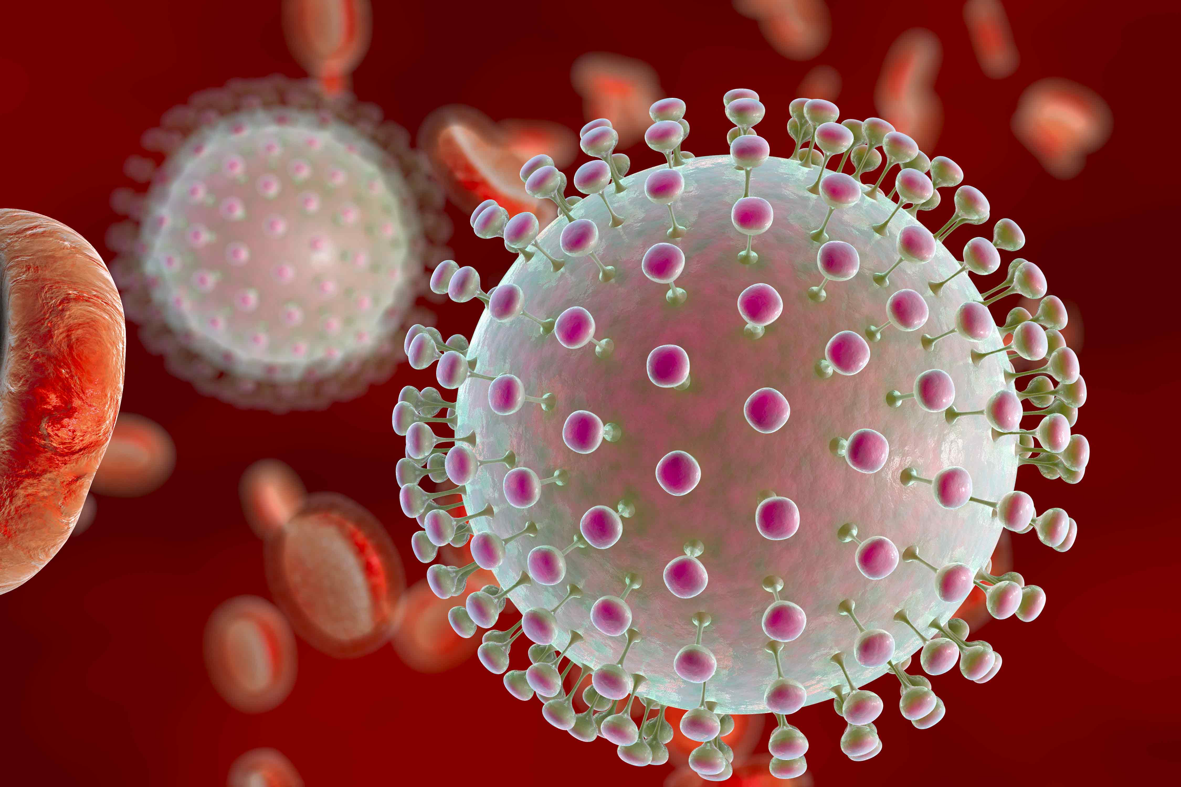
(Liège-Paris, December 11, 2017) –  Epidemiological studies show that in utero fetal infection with the Zika virus (ZIKV) may lead to microcephaly, an irreversible congenital malformation of the brain characterized by an incomplete development of the cerebral cortex. However, the mecha- nism of Zika virus-associated microcephaly remains unclear. An international team of researchers within the European consortium ZIKAlliance (coordinated by Inserm in France) has identified a specific mechanism leading to this microcephaly. Their findings are published this week in Nature Neuroscience.

To understand this mechanism, the scientific team led by Dr. Laurent Nguyen (frs-F.N.R.S., GIGA Neuroscience, University of Liège) and Prof. Marc Lecuit (Institut Pasteur, Inserm, University Paris Descartes, Necker Children’s Hospital, AP-HP) combined analysis of human fetuses infected with Zika virus, cultures of human neuronal stem cells and mice embryos. They showed that ZIKV infec- tion of cortical progenitors (stem cells for cortical neurons) controlling neurogenesis triggers a stress in the endoplasmic reticulum (where some of the cellular proteins and lipids are synthe- tized) in the embryonic brain, inducing signals in response to incorrect protein conformation (re- ferred to as “unfolded protein response”).

When it reaches the brain, Zika virus infects neuronal stem cells, which will generate fewer neu- rons, and by inducing chronic stress in the endoplasmic reticulum, it promotes apoptosis, i.e. the early death of these neuronal cells. These two combined mechanisms explain why the cerebral cortex of infected fetuses becomes deficient in neurons and is therefore smaller in size.

“These discoveries demonstrate a hypothesis that we had made following a basic research study we had just carried out in our laboratory, and thus confirm the physiological importance of the unfolded protein response in the control of neurogenesis,”  says Laurent Nguyen.

Researchers continued their studies on mice by administering inhibitors  of protein-folding  re- sponse in cortical progenitors and found that this inhibited the development of microcephaly in mice embryos infected with Zika virus.

Furthermore, the defects observed are specific to an infection by ZIKV, as other neurotropical vi- ruses of the flavivirus family (West Nile virus, yellow fever,…) did not cause microcephaly, in con- trast to Zika virus.

According to Prof. Marc Lecuit, “these results illustrate how studying fundamental biological pro- cesses is an essential step in understanding the mechanisms of infections, and lead to novel thera- peutic strategies.”

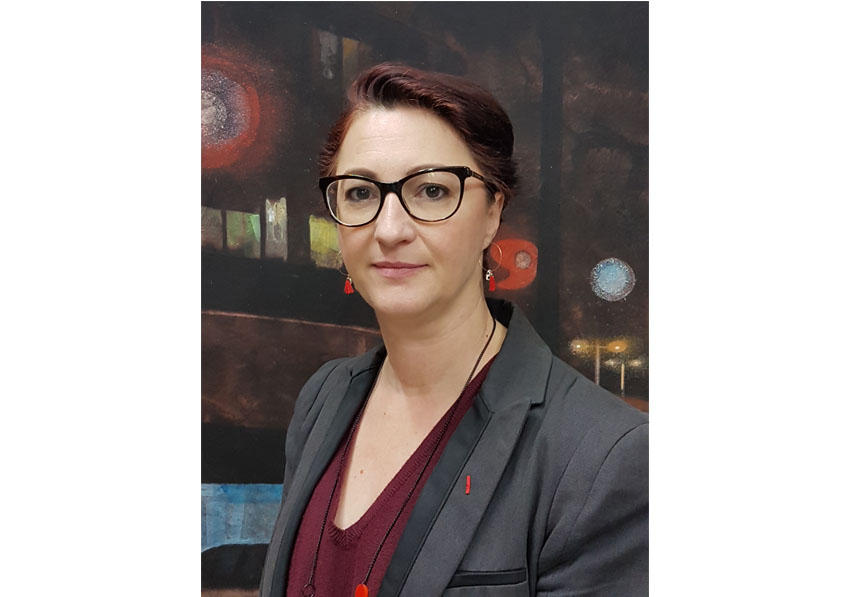
Claire Giry becomes Deputy Director General of Inserm

©DR

She replaces Thierry Damerval, who has been appointed Chairman and CEO of the French National Research Agency (ANR).

Since July 2016, Claire Giry had been in charge of the “Centers of Excellence” program run by the Commissariat général à l’investissement (CGI – General Investment Commission).

She was previously Deputy Director for fundamental research at the CEA and Director of the Fontenay-aux-Roses research center primarily dedicated to life sciences. She had a number of roles relating to communication, European affairs and partnerships within the organization.

She led Inserm’s National and Foreign Affairs Department (national, European and international partnerships) between 2012 and 2014, and had previously set up the Ministry of Higher Education and Research’s Strategic and regional coordination service, shared by the DGESIP (Department for higher education and workplace integration) and the DGRI (Department for research and integration), responsible, in particular, for Investments for the Future and regions.

Between 2007 and 2009 Claire Giry was a technical advisor to the Prime Minister for higher education and research.

Born in 1970, she is a graduate of the ENS school in Lyon and holds a doctorate in molecular and cellular biology from Claude Bernard University in Lyon.

She was named “Chevalier de la Légion d’honneur” and “Chevalier dans l’Ordre national du mérite” (French National Order of Merit).

Thierry Damerval is moving on to a new role after 10 years with Inserm’s Directorate General.

©Inserm/Heidinger, Jean-Marie

Thierry Damerval was appointed President and CEO of the French National Research Agency (ANR) by presidential decree on December 8, 2017, having been proposed for the role by the Minister for Higher Education, Research and Innovation.

Having joined Inserm in December 2007 as Deputy Chief Executive Officer for Strategy, Thierry Damerval became Deputy Director General in 2011, alongside André Syrota and subsequently Yves Lévy.

Over a ten-year period, Thierry Damerval supported the organization’s strategy through some of Inserm’s key milestones in a context of a rapidly changing research landscape: creation of the Aviesan Alliance, coordinated by Inserm in 2009; evaluation of Inserm by an international committee in 2008 and 2015, definition of the 2010-2015 and 2016-2020, strategic plans, as well as the associated performance agreements; participation of Inserm in the French Investissements d’avenir [Investments for the Future] program since 2010.

Thierry Damerval also represented Inserm on numerous bodies essential to extending the organization’s reach, both nationally, in the context of the site policy, and on a European level, within the EMRC (European Medical Research Council), for example, or as France’s representative in the States Representatives Group (SRG) of the European Union’s IMI (Innovative Medicines Initiative).

Thierry Damerval helped support Inserm’s new structure in the form of theme-based institutes, enabling the organization to be reactive in its response to major research challenges in key scientific areas. He worked closely with major bodies and, in particular, the chairs of Inserm’s various scientific advisory boards.

He also supported the implementation of structural changes within the organization, coordinating the actions of departments and regional offices since 2011. Finally, he oversaw the reinforcement of social dialog, either within the context of the CHSCT (Health, Safety and Working Conditions Committee), which he chaired, or in liaison with Inserm’s Social support and mutual assistance committee (CAES).

Yves Lévy confides that he “greatly appreciated Thierry Damerval’s professional and human qualities. His commitment and personality will leave their mark at Inserm. I wish him every success in his new role as he continues to serve research.”

Combining Administration Routes for Tailor-made Vaccination

© fotolia

Combining multiple vaccine administration routes achieves a better immune response. This is the finding of a recent study conducted as part of the European CUT’HIVAC project, coordinated by Béhazine Combadière, Inserm Research Director at the Center for Immunology and Infectious Diseases (CIMI-Paris, Inserm/Université Pierre et Marie Curie/ CNRS). This research opens new perspectives for “personalized” vaccination in which the immune system’s response to infection can be adapted. This research, performed as part of a candidate DNA vaccine against HIV, was published in Scientific Reports in October 2017.

Since the development of the very first vaccine, against smallpox, in the 18th century, some twenty vaccines have been created. Nowadays, researchers worldwide are working on the development of new vaccines capable of eradicating certain viruses that continue to cause the deaths of thousands of people, such as the human immunodeficiency virus (HIV) or, more recently, Zika and Ebola.

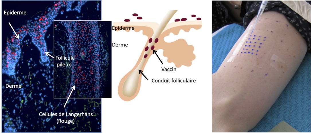
Based on the innate ability of human beings to develop “memory” immune responses, vaccination consists of administering an attenuated or inactivated form of an infectious agent. Vaccines can be brought into contact with the body via various routes of administration. At present, vaccination is carried out mainly via the intramuscular or subcutaneous routes. A new route, called the transcutaneous route (through the skin), is currently being studied and is still undergoing clinical trials. This route is rarely used for vaccine delivery because it requires a high level of expertise. The benefits are, however, that it uses low doses and can be performed without needles by depositing the vaccine in hair follicles.

As part of a collaborative project called CUT’HIVAC (acronym for “Cutaneous and Mucosal HIV vaccination”), Combadière and other researchers are seeking to develop new vaccines, in particular against HIV. One of the aspects being studied is the quality of immune responses induced depending on the various vaccine administration routes, including the transcutaneous route. In addition, transcutaneous administration more particularly targets the antigen-presenting Langerhans cells located in the epidermis, which play a fundamental role in initiating the proper cellular immune response.

Three groups of healthy subjects were formed:

  • Group 1, vaccinated via a combination of intramuscular and intradermal administration;
  • Group 2, vaccinated via a combination of intramuscular and transcutaneous administration;
  • and Group 3, vaccinated via intramuscular injection administered with electroporation, a microbiology technique used to increase cell membrane permeability – in this case to the DNA contained in the vaccine – by applying an electric field lasting one millisecond.

This recent research reveals that the group that received the intramuscular injection with electroporation obtained the strongest immune response, as reflected by the production of large quantities of interferons (cytokines secreted by the immune system) in contrast to the other groups. However, this response, albeit strong, was of insufficient quality. Interestingly, the group that received the transcutaneous administration presented an immune response in which a variety of cytokines was produced, which reflects a better immune response.

While preliminary, these results offer new perspectives for differential shaping of desired cellular immunity, required to fight the vast array of infectious diseases and cancers.  Combining multiple administration routes achieves a more specific improved immune response, able to adapt to any infection. “These are encouraging results in that they help to further research into DNA vaccines, particularly for HIV. The next step will be to determine whether this vaccination approach is able to modify the immune responses of individuals infected with HIV,” state the researchers having conducted this study.

The CUT’HIVAC project received European Union funding through the 7th Framework Programme, an international collaboration involving research groups located in the United Kingdom, Germany, France and in two biotech companies.

What if meditation allowed us to age better?

© vege/fotolia

And what if meditation enhanced the aging process? This is suggested by the results of a pilot study, conducted by Inserm researchers based in Caen and Lyon. 73 individuals, with an average age of 65 years, underwent brain imaging tests. Among these individuals, “meditation experts” (with 15,000 to 30,000 hours of meditation to their name) showed significant differences in certain regions of the brain. By reducing stress, anxiety, negative emotions and sleep problems, which tend to become more pronounced with age, meditation could reduce the harmful effects arising from these factors and have a positive effect on brain aging.

These results have been published in the journal Scientific Reports.

With age, brain volume and glucose metabolism gradually decrease, resulting in declining cognitive function. These physiological changes may be exacerbated by stress and poor sleep quality. The latter two parameters are considered to be risk factors for Alzheimer’s disease. Acting on stress and sleep could therefore be one of many helpful approaches in order to delay the onset of the disease as far as possible. One line of research, notably conducted at Inserm, focuses on the role of meditation to achieve this goal.

Hence, a pilot study conducted by Inserm researchers from Caen and Lyon explored the possibility that meditation is able to delay the age at which brain changes conducive to the development of Alzheimer’s disease appeared, by a few years. They thus studied brain function in 6 individuals who practiced meditation. “The ‘experts’ taking part in the study had an average age of 65 years and had accumulated between 15,000 and 30,000 hours of meditation. We selected these subjects because they practice meditation based on different Buddhist traditions, which allowed us to create a representative panel,” explains Gaël Chételat, Inserm researcher and lead author for these studies. The researchers then compared their brain function with 67 control subjects, who did not practice meditation, also with an average age of 65 years. A broader group comprising 186 individuals aged 20 to 87 years was also included, to evaluate the classic effects of aging on the brain, and to shed light on the specific effects of meditation.

All individuals taking part in this study underwent neurological MRI and PET examinations at the Cyceron biomedical imaging platform in Caen. Significant differences were evidenced in the volume of gray matter and glucose metabolism. The detailed results of the tests show that the frontal and cingulate cortex and insula in the individuals who practiced meditation were larger in volume and/or had a stronger metabolism than the control subjects, even when differences in terms of level of education or lifestyle were taken into account. “The brain regions detected with a larger volume or stronger metabolism in the individuals who practiced meditation are specifically those which decline the most with age,” explains Gaël Chételat. The effects of age evaluated in this study among individuals who do not meditate, aged 20 to 87 years, were effectively concentrated in certain highly specific regions – the same as those which were preserved among elderly individuals who practiced meditation.

These initial results suggest that meditation could reduce the harmful effects of these factors on the brain, and have a positive effect on brain aging, possibly by reducing stress, anxiety, negative emotions, and sleep problems which tend to become more pronounced with age.

Evidently, this is a pilot study, hence these findings will need to be repeated on larger population samples in order to obtain more robust results. Furthermore, the researchers are also endeavoring to shed light on the mechanisms allowing mediation to have this positive impact on brain aging.

The researchers behind this study were awarded EUR 6 million in funding by the European Commission to complete a larger scale study on aging well, known as the Silver Santé Study (https://silversantestudy.fr/). This project will shed light on the lifestyle factors for aging well, and will test the benefits of brain training in meditation or learning English with regard to mental health and wellbeing among seniors. It is coordinated by Inserm (Gaël Chételat, U1237, Caen) and brings together ten partners in 6 European countries (France, Switzerland, the United Kingdom, Germany, Belgium, and Spain). The initial results should come to light in 2019.

3D Objects of Unequaled Precision Made from DNA

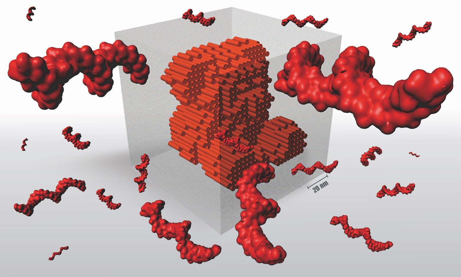
A revolution in the field of nanotechnology! An Inserm researcher[1] in collaboration with Harvard University has succeeded in creating 3D shapes of unprecedented sophistication, thanks to the four DNA bases A, T, C and G. In practice, these researchers can create nanoscopic (10-9 m) objects from 30,000 DNA sequences that fold and self-assemble like LEGO® bricks. In time, this will make it possible to manufacture new tools adapted to the size of our cells. These results have been published in Nature.

Nanotechnology represents a rapidly expanding scientific field, particularly when it comes to creating materials with increasingly specific properties. This is the case of carbon nanotubes, for example, which are light but solid and possess very high thermal and electrical conductivity. However, a slightly less well-known field of research exists: that of DNA-based nanotechnology,  whose objective is to model living matter to use as a therapeutic tool on a scale compatible with that of a human cell. However, this technology, which was developed in 2012 and is called DNA LEGO® bricks, encountered challenges in programming DNA sequences sufficient to create increasingly complex objects.

In this paper published in Nature, the researchers have reached a turning point. Their objects, manufactured according to the LEGO® brick method, use one million DNA bases, a size comparable to the genome of a bacterium, whereas until then the objects comprised only one thousand bases.

So, how does it work?

The method uses bricks, like LEGOs®, each comprising 52 DNA bases. One of the properties of DNA is based on the fact that the nucleobases of a DNA strand (A, T, C and G) can interact with those of another strand by always pairing in the same way: A with T, and C with G. Like LEGOs®, these units all have the same general shape but the internal order of the 52 bases determines which bricks will be able to interlock with which and at what level.

Next comes the choice of shape, which is either designed or selected from a database of 3D forms (cube, teddy bear, rabbit, Möbius strip, etc.). Then, each “voxel” [2] of the design is translated into DNA bricks using software developed by the researchers, called Nanobricks. “Nanobricks ‘codes’ the DNA by specifying in advance the order of the 52 bases of each brick that will subsequently be used. This step determines how the 30,000 initial sequences will fit together to produce one final 3D structure,” explains Gaëtan Bellot, Inserm researcher and co-author of this paper.

Once the IT procedures are complete, the 30,000 sequences are synthesized in a laboratory and mixed in a tube. The 30,000 DNA sequences are then completely destructured by means of a denaturing step performed at 80°C. The mixture is then gradually cooled to 25°C at a rate of 0.5°C/hour, which is when the self-assembly takes place. The molecules spontaneously fold and adopt a final shape in accordance with the 3D model selected. In this paper, the researchers produced 13 different objects.

To make objects from 30,000 sequences, they had to increase the diversity of the DNA brick sequences. By exploring various brick sizes, the research teams were able to define an optimal brick size (52 bases) making it possible both to maintain a 3D geometry similar to that of a LEGO brick and increase the diversity of individual bricks to 67 million.

This means that it is possible to obtain increasingly sophisticated objects with a greater diversity of individual bricks. The researchers have succeeded in creating objects with cavities. This level of precision is necessary if we are to design useful and effective tools. “With a key, you can open a car. With a DNA tool you can, for example, build a capsule into which you can place a medicine. And if this object has cavities, you can create a biological chain reaction depending on the products present in each cavity. Taking inspiration from life, this approach will enable the reproduction, on a nanometer scale, of solutions and inventions that occured after millions of years of evolution,” explains  Bellot.

This method offers two advantages. The first is that unlike industrial assembly processes, such as car production lines, this technology compresses all the stages into one. Imagine placing a car’s various components in the presence of each other for them to spontaneously self-assemble! The second resides in its rapidity: 30,000 components self-assemble within several hours into an object duplicated a billion times inside the same tube.

Unlike carbon nanotubes, DNA nanotechnologies are biocompatible and can be rapidly eliminated from the human body and the environment.  Nevertheless, even if the DNA molecules used are synthetic and as such not biologically active, potential interaction with the DNA present in living organisms cannot be ruled out.


[1] From the Institute for Functional Genomics (Inserm/CNRS/Université de Montpellier)

[2] Voxel, a contraction of the words “volume” and “element”, is a 3D pixel.

The Conscious and Unconscious Work in Unison to Sort Images in Our Brains

©CEA

Our brains are constantly bombarded with sensory information. Far from being overloaded, the brain is an expert in managing this stream of information. Researchers from Neurospin (CEA/Inserm) have discovered how the brain incorporates and filters information. By combining high temporal resolution brain imaging techniques and machine learning algorithms, the neurobiologists were able to determine the sequence of neuron operations which allows the brain to specifically select the relevant information. The key part of this information is processed and filtered unconsciously by our brains. The relevant information within this stream is selected by a three-step operation, and sent to the association areas of the brain to be memorized. These findings were described in Nature Communications on December 5, 2017.

The researchers measured the brain activity of 15 participants, while they attempted to locate a “target” image in a stream of 10 images per second1. The neurobiologists were thus able to observe three successive operations allowing the participants to process and sort the image stream:

► While ten or so images are shown each second, each image is analyzed by the sensory regions of the brain for approximately half a second. This represents the first phase of automatic processing, unconscious and effortless.

► When the participants are asked to pay attention and memorize a specific image, it is not only the ‘target’ image that is selected, but all images still being processed in the sensory regions. The subject’s concentration will have the effect of amplifying the neuronal responses induced by these images.

► The third phase of processing corresponds to the subject’s conscious relationship. Only one of the selected images induces a prolonged cerebral response involving the parietal and frontal regions. This is the image which the subject will claim to have perceived.

In this study, we demonstrate that the human brain is capable of processing several images simultaneously, unconsciously,” explains researcher Sébastien Marti, the author of this study with Stanislas Dehaene, Director of Neurospin (CEA/Inserm). “Concentration boosts neuronal activity and enables a specific image to be selected, which is relevant to the task the subject is in the process of performing. Only this image will be consciously perceived by the subject,” the researcher continues.

Bombarded with an ever-increasing amounts of information, our brains, in spite of everything, are thus able to manage surplus data by effortless automatic filtering, and a three-phase selection process.

Technological advances in brain imaging and information science have led to a spectacular acceleration in neuroscience research, and this study is a fine example of that.

To further explore the way in which the brain processes images

► Several studies have shown that the brain was potentially capable of analyzing up to 70-80 images per second.

► The faster the image stream, the less capable our brains are at distinguishing these images.

► Visit the CEA website to discover the brain, its function, and the major research challenges explored in the laboratory: http://www.cea.fr/comprendre/Pages/sante-sciences-du-vivant/Essentiel-sur- le-cerveau.aspx

An antioxidant protein to fight changes to the intestinal microbiota and control inflammation

©AdobeStock

Teams from Hôpital Paul-Brousse AP-HP, Inserm and Paris-Sud University have recently evidenced a mechanism which modulates the intestinal microbiota, involving a molecule with antioxidant and anti-inflammatory properties, known as REG3A. The latter is thought to protect the intestinal barrier and the bacteria most sensitive to oxygen forming the microbiota, thus improving “good” bacterial survival and growth. Transplantation of fecal microbiota in mice models of severe colitis or administration of a REG3A recombinant protein to wild type mice evidences a marked reduction in their susceptibility to the disease. These results have been published in the journal Gastroenterology and represent a new approach to manipulation of the intestinal microbiota for therapeutic purposes, restoration of host-microbiota symbiosis, and alleviation of intestinal inflammation.

One of the key factors for an imbalanced microbiota composition or “dysbiosis” is intestinal oxidative stress. Combined with immune responses, this is capable of amplifying the production of free radicals, activation of inflammatory cells (macrophages), imbalances of microbiota composition in favor of aerotolerant bacteria, and lesions in the intestinal barrier.

Dr. Jamila Faivre from the Department of Oncology-Hematology at Hôpital Paul-Brousse, AP-HP, and her team from Unit 1193 “Physiopathogenesis and Treatment of Liver Disease” at the Hepatobiliary Center (Inserm/Paris-Sud University) are studying oxidative stress as a therapeutic target to prevent or treat diseases and/or disorders related to dysbiosis.

In this study, the researchers show that a recombinant human protein known as REG3A is capable of modifying the intestinal microbiota by reducing the levels of free radicals. This regulatory mechanism is based on the antioxidant activity of this molecule.

REG3A protects commensal gut bacteria from oxidative stress by trapping free radicals and improving the survival and growth of “good” gut bacteria known to be highly sensitive to oxygen.

In keeping with the data obtained from in vitro bacterial cultures, the molecule delivered into the gastrointestinal lumen of transgenic mice modifies intestinal microbiota composition with the over-representation of Gram-positive symbionts, such as Clostridiales, and improves barrier function and resistance of the mice in two models of severe experimental colitis.

With further investigation, the researchers observed that transplantation of fecal microbiota originating from transgenic mice strongly expressing REG3A protects conventional wild type mice, together with germ-free mice colonized with induced severe colitis. Furthermore, intrarectal administration of REG3A recombinant human protein to wild type mice significantly reduces their susceptibility to induced colitis.

These results suggest that biological therapy based on administration of REG3A recombinant protein is a novel approach to (re)modeling the intestinal microbiota, alleviating intestinal inflammation, and, indeed, to preventing colorectal cancer.

Compared to current strategies, this approach is innovative in two respects: using a human protein produced endogenously in the intestine, and increasing the proportion of gut bacteria with anti-inflammatory potential by raising the intraluminal concentration of REG3A to preserve host-microbiota symbiosis and thus fight intestinal, or, indeed, extra-intestinal inflammation more effectively.

A New Look for www.inserm.fr

A new version of the Inserm website is now online. Designed and created by Inserm and the W* agency, the site has been rethought, not only in terms of graphics, but also from an ergonomic, technical, and editorial perspective. Objective: to consolidate the role of the Institute as an outlet for information on health and biomedical research, and to enhance the visibility of Inserm on the web.

New features include:

  • A home page for all of Inserm’s audiences 

From the home page, web users can discover not only leading new items of the moment (discoveries, events, etc.), but also have access to information about the institute, or more specifically intended for research professionals.

  • A new “Research at Inserm” section 

A “general public” section which describes the different sectors of biomedical research in which Inserm operates, from fundamental research to value creation for new discoveries, from the laboratory to the patient’s bedside. This section also puts the spotlight on Inserm laboratories and researchers, through photo reports (issues of Science&Santé magazine) and “Meet our researchers” features.

  • A new layout for “Health and Research From A-Z” 

“Health and Research From A-Z”, dossiers on multiple sclerosis, drug development, intestinal microbiota, and more, are the most frequently viewed pages on the site. A new layout has been designed to make them even more attractive. It notably offers a new reading level for the busiest web users! These pages will soon be embellished with scientific computer graphics very shortly.

  • The international visibility of Inserm on the web is being developed via the English-language version of the site, with extensive content: https://www.inserm.fr/en/

Furthermore, Inserm is simplifying its communications strategy to achieve greater visibility in an ever-changing partnership-based context. This approach notably relies on optimizing its logo, which has now been “streamlined” by removing its original text components. At the same time, Inserm is adopting a signature which symbolizes the missions of the Institute and its values: “Health through science, and its international vocation “From science to health“.

* W is a brand-focused strategic and creative agency (brand management, retail, integrated communications, digital, editorial). Since it was founded by Denis Gancel and Gilles Deléris in 1997, W has received national and international creative awards in all disciplines.

For more information: www.wcie.fr/leblog.wcie.fr

Agence W press contacts: Marion Weill – rf.eicw@lliew.m – +33 (0)7 70 44 71 59

fermer