Menu

Influence of the Gut Microbiota on Tryptophan Metabolism and our Health

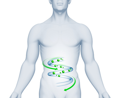
Crédits: Fotolia

Numerous compounds are involved in the complex interactions that exist between our body and its microbiota. One of these is the essential amino acid, tryptophan. On June 13, 2018, in the journal Cell Host and Microbe, a team from Inra, AP-HP, Sorbonne Université and Inserm gathered the most recent advances concerning the central role of tryptophan in the dialog with our gut microbiota. Data which opens opportunities for research and future therapeutic applications, particularly in the treatment of inflammatory bowel disease.

Back in 2016, a team from Inra, AP-HP, Inserm and Sorbonne Université studied the role of the CARD9 gene in susceptibility to inflammatory bowel disease (IBD)[1]. Without this gene, the mice studied were more sensitive to gut inflammation and their gut microbiota did not make effective use of tryptophan, an essential amino acid supplied only through diet whose metabolites are particularly implicated in the immune pathways. In addition, the researchers also showed that transferring the microbiota of mice without CARD9 to genetically normal mice also transmitted this susceptibility to gut inflammation. How is the microbiota capable of modulating immune response? How are genetics, immunity and gut bacteria linked? And, more specifically, what is the role of tryptophan in the dialog between the host and its microbiota?

To answer these questions, the same team two years later has gathered the most recent scientific advances on the role of tryptophan and its metabolites in the dialog with the gut microbiota. In their synthesis they address two aspects in particular: 1) the effects of the tryptophan derivatives produced directly by the microbiota bacteria, and 2) the indirect control of the host tryptophan metabolism by the gut microbiota. The researchers describe and analyze the three major pathways of tryptophan metabolism in the gut:

  • The aromatic hydrocarbons (AhR) pathway:

Tryptophan is transformed by certain microbiota bacteria into indole derivatives which are capable of activating AhR, a receptor present on immune cells and epithelial cells in the gut. By activating this receptor, the immune cells notably produce interleukin-22, which has an anti-inflammatory action and a protective role of the mucosa.

  • The serotonin pathway 

This neurotransmitter is produced from tryptophan and affects all parts of the body.  It is involved in various biological processes and in many pathologies. Yet more than 80% of the serotonin of our body is produced in the gut by specialist cells and under the influence of the microbiota.

  • The indoleamine 2,3-dioxygenase (IDO) pathway:

From tryptophan, the IDO pathway leads to the production, notably of kynurenine but also of many other metabolites which are involved in immune, metabolic and even neurological processes.

In decoding the complex balance between these various metabolic pathways, this research allows a better understanding of the development of intestinal and extra-intestinal diseases. They reveal new connections between the microbiota and our health and open opportunities for new therapies.

[1] Read the press release “Genetics and the Gut Microbiota together contribute to IBD”: http://presse.inra.fr/en/Press-releases/Genetics-and-the-gut-microbiota-together-contribute-to-IBD
Hypertension at Age 50 is Said to Increase the Risk of Developing Dementia

©JORGE LOPEZ on Unsplash 

And what if, depending on the age at which it develops, hypertension had more or less significant consequences on maintaining our cognitive function? This was suggested by a study conducted by an Inserm team in partnership with the Department of Epidemiology and Public Health at University College London which has been monitoring changes in blood pressure and the onset of dementia in more than 10,000 volunteers since 1985. This research, published in the European Heart Journal, suggests that, at the age of 50, high blood pressure, although still below the diagnostic threshold for hypertension, could be linked to a higher risk of developing dementia later in life, even for individuals with no other cardiovascular disorders.

Although studies linking blood pressure to an increased risk of dementia at an advanced age already exist, they focus on blood pressure values in a large population segment ranging from age 35 to 68, and have never been conducted in specific age groups.

With the Whitehall II study, Inserm researchers, in partnership with the Department of Epidemiology and Public Health at University College London (UCL), embarked upon the long-term follow-up of a population of 10,000 volunteers aged 35 to 55 when the study began in 1985, in order to study the link between age, hypertension, and dementia. The researchers measured the participants’ blood pressure in 1985, 1991, 1997, and 2003. Participants were monitored until 2017 in order to detect the possible onset of dementia.

Less than 5% of participants developed dementia as they aged, and the mean age at diagnosis was approximately 75 years.

The research team studied two different types of blood pressure values: systolic pressure – measured as the heart contracts to eject blood into the arteries (systole) – and diastolic blood pressure – measured as the heart relaxes and fills with blood (diastole).

While diastolic pressure does not appear to have an impact on the risk of developing dementia, the researchers nonetheless observed that fifty-year-olds with a systolic pressure of 130 mmHg or over (according to the European Society of Cardiology, the limit value for diagnosing hypertension is 140 mmHg) had a 45% higher risk of developing dementia compared to individuals with a lower systolic pressure at the same age. No increase in this risk was observed among individuals with hypertension at age 60 or 70.

Furthermore, the higher risk associated with blood pressure above 130 mmHg is also observed among individuals not developing cardiovascular disorders during the follow-up period: their risk was 47% higher compared to individuals without cardiovascular disorders, with a systolic pressure below 130 mmHg.

According to Archana Singh-Manoux, Inserm research director in charge of the research project and professor emeritus at UCL, these analyses “suggest that the impact of blood pressure on brain health is dependent on the duration of exposure; hence, individuals with high blood pressure at the age of 50 would be more likely to develop dementia than those who develop hypertension at 60 or 70.”

This could be explained by the fact that high blood pressure causes ministrokes which, although often undetected, are harmful to the brain and may ultimately lead to a decline in function.

“In this study, we were able to evidence different patterns of association according to the age groups studied,” clarified Jessica Abell, the lead author of the article, postdoctoral researcher at Inserm and associate researcher at UCL, who adds that “these results could thus help redefine the age groups to be studied in order to assess the impact of hypertension on health.” She concludes: “it is important to emphasize that these results originated from an observational study on a population sample, and cannot be directly used as predictive instruments for each individual. Defining the optimum limit value for diagnosing hypertension is currently the focus of the debate.”

Pandoravirus: giant viruses invent their own genes

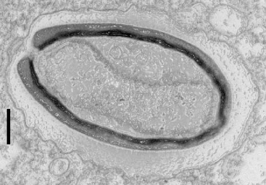
Pandoravirus quercus, found in Marseille, France. Thin section, viewed via electron microscopy. Scale bar: 100 nm.  ©IGS- CNRS/AMU.

Three new members have been isolated and added to the Pandoravirus family by researchers at the Structural and Genomic Information Laboratory (CNRS/AixMarseille Université), working with partners at the Large Scale Biology Laboratory (CEA/Inserm/Université GrenobleAlpes) and at CEA-Genoscope. This strange family of viruses, with their giant genomes and many genes with no known equivalents, surprised the scientists when they were discovered a few years ago. In the 11 June 2018 edition of Nature Communications, researchers offer an explanation: pandoviruses appear to be factories for new genes – and therefore new functions. From freaks of nature to evolutionary innovators, giant viruses continue to shake branches on the tree of life!

In 2013, the discovery of two giant viruses unlike anything seen before blurred the line between the viral and cellular world. Pandoraviruses are as big as bacteria, and contain genomes that are more complex than those found in some eukaryotic organisms[1]. Their strange amphora shape and enormous, atypical genome[2] led scientists to wonder where they came from. 


The same team has since isolated three new members of the family in Marseille, continental France, Nouméa, New Caledonia, and Melbourne, Australia. With another virus found in Germany, the team compared those six known cases using different approaches. Analyses showed that despite having very similar shapes and functions, these viruses only share half of their genes coding for proteins. Usually, however, members of the same family have more genes in common.

Furthermore, these new members contain a large number of orphan genes, i.e. genes which encode proteins that have no equivalent in other living organisms (this was already the case for the two previously discovered pandoraviruses). This unexplained characteristic is at the heart of many a debate over the origin of viruses. What most surprised researchers was that the orphan genes differed from one pandoravirus to another, making it less and less likely that they were inherited from a common ancestor!

Bioinformatic analysis showed that these orphan genes exhibit features very similar to those of non-coding (or intergenic) regions in the pandoravirus genome. Findings indicate the only possible explanation for the gigantic size of pandoravirus genomes, their diversity and the large proportion of orphan genes they contain: most of these viruses’ genes may originate spontaneously and randomly in intergenic regions. In this scenario, genes “appear” in different locations from one strain to another, thus explaining their unique nature.  

If confirmed, this groundbreaking hypothesis would make these giant viruses craftsmen of genetic creativity – a central, but still poorly explained component of any understanding of the origin of life and its evolution.


[1] Organisms whose cells contain nuclei, unlike the two other kingdoms of living organisms, bacteria and archaea.

[2] Up to 2.7 million base pairs.


This research received funding from the Bettencourt Schueller Foundation, through the “Coup d’Elan Prize for French Research” awarded to Chantal Abergel in 2014.


Credit: IGS- CNRS/AMU

A Computer Program Able to Automatically Detect and Identify Brain Lesions

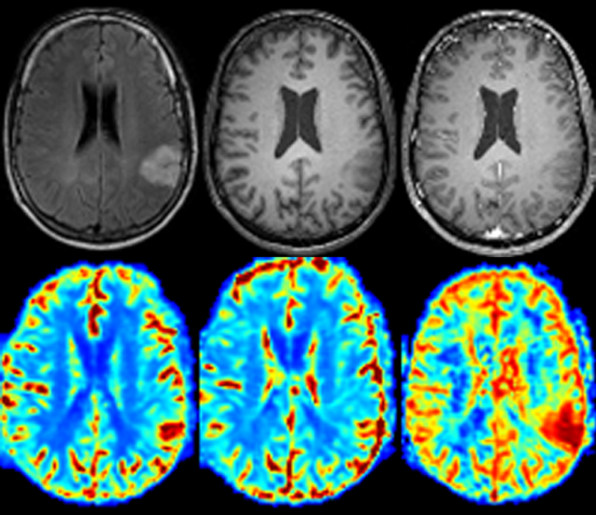
 ©Emmanuel Barbier – Inserm/Inria/Univ. Grenobles Alpes  – Figure d’IRM chez l’homme obtenue ici en présence d’une tumeur cérébrale. En gris, des IRM classiques, en couleur, des IRM quantitatives.

Will the radiology of the future come from machine learning? That is the view of Inserm and Inria researchers working in collaboration at the Université Grenoble Alpes who have developed a program able to localize and diagnose various types of brain tumors via MRI image analysis. These analyses have produced highly reliable results, with tumor localizations and tumor-type diagnoses accurate in 100% and over 90% of cases, respectively. This innovative method and its results are the subject of a study published in IEEE-TMI.

MRI – or magnetic resonance imaging – with its ability to reveal various brain tissue characteristics is the medical imaging technique of reference when it comes to obtaining highly-detailed images of the brain. It can produce what is known as “quantitative” images, which each map a measurable brain parameter (such as blood flow or blood vessel diameter). Although the quality of these quantitative images is less dependent on the calibration of the measuring apparatus than that of the standard images obtained with MRI – and so is more reliable – this type of technique is still infrequently used in the clinical MRI setting.

Inserm researchers have been working in conjunction with a research team from Inria on the analysis protocols of these quantitative images at the Université Grenoble Alpes.  The researchers combined various innovative mathematical tools in order to teach a computer program how to analyze quantitative brain MRI images and diagnose any tumors present.

First of all, the program learned how to recognize the characteristics of a healthy brain. Then, when it was shown images of brains with cancer, it became able to automatically localize the regions whose characteristics diverge from those of healthy tissues and to extract the distinguishing characteristics.

Finally, in order to teach the artificial intelligence how to discriminate between the different types of tumor, the researchers then gave it the diagnosis associated with each of the pathological brain images which had been presented to it.

In order to test the ability of the program to differentiate healthy from diseased tissue, the research team provided it with images that it had not seen before – sometimes of healthy brains, sometimes of pathological brains. The program had to indicate whether a tumor was present in these images and, if so, be able to characterize it. And, by succeeding in localizing the lesions perfectly (100%) and diagnosing them very reliably (over 90%), the artificial intelligence turned out to be a very quick study.

“At present, the acquisition of quantitative images does not correspond to what is happening in routine clinical practice in the MRI departments”, specifies Emmanuel Barbier, Inserm researcher leading the study. “But this research shows the value of acquiring these types of images and informs radiologists of the analytical tools that could be available to them in the near future to aid their interpretations. “

In the meantime, the research team will focus on the most relevant images to acquire in order to diagnose brain tumors as precisely as possible and with the greatest possible reliability. It will therefore continue to develop mathematical tools with the aim of improving the program’s self-learning abilities, with the ultimate objective being to extend the diagnostic potential of this artificial intelligence to other brain diseases, such as Parkinson’s.

These quantitative MRI machine learning tools applied to brain tumors are currently being evaluated as part of the Cancer Plan driven by Inserm, within the Tumor Heterogeneity and Ecosystem program.

Their development in the context of Parkinson’s disease diagnosis is also underway via the NeuroCoG multidisciplinary project funded by the Université Grenoble Alpes IDEX.

Our Food Choices Predicted by our Brain Anatomy

©Katherine Chase – Unsplash 

Do you tend to go for cake or vegetables? If you sometimes find it difficult to eat healthily, a study conducted by a team of researchers from Inserm, CNRS and Sorbonne Université, including Liane Schmidt and Hilke Plassmann, at the Brain & Spine Institute (ICM) has established a link between the anatomy of certain regions of our brain and our ability to control our food choices. These results were published in The Journal of Neuroscience on June 4, 2018.

For many people, eating healthily is no easy feat. Individuals differ greatly in their ability to maintain a balanced diet and make healthy food choices.

From the cognitive perspective, making a choice involves two principal mechanisms – the first of which consists of attributing a value to each option. In the case of food, its taste and nutritional quality can, for example, determine its value. The second mechanism for our brain consists of analyzing the value given to each option in order to choose the most suitable – i.e. the food item to which we attribute the highest value.

How are these decisions expressed in the brain? To answer these questions, Liane Schmidt, Inserm researcher, Hilke Plassmann, and their colleagues Anita Tusche from the California Institute of Technology (USA), Cendri Hutcherson from the University of Toronto (Canada) and Todd Hare from the University of Zurich (Switzerland), pooled brain imaging data taken from four studies on food choices.

In three of these studies, the participants performed the same task which involved evaluating their appetite for a specific food according to three criteria: its taste, health benefits and their own natural preference. As such, the participants could base their choice either on taste or on nutritional value.

In the fourth study, the participants were asked to use a method of their choice (save money, eat organic, or lose weight) to reduce their desire for tasty products devoid of nutritional value. This last study “involved a more flexible control strategy not specifically focused on attributes of taste or health but on the various means of distancing oneself from a food or resisting desire” specifies Liane Schmidt, lead author of the study and Inserm researcher.

The researchers studied the variations in the quantity of gray matter in the brains of the participants using imaging data from the first three studies.

As such, they revealed a correlation between food choices and the quantity of gray matter in two regions of the frontal lobe: the dorsolateral prefrontal cortex (dlPFC), which governs the regulation of decisions, and the ventromedial prefrontal cortex (vmPFC), which is in charge of value attribution. They observed that people with more gray matter in these regions had a greater appetite for the foods they considered healthy.

The research team then sought to predict the food choices of the participants in the fourth study based on the quantity of gray matter detectable in the two regions identified previously. “The idea here was to see whether the correlations established in a context in which the control strategies are very clear – focus on taste or health – can be generalized to a situation in which the control strategies are more vague. ” continues Hilke Plassmann.

The researchers confirm these results, thereby establishing for the first time that neuroanatomical differences in the dlPFC and vmPFC regions play a role in individual food choices. These results open up avenues which should ultimately lead to the treatment of eating disorders associated with disrupted food control, such as bulimia nervosa or anorexia nervosa.

Reducing the Impact of Emergency Room Stress

Crédits: AdobeStock

A visit to the emergency room is no picnic. Irrespective of the reason for their consultation, around 1 in 5 people will experience various symptoms (headache, difficulty concentrating, irritability, sensory disturbances, etc.) for several months afterwards. To address this phenomenon, Inserm researchers from Unit 1219 Bordeaux Population Health Center have demonstrated the benefits of an EMDR session performed within 6 hours of the event responsible for the visit. Such sessions are effective in reducing post-concussion syndrome and post-traumatic stress disorder by up to 75%. These results have been published in The Journal of Psychiatric Research.

According to a study performed in 2012, more than 10 million people come or are taken to the emergency room (ER) in France each year. When multiple visits are taken into consideration, this represents 18 million annual visits. Around 10 to 20% of patients will, for several months afterwards, experience very diverse, persistent symptoms (headache, difficulty concentrating, irritability, sensory disturbances, etc.) which significantly impact their quality of life. With approximately 1 million people affected each year, this represents a major public health issue. Because these symptoms were initially described in people with concussion, their persistence over several months is known as post-concussion syndrome (PCS). Roughly 5% of these patients also suffer from a similar disorder called post-traumatic stress disorder (PTSD), whose symptoms are traditionally described in people having experienced a situation in which their physical or psychological integrity or that of those close to them has been threatened or harmed.

Inserm researchers from the Bordeaux Population Health Research Center, Bordeaux University Hospital and Cadillac Hospital have noted, during investigations conducted since 2007, that the stress levels of these patients were particularly high. They wondered whether it was possible to reduce these levels, with the ultimate objective being to prevent the onset of these invalidating symptoms.


An ER-specific treatment protocol

 To do this, they conducted a new study to evaluate the efficacy and feasibility of a recognized therapeutic approach, called Eye Movement Desensitization and Reprocessing (EMDR), for which they adapted a protocol that can be administered in an ER setting. During these EMDR sessions in the ER, the patient was asked to recall stressful elements and focus on their emotions and sensations.

 At the same time, the therapist performed sets of alternate bilateral stimulations, consisting of the patient moving their eyes from side to side or up and down or, if their clinical condition did not permit this, the therapist making alternate tapping movements on the patient’s knees or shoulders.

This psychotherapeutic model is one of two therapies recommended by the French National Authority for Health (HAS), the World Health Organization (WHO) and Inserm in the management of PTSD. The duration of treatment varies, usually requiring 2 or more sessions. In the present study, the aim was to prevent the onset of PCS and PTSD, using a protocol in which the intervention was brief (no longer than 1 hour), early (within 6 hours of the event leading to the ER visit) and performed just once. The patients enrolled in the study were evaluated as being at high risk of developing these two disorders (the main predictive factors being a history of antidepressant use, poor health over the previous year and female sex).


EMDR-treated patients much less subject to PTSD

 A total of 130 consenting patients were selected and randomized into 3 groups: the first receiving a 60-minute session of EMDR, the second a 15-minute interview with a psychologist, and the third no psychological treatment.

The patients were then contacted by telephone three months later, in order to identify those who had developed PCS or PTSD.

In these three groups, the proportions of patients suffering from PCS three months later were 15%, 47% and 65%, respectively. The proportions of those presenting PTSD were 3%, 16% and 19%.

“This is the first randomized controlled trial worldwide to show that brief and ultra-early EMDR intervention is not just achievable in an ER setting but also potentially effective,” considers Inserm Research Director, Emmanuel Lagarde. These results remain to be confirmed by a new, larger, study. Such a study was launched in January 2018 by the same team at the University Hospitals of Lyon and Bordeaux, involving more than 400 patients. Its results will be available by the end of 2018.

Immunoscore: a test to improve the care and treatment of colon cancer

Crédits: Inserm/Sénégas-Balas, Françoise

With Immunoscore, a test devised by a team of researchers from Inserm and Université Paris Descartes and doctors from the Paris AP-HP hospitals, disease progression in patients with colon cancer can now be defined more accurately. According to an international study conducted in more than 2,500 patients, Immunoscore has proved effective in predicting which patients are at high risk of tumor recurrence and, as such, would benefit from intensified treatment following surgery. These results have been published in The Lancet.

The seriousness of cancer, and particularly that of the colon, is essentially estimated according to the extent to which it has spread within the affected organ and the presence of metastasis. This estimation of the aggressiveness of the cancer and its risk of recurrence following treatment must however be improved.

For decades, it has been thought that the immune reaction developed by the patient against his or her cancer has a beneficial influence. Researchers from Inserm and doctors from the Paris AP-HP hospitals have demonstrated in recent years that infiltration of the cancerous tumor by immune cells is a good indication of the way in which colorectal cancer might develop, thereby representing a potential prognostic tool. The immune cell populations which provide the most information on disease progression were identified and the method of evaluating these populations optimized.

This method has led to the creation of an immunological test, applicable in clinical practice, called “Immunoscore”. It works by quantifying the density of two types of immune cells in the tumor and its invasive margin: total T-cells (CD3+) and killer T-cells (cytotoxic CD8+).

The objective of this international study published in The Lancet was to evaluate the prognostic value of Immunoscore in patients with colon cancer on a very large scale. For this, an international consortium of 14 immunology and pathology centers in 13 countries was formed.  A total of 2,681 patients from these centers were included in this analysis. The prognostic performance of Immunoscore, in which patients are classified into 3 groups (high, intermediate and low), was evaluated on the basis of recurrence risk (evaluated during the 5 years following the surgery) and survival. The statistical analyses were all performed by a group of external biostatisticians from the Mayo Clinic in the USA.

The results show that patients with a high Immunoscore present the lowest recurrence risk and prolonged survival. 

In the test group comprising 700 patients, only 8% of those with a high Immunoscore presented a recurrence after 5 years. However, the recurrence rate increased significantly in patients with intermediate and low Immunoscores, reaching 19% and 32%, respectively.  These findings were confirmed in the two other patient groups analyzed, representing 1,981 patients. Furthermore, Immunoscore had a stronger bearing on patient survival than the tumor criteria which are currently used to guide therapy. 

These findings show that Immunoscore provides an accurate and reliable estimation of recurrence risk in patients with colon cancer. The researchers consider that these results support the use of Immunoscore as a new component in the classification of cancer, in which recurrence risk is used to improve individual patient treatment strategies, particularly the modulation of chemotherapy.

In view of the highly positive results of this test in colon cancer, researchers are currently evaluating Immunoscore in other types of cancer and are studying its ability to predict patient response to the immunotherapies which are currently revolutionizing the treatment of cancer.

The search for the origin of mast cells

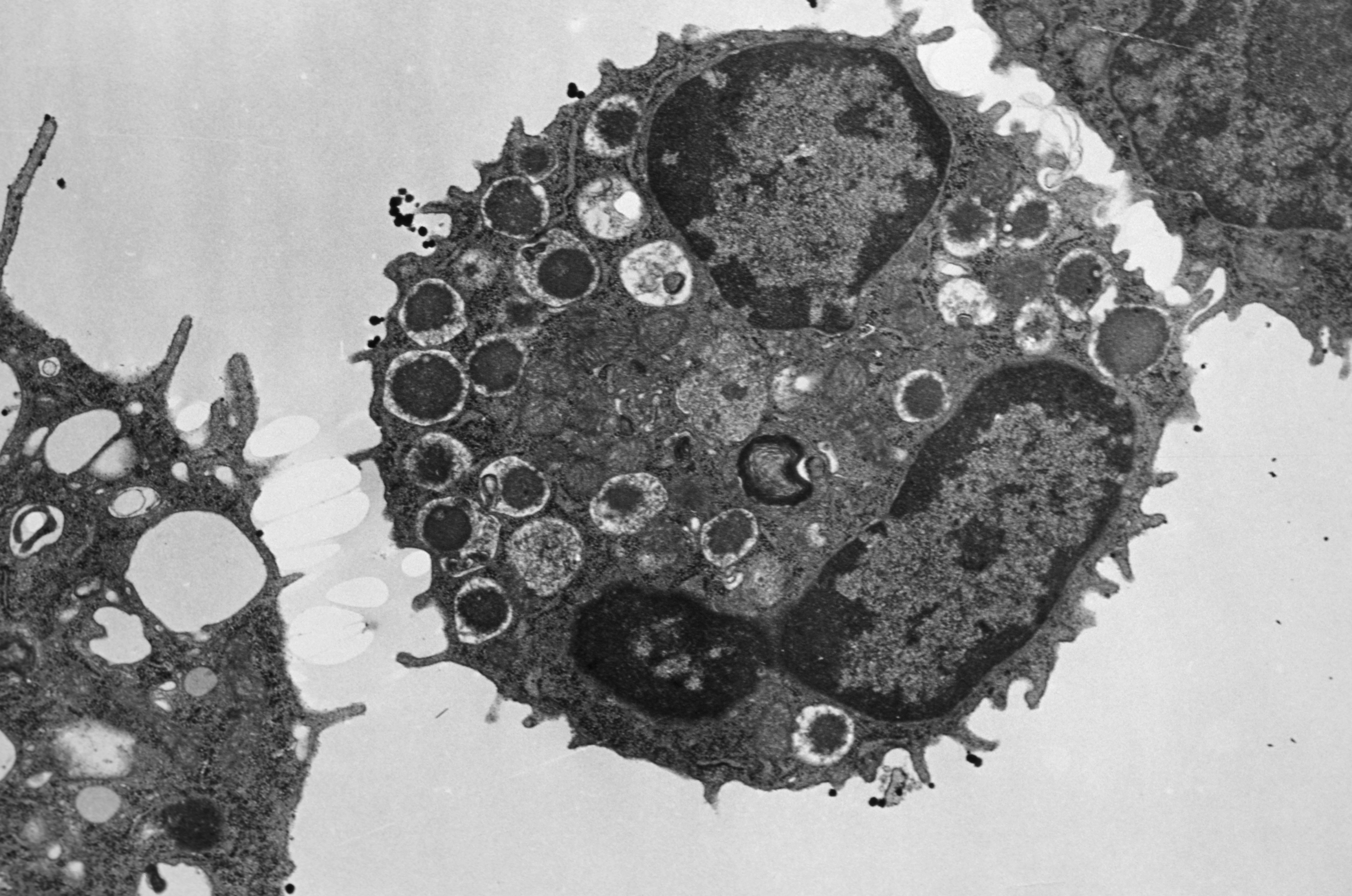
©Inserm/Nabarra, Bernadette, 1989

A team of researchers from CNRS, INSERM and Aix-Marseille Université (AMU) at the Centre of immunology (Marseille-Luminy (CIML), together with the Singapore Immunology Network (SIgN)1, has proven that not all of the immune system’s important mast cells are produced in bone marrow, as was previously thought. Scientists found embryonic mast cells in mice with functions that are likely to be different than the mast cells found in adults. The study appears in the June 2018 edition of Immunity.

Mast cells are immune system cells that play a role in inflammatory processes in the body. They provide an initial line of defence against certain pathogens and play a vital role in allergic reactions. Until now, research on these cells has shown that they are produced in bone marrow, as are most cells found in blood.

In this study, scientists from CNRS, INSERM and AMU identified other mast cells generated in the embryonic stage of life. Called “primitive mast cells”, these cells are produced in the yolk sac, an extra-embryonic organ known to supply nutrients to the embryo and generate certain blood cells. In mice, these primitive mast cells are generated after the 8th day of embryonic development. They then migrate to what will be the dermis and remain there until birth, gradually disappearing as the embryo begins to produce the other “definitive” mast cells.

Given that an embryo is already protected by the physical and immune barriers of its mother, researchers concur that primitive mast cells do not serve an immune-related purpose. The next step for researchers is to understand why the body produces two successive waves of mast cells with what appears to be same genetic origin but different functions. The study is also an opportunity for the scientific community to approach the subject from a different angle.

1The Singapore Immunology Network (SIgN) is part of the Agency for Science, Technology and Research of Singapore (A*STAR)

fermer