Menu

How the Zika Virus Persists in Semen and Alters the Spermatozoa

Crédits: AdobeStock

While we already knew that the Zika virus is sexually transmitted and that it remains in the semen for several months after infection, what we did not know was why it persists and why there is a decrease in the number of spermatozoa in infected men. A team of Inserm researchers led by Nathalie Dejucq-Rainsford (Unit 1085 “Research Institute for Environmental and Occupational Health”, Irset), has shown that the virus infects several types of testicular cells –including the germ cells responsible for spermatozoa– which could alter the process of sex cell production. In addition, the low levels of immune defense of the testicle against Zika could be detrimental to eliminating the virus in the organ and as such contribute to its persistence in semen. This research was published in Journal of Clinical Investigation.

The mosquito-borne Zika virus leads to birth defects and can also be sexually transmitted from men to women. The virus can persist in semen for several months and be transmitted more than 40 days after symptoms disappear, which suggests infection of the male reproductive system. Animal models suggest that sexual transmission could facilitate infection of the fetus in pregnant women. Decreases in the number of spermatozoa and increases in abnormal spermatozoa have also been observed in the semen of infected men. However, the organ responsible for the persistence of the virus in semen remained to be elucidated, in addition to the origin of the modifications of the semen.

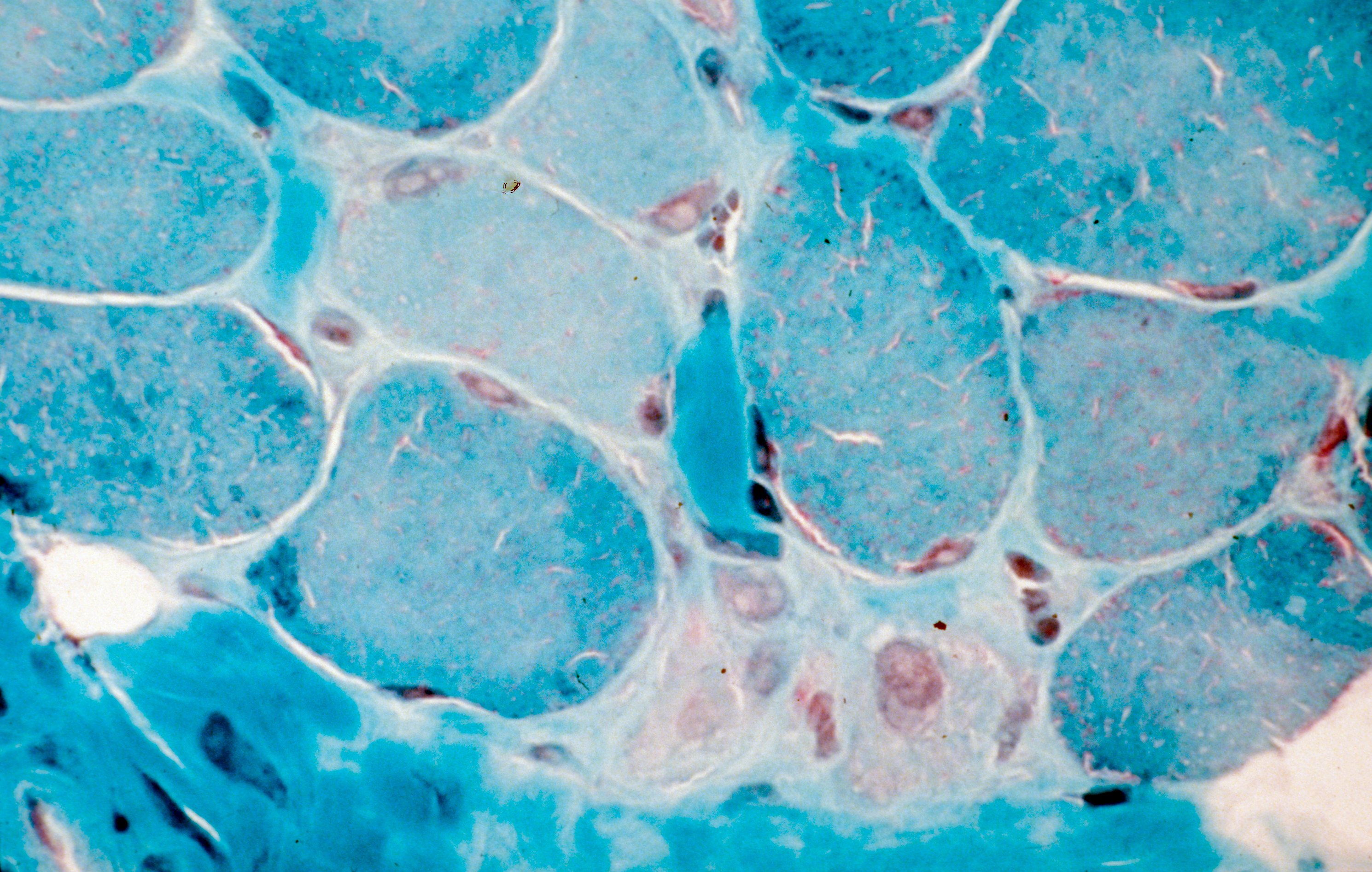
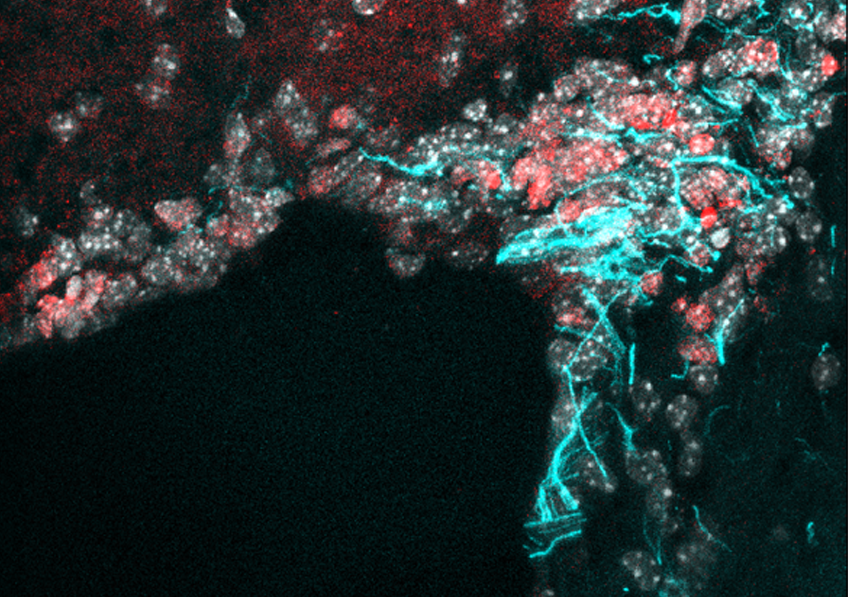
Using a novel human testicular culture model, the team of Dr. Dejucq-Rainsford at Inserm (Inserm Unit 1085 “Research Institute for Environmental and Occupational Health”, Irset) has shown that the Zika virus infects a number of testicular cell categories, including the germ cells responsible for spermatozoa. They did this by exposing to the virus testicular fragments from uninfected men and maintaining them in culture for 9 days to track the development of the infection. These findings were confirmed in Zika patients with the demonstration of infected testicular germ cells and spermatozoa in semen.

Following further analysis, the researchers suggest that the testicle could constitute a reservoir for the virus, which would explain why it is found in semen long after having disappeared from the rest of the body. Indeed, this organ has low levels of defense against the infection because it does not produce the proteins essential for an antiviral response (interferons) and that it produces only very small quantities of the molecules tasked with signaling the presence of pathogens to the immune system (pro-inflammatory cytokines).

In addition, and unlike other organs, the infected testicular cells do not die – they persist in the testicle. These various elements could enable the virus to be produced in the testicle for long periods without it being eliminated by the immune system.

Finally, infection of the germ cells themselves as well as of other testicular cells involved in the spermatozoa production process (Sertoli cells, Leydig cells) and in their expulsion from the testicle (peritubular contractile cells) could affect semen production. A hypothesis supported by the observation of modifications of semen recorded in infected men up to two months after contact with the Zika virus.

This study provides essential information on the manner in which the Zika virus can persist in semen and modify its parameters, as well as a valuable tool for testing the efficacy of antiviral agents on testicular infection ex vivo.

Myositis: A New Classification Representing a Decisive Step Towards Improved Diagnosis and Personalized Treatment

Coupe transversale de muscle humain, régénération de fibres musculaires après un traitement de la myopathie de Duchenne. Crédits: Inserm/Fardeau, Michel

Prof. Olivier Benveniste’s Inflammatory myopathies and innovative targeted therapies team at the Institute of Myology has produced a new classification of the different forms of myositis (rare inflammatory muscle diseases). Four new types of myositis taking into account the various clinical criteria of patients have now been defined. This research, involving teams from the Institute of Myology, Inserm, the Paris public hospitals system (AP-HP) and Sorbonne Université, was published in September in JAMA and paves the way for reliable diagnosis and personalized treatments.

Myositis (rare inflammatory muscle diseases) is a group of rare autoimmune muscle diseases in which the immune system, in charge of protecting the body from external aggressions (bacteria, viruses…), dysfunctions and attacks the body (in this case, the muscle). These diseases affect between 3,000 and 5,000 adults and children in France.

While the different forms of myositis all have an autoimmune component, each has its own specific triggering mechanisms. Until now, three types of myositis (polymyositis, dermatomyositis, inclusion body myositis) had been identified according to a classification system established in 1975 and updated in 2017 (ACR/EULAR rheumatologist criteria) based essentially on clinical and histological criteria. Prof. Olivier Benveniste, head of the Inflammatory myopathies and innovative targeted therapies team at the Institute of Myology who has been monitoring patients daily at Pitié-Salpêtrière Hospital AP-HP over the past 20 years, had identified major diagnostic errors related to this incomplete and, as a consequence, non-homogeneous, classification which would sometimes even lead to errors in patient treatment. Some patients mistakenly diagnosed with inclusion body myositis were given high-dose steroids which made their condition worse.

That is why, together with his team and the Center of Reference for Neuromuscular Diseases of the Institute of Myology, Prof. Benveniste launched a study on 260 patients in whom he collected and analyzed the various clinical characteristics –and particularly the presence of autoantibodies, which are sometimes causes or consequences of the disease. Using innovative statistical methods, without a priori, in which the mathematical algorithm works unsupervised to aggregate similar patients into subgroups (cluster analysis), the researchers revealed a new classification with four major types of myositis: inclusion body myositis, dermatomyositis, immune-mediated necrotizing myopathy, anti-synthetase syndrome (with polymyositis no longer forming a type of myositis as such).

Characteristics of the four forms of myositis:

Inclusion body myositis: This form of myositis more often affects men over 60 years of age. It progresses slowly but ultimately leads to a highly disabling motor deficit. It particularly affects the quadriceps (thigh muscles used to climb stairs, get up from a chair, maintain stability when walking…), the muscles used to close and shake hands and the muscles used for swallowing. This disease is resistant to standard immunosuppressive treatments such as steroids. It is due to the presence of an inflammatory reaction (myositis) in the muscle and a neurodegenerative process related to Alzheimer’s disease (presence of inclusions).

Dermatomyositis: This form more often affects women. Children can also be affected. There is an associated cancer risk in the most elderly subjects (usually after 60 years). In addition to myositis, which causes predominant muscle weakness in the shoulders, this disease is characterized by the presence of typical dermatological lesions. Dermatomyositis is due to an imbalance of the immune system involving type 1 interferon that helps protect against viruses. New therapies specifically targeting this interferon pathway are under development. Dermatomyositis-specific antibodies are anti-Mi2, anti-SAE, anti-NXP2, and anti-TIF1 gamma.

Immune-mediated necrotizing myopathy: This is characterized by muscular weakness affecting patients of all ages. In the absence of treatment, this type of myositis leads to the most severe and disabling muscle atrophy. This disease is related to the presence of two specific anti-SRP or anti-HMGCR antibodies which directly attack and destroy the muscles. Anti-HMGCR may appear after taking statins. Treatment aims to remove these antibodies.

Anti-synthetase syndrome: This disease affects muscles but also joints (leading to rheumatism), and the lungs (leading to shortness of breath that is sometimes severe). Here too, certain antibodies appear to be responsible: anti-Jo1, anti-PL7 and anti-PL12.

This new classification is decisive in establishing a diagnosis and offering personalized treatment to patients.

“We realized that the current myositis classification was unsuitable and could often lead to the failure of a potential treatment due to non-homogeneous patient groups within a given trial. So our aim was to define a classification based on phenotypic, biological and immunological criteria in order to better diagnose the different types of myositis and ultimately find suitable treatments for patients. This new classification is becoming a reference because even the FDA, which up until then was using the US classification, recommends using our research as a basis. ” explains Prof. Benveniste.

Using Light to Switch Off Nicotine Addiction?

Crédits: AdobeStock

Researchers from Inserm, CNRS and Sorbonne Université at the Paris-Seine Neuroscience laboratory1 in collaboration with Institut Pasteur2, New York University (NYU) and University of California Berkeley (UC Berkeley) have succeeded in controlling the activity of nicotine receptors in the brains of mice. To do this, they developed an optogenetic pharmacology strategy in which light is used to block the nicotine receptors, with the result being the possibility to control the addictive effects of nicotine. This study was published in eLIFE on September 4, 2018.

Every year, more than 7 million people die from smoking worldwide. Nicotine, the main addictive substance in tobacco, acts on the brain by binding to the nicotine receptors. At present, standard pharmacology techniques do not enable precise and reversible action on these receptors. That is why researchers had the idea of producing molecular tools able to interrupt the functioning of these receptors in the brain through the use of light.

In this study, the researchers modified the nicotine receptor in mice in order to attach a chemical nanoswitch which reacts to light. Under the effect of violet light, the switch folds away, preventing the nicotine from binding: the receptor is “off”. Under the effect of green light, or in darkness, the switch unfolds and allows the nicotine to act: the receptor is “on”.

For this study, the researchers focused on a specific nicotine receptor –type β2– and on a key area of the reward circuit, which delivers dopamine. When nicotine is injected intravenously, the dopamine neurons respond with an increase in their electrical activity, with the resulting dopamine release being key to acquiring the addiction. In this research, this effect of nicotine was found to be greatly reduced when the nanoswitch was triggered by the violet light but was rapidly restored under green light.

The researchers then demonstrated that it was possible to inhibit the attraction for nicotine by triggering this switch.

To do this, they compared the time spent by mice in two compartments, with and without nicotine. Under green light, when nicotine could exert its effect, they observed that the animals preferred the compartment with nicotine. However, under violet light, the mice spent equal amounts of time in each compartment, proving that they were no longer attracted by the nicotine.

Crédits : A.Mourot/S.Mondoloni/R.Durand de Cuttoli

This study proves that it is possible to manipulate the attraction for nicotine in mice, both rapidly and reversibly. Alexandre Mourot, Inserm researcher in charge of the study, states: “This innovative technology provides us with a better understanding of the role of the various nicotine receptors and neuron pathways in acquiring and maintaining nicotine addiction, and also in the processes of withdrawal and relapse. This step is particularly important when it comes to identifying suitable new therapeutic targets for fighting nicotine addiction“.

1 Laboratory located on the Pierre et Marie Curie campus of Sorbonne Université, shared with CNRS and Inserm

2 At the Genes, synapses and cognition laboratory (Institut Pasteur/CNRS).

Syndecan-1: A Candidate to Activate Neural Stem Cells and Regenerate the Brain

Crédits: M-A.Mouthon et al.

The adult brain has a very limited capacity for regeneration. However, researchers have updated the role of a key membrane molecule in the proliferation of stem cells in the adult mouse brain: Syndecan-1. CEA, Inserm and the Paris-Sud and Paris Diderot universities have joined forces to demonstrate that Syndecan-1, present in the membrane of the neural stem cells, permits their proliferation. Syndecan-1 therefore becomes a possible tracer of brain regeneration, opening up new perspectives in the field of regenerative medicine. This research, for which a patent is pending, was published on August 14 in Stem Cell Reports.

The adult brain has a very limited capacity for regeneration. It contains a small number of neural stem cells in specialized niches. These cells, which have the capacity to produce new functioning neurons, lie dormant for the most part but can be activated in response to stress.

Thanks to an innovative cell sorting technique and comparative transcriptome analysis, researchers at the Institute of Cellular and Molecular Radiobiology (IRCM)[1] have revealed some of the molecular modifications that accompany the activation of dormant neural stem cells, in response to the brain’s exposure to ionizing radiation. Through comparative mapping of the genes expressed, they identified the membrane molecule Syndecan-1 as being specifically expressed in the neural stem cells activated. By means of functional studies, the team also demonstrated that this molecule played a direct role in the proliferation of these cells.

Knowledge of the molecular signals that regulate the activity of the neural stem cells present in the adult brain could make it possible to stimulate the regenerative capacities of the brain and have many therapeutic applications.  Syndecan-1, as well as the various molecular pathways identified in this research, open up new perspectives in this field of medical research.

[1] Joint Research Unit 967, François Jacob Institute, CEA Paris Saclay; Inserm; Université Paris-Sud / Paris Saclay; Université Paris Diderot.

Genetic control of human thymic function: Needle in a haystack

A study conducted by a group of researchers from Paris Diderot University, INSERM and the Institut Pasteur reveals the existence of a genetic factor influencing the function of the human thymus. The results of the study, part of the Laboratories of Excellence project Milieu Intérieur coordinated by the Institut Pasteur, are published in the journal Science Translational Medicine on September 5, 2018.

Our immune system has developed different strategies to respond to external pathogens and to destroy emerging cancer cells while keeping autoimmunity in check. Among those strategies, adaptive immunity enables to keep a memory of an initial encounter, with increased recall responses after subsequent antigenic challenges. This is the basis of vaccination. Adaptive immunity is dependent on T lymphocytes present in lymphoid organs (spleen, tonsils, lymph nodes and mucosa associated lymphoid tissue) which patrol the body. Their name, « T » cells, derives from the thymus gland, the organ where they are produced.

The thymus is therefore a critical organ in health and disease, acting throughout life for the generation of new T cells. Its function and output is high in newborns and children but decreases with age, accounting in part for higher infection rates and cancer incidence in the elderly, both major public health issues in an ageing world. However, apart from age, environmental or genetic factors that may govern thymic function in humans remain unknown.

The objective of the Milieu Intérieur Consortium (http://www.milieuinterieur.fr) is to describe in a large-scale multidisciplinary approach the immune variation within the French population. The consortium enrolled 1000 healthy adults (500 men, 500 women aged 20 to 69 years) and gathered an expansive collection of biological specimens together with extensive data on medical history, vaccination and lifestyle. The scientists leveraged samples from this cohort to see how thymic output, known to decrease over time, is affected by other factors.

In addition to seeing sex-dependent differences, thymic function being higher in women of all ages, the most striking result from the study was to identify a genetic factor influencing the thymic function in the general population. A genome-wide association study revealed variants that were associated with thymic output, which was confirmed in an independent cohort of 612 individuals (MARTHA cohort), as well as in a mouse model. This common genetic variation is located in the T cell receptor locus itself and associates with marked differences in the levels of thymic function among individuals. Taking into account age, sex and this genetic variation, we could define a “biological age” of the thymus in men and women. Between individuals of the same chronological age, a difference of up to 18.5 years in “thymic age” depending on sex and genetics could be calculated. For instance a 58.5 year old women and a 40 year old man of different genotypes may have a similar “thymic age”, possibly explaining some of the differences in immune responses between healthy individuals.

This discovery may have a direct impact for the advancement of precision medicine in all situations where T cell generation is key, such as in the context of allogeneic stem cell transplantation, HIV disease or vaccine development. It would be also interesting to study the association of this genetic variation with the incidence of autoimmune diseases, which is higher in women.

Adolescent depression: early signs and changes in brain development underscore the need for new preventive approaches

Photo by Abbie Bernet on Unsplash

Teams of AP-HP, Inserm and Université Paris Sud, studied as part of an international research group, depressions called “subsyndromal” among young teenagers as they have a high risk to progress to depression in adolescence and later in adulthood.

This research demonstrates the existence of deviations of the microstructure of white matter in the prefrontal beams that provide connections between brain regions. The study in nearly 100 adolescents from 14 years of school, with only some depressive symptoms without apparent seriousness, and compared to a control population of more than 300 adolescents without symptoms recruited at the same time is published in the journal The American Journal of Psychiatry.

This variation of the normal maturation individual predictive value of a diagnosis of depression two years later. The identification of these adolescents at risk could improve the prevention of depression.

Adolescence is a particularly vulnerable period for the onset of depressive disorders. Authentic depressive episodes can occur, affecting about 12% of teenagers, but about 20% of teens will present depressions called subclinical or subsyndromal, that is to say that do not have visible symptoms.

Brain areas rearrangements such as cerebral cortex and white matter occur at this age, but the predictive factors of cerebral transition to depression in adolescents are not known.

Recently changes in prefrontal gray matter associated with the risk of depression in adolescence have been reported. The Child Psychiatry Service of the research teams and Adolescent to Pitie-Salpetriere Hospital, AP-HP, INSERM, Université Paris Descartes and Paris-Sud University (unit Neuroimaging and psychiatry 1000) have investigated the changes in the white matter underlying the subsyndromal emotional states, commonly observed in young characterized without psychiatric disorders.

Comparing a group of teenagers from 14 years of school, with only some depressive symptoms without apparent gravity (96 individuals) to a control group (336 individuals), the researchers demonstrate the existence of deviations of the microstructure of white matter bundles prefrontal among teenagers in the first group.

These deviations relate to areas usually involved in major depressive episodes involved in the regulation of emotions and motivation.

In detail, the results suggest a delayed development of the myelin and a different maturation in these adolescents compared to control teenagers.

In addition, the research team found that these deviations have individual predictive value of a diagnosis of depression two years later.

These deviations from normal adolescent development constitute a vulnerability factor. Through these results, the authors encourage the development of preventive strategies for adolescents at risk.

To Vaccinate is to Protect

Crédits: AdobeStock

Epidemics have no concept of borders! That is why, via Inserm, France is making a commitment to the health of European citizens by creating a European platform to intensify national responses to the challenges posed by vaccination. This European Joint Action on Vaccination (EU-JAV), coordinated by Inserm and backed by the French health ministry, was launched on September 4 with the participation of 19 other European countries.

Vaccines save between 1 and 3 million lives each year. Despite this, a phenomenon of reticence towards vaccination has been emerging over the previous decade, with many European countries experiencing insufficient vaccine cover for infectious diseases which would normally be eliminated.

With record levels of measles being registered in Europe – three times more cases in the six first months of 2018 than in the whole of 2017 – no fewer than 20 countries represented by their health ministries, public health institutes, research bodies, or universities are mobilizing to combat the re-emergence of diseases that can be avoided thanks to vaccines. This action is supported by the large international and European organizations: WHO, OECD, ECDC, EMA, as well as numerous stakeholders representing health professionals, civil society and the vaccines manufacturers.

By building on existing initiatives, the objective of JAV is to create a joint platform for the participating countries, whether or not they are EU Member States, to strengthen such cooperation mechanisms as:

– tools to share digital data for more precise epidemiological monitoring of vaccine cover

– shared methods for the systematic review of clinical trials to evaluate vaccine efficacy and safety prior to approval

– an updatable inventory of each country’s vaccine stocks and requirements for the prevention of shortages

– strengthened dialog between the various stakeholders to establish joint priorities and harmonize research and development strategies

And finally, to overcome the crisis of public confidence in vaccinations: experience-sharing with countries with high or restored levels of confidence, and good practices for the dissemination of information.

fermer