Menu

Coronavirus: A Model Estimating Importation Risk to Europe

 ©chuttersnap

Could the coronavirus epidemic that has hit China spread to Europe? A pertinent question given the new cases being announced by the Chinese authorities, of which eight have already been exported to other countries. An Inserm team led by researcher Vittoria Colizza at Pierre Louis Institute of Epidemiology and Public Health (Inserm/Sorbonne Université) has modeled the potential spread of 2019-nCoV in order to orient prevention and surveillance policies. A model which comes with one caveat: derived from research, its purpose is not to make predictions but rather be used as a theoretical tool to aid public decision-making.

Update on 31 January 2020 :

As the situation is unfolding quickly, the following figures may also change, depending on the number of confirmed cases in and outside China. Vittoria Colizza’s model, based on the most recent figures up to 30 january, is now published on Eurosurveillance

To follow the evolution of the number of cases, you can visit  GISAID.

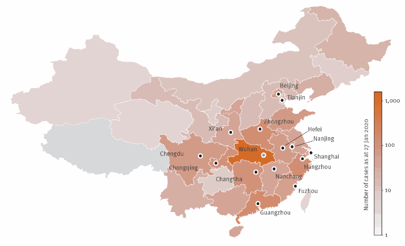
Just two weeks after announcing the discovery of a new virus from the coronavirus family responsible for severe pneumonia, there were 571 confirmed cases in China. In an effort to contain this epidemic whose death toll currently stands at 18, the Chinese authorities have taken drastic measures – particularly restrictions on travel from the province of Hubei, where the city of Wuhan is located.

Many questions continue to remain unanswered as to the origin of this new virus, 2019-nCoV, and about the capacity of the epidemic to spread to other regions of the world – particularly Europe. In the space of two weeks, eight cases have already been exported from China to Japan, South Korea, USA, Thailand and Taiwan.

From the start of the epidemic, Inserm researchers under the aegis of the research group REACTing have been working to model its potential dissemination.

Led by Inserm researcher Vittoria Colizza at the Pierre Louis Institute of Epidemiology and Public Health (Inserm/Sorbonne Université), a team is now able to propose a model to anticipate a potential arrival of the epidemic in Europe in order to guide surveillance and prevention measures. However, it is important to note that this model in no way constitutes a prediction of the future number of cases in France and Europe, but rather a theoretical tool to aid public decision-making.

Air traffic flows from China

When developing their model, the researchers focused on those Chinese provinces reporting more than ten cases. Their estimations of the risks of these cases being exported are based on January 2019 data on air traffic from these regions to Europe produced by the OAG – a world leader in flight data collection.

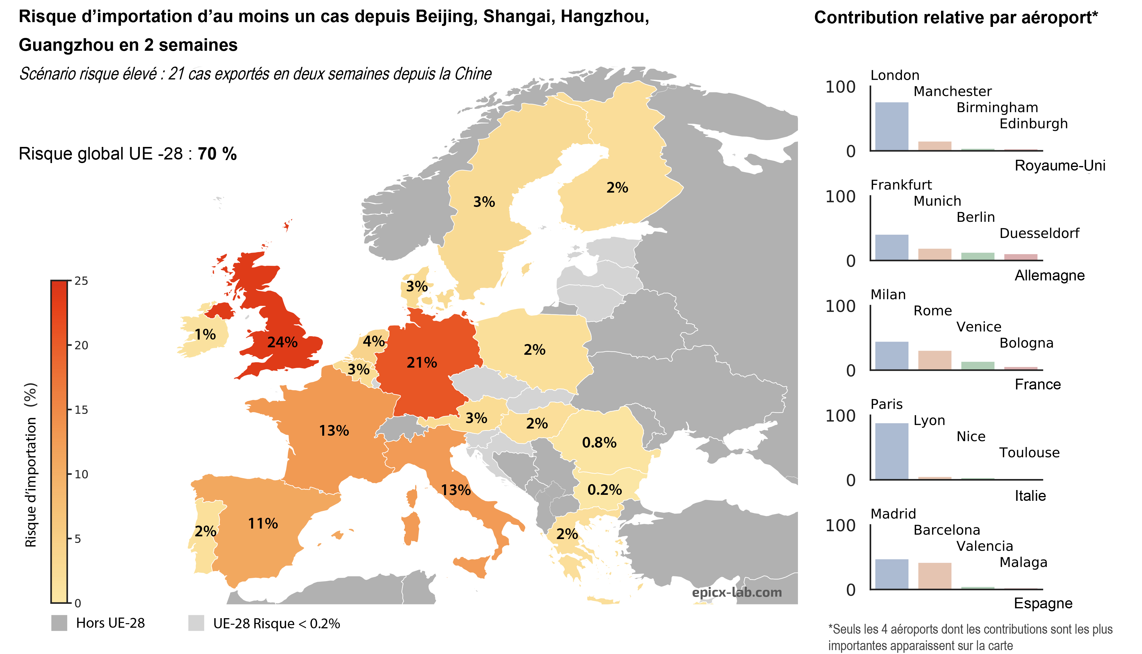
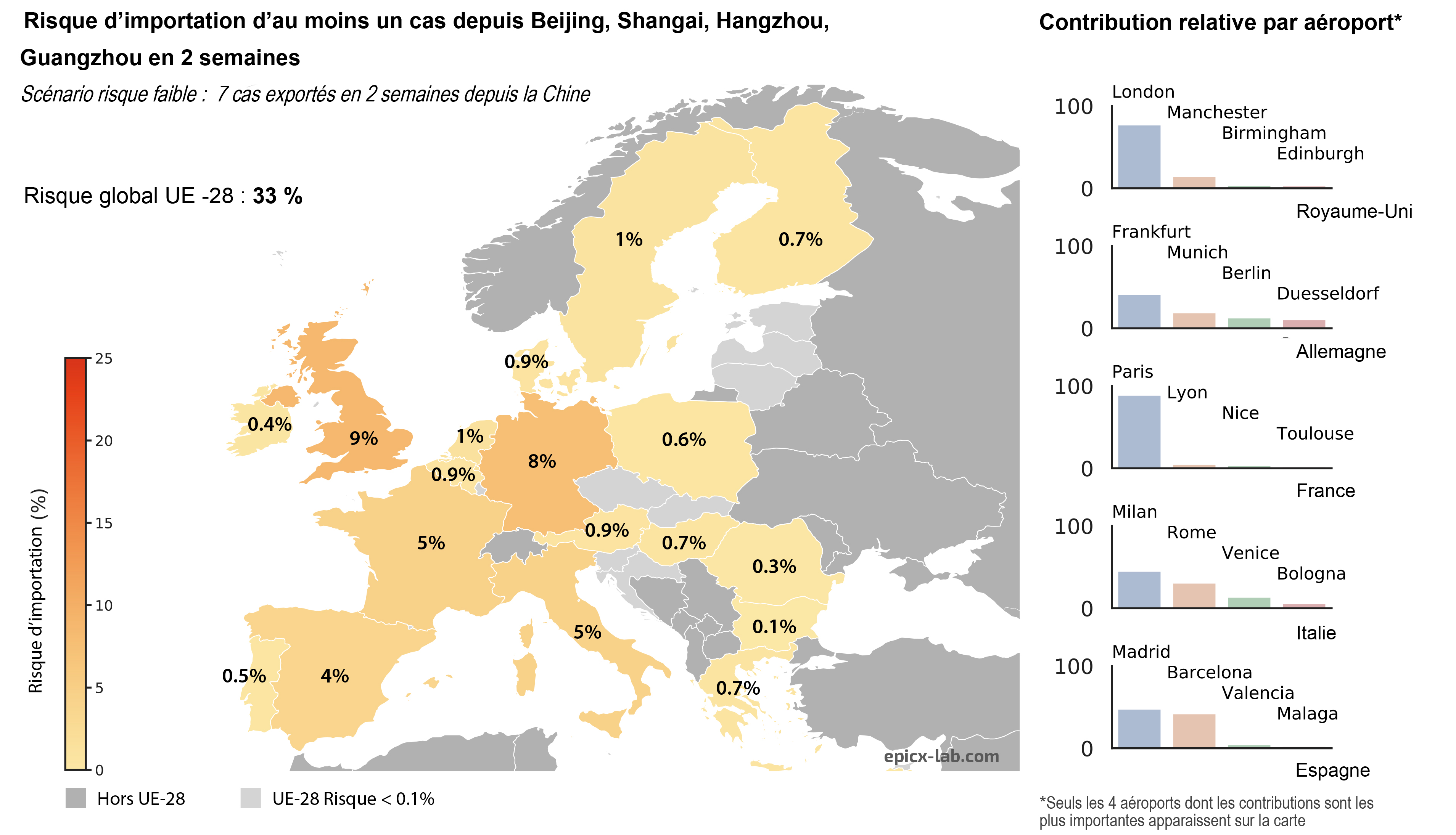
What is the risk of at least one case being imported to Europe in the next two weeks? The team addressed this question by preparing two scenarios: one of a low risk of dissemination and the other of a high risk.

The low-risk scenario is based on the situation (seven cases exported from China) prior to the flight ban by the Chinese government. It estimates the risk of at least one case being imported to Europe if seven cases are exported from the Chinese provinces affected in the next two weeks.


The high-risk scenario proposes an estimation of the same risk if three times more cases are exported from China. “It is an arbitrary choice but one which reflects the fact that the number of Chinese cases is on the increase, making it possible to anticipate the case of a greater number of infected people being exported”, emphasizes Colizza.


According to the researchers, the risk of at least one case being imported to Europe is 33% in the first scenario and 70% in the second. Given the flows of air traffic, the countries most exposed would be Germany and the UK. The risk of an infected passenger arriving in France is 5% in the first scenario and 13% in the second and would mainly be concentrated around the airports serving Paris. “Our findings are not predictions, they simply make it possible to pinpoint where the risk is located and where prevention and surveillance need to be stepped up”, states Colizza.

Deadly outbreak in China linked to a novel form of coronavirus


Map of Chinese provinces colour coded according to the number of cases of 2019-nCoV [4] as at 27 January 2020. Credits Vittoria Colizza

 
Inserm is a signatory to the following international joint statement issued by the Wellcome Trust [1] :

Sharing research data and findings relevant to the novel coronavirus (nCoV) outbreak
 
The outbreak of the novel coronavirus in China (2019-nCoV) represents a significant and urgent threat to global health.
 
We call on researchers, journals and funders to ensure that research findings and data relevant to this outbreak are shared rapidly and openly to inform the public health response and help save lives.
 
We affirm the commitment to the principles set out in the 2016 Statement on data sharing in public health emergencies, and will seek to ensure that the World Health Organization has rapid access to emerging findings that could aid the global response.
 
Specifically, we commit to work together to help ensure:
 
all peer-reviewed research publications relevant to the outbreak are made immediately open access, or freely available at least for the duration of the outbreak
research findings relevant to the outbreak are shared immediately with the WHO upon journal submission, by the journal and with author knowledge
–  research findings are made available via preprint servers before journal publication, or via platforms that make papers openly accessible before peer review, with clear statements regarding the availability of underlying data
–  researchers share interim and final research data relating to the outbreak, together with protocols and standards used to collect the data, as rapidly and widely as possible – including with public health and research communities and the WHO
–  authors are clear that data or preprints shared ahead of submission will not pre-empt its publication in these journals
 
We intend to apply the principles of this statement to similar outbreaks in the future where there is a significant public health benefit to ensuring data is shared widely and rapidly.
 

Coronaviruses, which are named after the crown shape of the proteins that surround them, are a family of viruses that have a traditionally animal reservoir.

In some cases, transmission from animal to human is possible, as the SARS, MERS and probably this new virus have shown.

While these viruses are mostly benign in humans, two death-causing epidemics have been reported in recent years in the Middle East (MERS-Cov, 2012-2013) and in China ten years earlier (SARS-Cov, 2002-2003).

For this third outbreak, the outbreak would come from a fish and seafood market in Wuhan, where live animals are also sold. The search for the reservoir and host is ongoing.

Inserm is already mobilized to respond to the epidemic, under the action of the Reacting consortium, which brings together French research groups of excellence. Several initiatives have been launched:

  • The consolidation of a «modelling group» set up at the request of Reacting, to model the possible spread of the epidemic in France and Europe, and anticipate its impact on populations.


  • The work entrusted to Bruno Lina (Inserm Unit 1111 – Centre International de Recherche en Infectiologie, CIRI) to propose a research project on potential therapeutic targets to identify molecules capable of acting on several types of coronavirus.


  • Support for clinical research, through the dissemination to Reacting’s French and international partners of best practices in research protocols/ standardization of data collection.

[1] List of signatories : Academy of Medical Sciences, UK; African Academy of Sciences; Assistant Secretary for Preparedness and Response / Biodefense Advanced Research and Development Authority, USA; Austrian Science Fund (FWF), Bernhard Nocht Institute for Tropical Medicine (BNITM); Bill & Melinda Gates Foundation; BioRxiv; Biotechnology and Biological Sciences Research Council (BBSRC), UK; Bulletin of the World Health Organization; The British Medical Journal (BMJ); Calouste Gulbenkian Foundation – Gulbenkian Science Institute; Canadian Institutes of Health Research; Centers for Disease Prevention and Control (CDC), USA; Chinese Centre for Disease Control and Prevention; The Department for International Development (DFID); DG Research & Innovation, European Commission; Dutch Research Council (NWO); eLife; EcoHealth Alliance; EMBO; Epicentre – MSF; European Respiratory Journal ; ERJ Open Research; F1000 Research Limited; Fondation Merieux; Food & Drug Administration, USA; French National Research Agency (ANR); The Global Health Network; Global Virus Network; GLOPID-R ; Inserm (Institut national de la santé et de la recherche médicale), France ; The Institut Pasteur; International Severe Acute Respiratory and emerging Infection Consortium (ISARIC); International Society for Infectious Diseases (ISID); Japan Agency for Medical Research and Development (AMED); Johnson & Johnson; The Lancet Luxembourg National Research Fund (FNR); Medical Research Council (UK); Médecins Sans Frontières/Doctors Without Borders (MSF); MedRxiv; Merck Research Laboratories; Microbide Limited National Academy of Medicine, USA; National Institutes of Health (NIH), USA; National Institute for Infectious Diseases Lazzaro Spallanzani, Italy; New England Journal of Medicine; Office of Global Affairs, Department of Health and Human Services, USA; Portland Press; PLOS; The Research Council of Norway; The Royal Society; Science Europe; Science Foundation Ireland ; ScienceOpen; South African Medical Research Council Takeda; Taylor & Francis; ZonMW – The Netherlands Organisation for Health Research and Development

A New Blood Component Revealed

Functional extracellular mitochondria revealed in the blood circulation. ©Alain R. Thierry/Inserm

Does the blood we thought to know so well contain elements that had been undetectable until now? The answer is yes, according to a team of researchers from Inserm, Université de Montpellier and the Montpellier Cancer Institute (ICM) working at the Montpellier Cancer Research Institute (IRCM), which has revealed the presence of whole functional mitochondria in the blood circulation. These organelles that are responsible for cellular respiration had hitherto only been found outside cells in very specific cases. The team’s findings, published in The FASEB Journal, will deepen our knowledge of physiology and open up new avenues for treatment.

Mitochondria are organelles that are found in the eukaryotic cells. A place of cellular respiration, they are the cells’ “batteries” and play a major role in energy metabolism and intercellular communication. Their particularity is to possess their own genome, transmitted solely by the mother and separate from the DNA contained in the nucleus. The mitochondria can sometimes be observed outside the cells in the form of fragments encapsulated within microvesicles. Under certain very specific conditions the platelets are also capable of releasing intact mitochondria into the extracellular space.

The work of a team led by Inserm researcher Alain R. Thierry at the Montpellier Cancer Research Institute (Inserm/Université de Montpellier/Montpellier Cancer Institute) has now revolutionized knowledge of this organelle by revealing that whole functioning extracellular mitochondria are in fact found in the bloodstream!

The researchers used previous findings which showed that the plasma of a healthy individual contains up to 50,000 times more mitochondrial DNA than nuclear DNA. They hypothesized that for it to be detectable and quantifiable in the blood in this manner, the mitochondrial DNA had to be protected by a structure of sufficient stability. In order to identify such a structure, plasma samples from around 100 individuals were analyzed.

This analysis revealed the presence in the blood circulation of highly stable structures containing whole mitochondrial genomes. Following examination of their size and density, as well as the integrity of their mitochondrial DNA, these structures observed using electron microscopy (up to 3.7 million per ml of plasma) were revealed to be intact and functional mitochondria.

Throughout the seven-year research period, the scientists used as many technical and methodological approaches as possible to validate this presence of circulating extracellular mitochondria in the blood.

“When we consider the sheer number of extracellular mitochondria found in the blood, we have to ask why such a discovery had not been made before, notes Thierry. Our team has built up expertise in the specific and sensitive detection of DNA in the blood, by working on the fragmentation of extracellular DNA derived from the mitochondria in particular”, he adds.

But what is the role of these extracellular mitochondria? The answer to that could be linked to the structure of the mitochondrial DNA, similar to that of bacterial DNA, which gives it the ability to induce immune and inflammatory responses. Based on this observation, the researchers hypothesize that these circulating mitochondria could be implicated in many physiological and/or pathological processes requiring communication between the cells (such as the mechanisms of inflammation). Indeed, recent studies have demonstrated the ability of certain cells to transfer mitochondria between themselves, such as the stem cells with damaged cells. “The extracellular mitochondria could perform various tasks as messenger for the entire body”, specifies Thierry.

In addition to its importance to our knowledge of physiology, this discovery could lead to improvements in the diagnosis, monitoring and treatment of certain diseases. In fact, the research team is now devoting its attention to evaluating the extracellular mitochondria as biomarkers in non-invasive prenatal diagnosis and cancer.

This research is supported by the Montpellier Integrated Cancer Research Site (SIRIC) (Inserm/CNRS/Université de Montpellier/Montpellier Cancer Institute/Montpellier University Hospital/Université Paul Valéry), funded by Inserm, the National Cancer Institute (INCa) and the Directorate General of Health Care Provision (DGOS).

Meningeal Lymphatic Network: A New Avenue in the Treatment of Brain Tumors

Glioblastomas are the most common tumors of the central nervous system Image taken using a Zeiss Axioimager Z1 widefield epifluorescence microscope. Inserm/Guichet, Pierre-Olivier

Glioblastomas are the most common type of brain tumor, and their prognosis is often highly unfavorable. A collaborative study by Jean-Léon Thomas, Inserm researcher at the Brain & Spine Institute (Inserm/CNRS/Sorbonne Université) and Pitié-Salpêtrière Hospital AP-HP, and Akiko Iwasaki (Department of Immunology, Yale University School of Medicine, USA), has revealed the beneficial role played by the meningeal lymphatic vascular network in treating these tumors – in the short and longer term. Their findings have been published in Nature.

Glioblastomas are not just the most commonly occurring type of brain tumor, they are also the most severe. With an estimated prevalence of 1/100,000, they mainly affect patients between 45 and 70 years of age. Treatment currently involves surgery combined with radiation therapy and chemotherapy. Therapeutic benefit, in terms of survival, remains modest (currently around 18 months), inciting researchers to continue to explore new avenues of potential treatment.

Eric Song (Yale University), first author of this study, Jean-Léon Thomas, Akiko Iwasaki and their colleagues studied the meningeal lymphatic network to see whether it regulates the immune system in response to the presence of a brain tumor. A veritable pipework of lymphatic vessels in the meninges surrounding the brain, the meningeal lymphatic network has been generating particular interest since the publication of studies over the previous five years showing its connection to the lymph nodes of the neck (where immune cells proliferate and differentiate), and its role of draining immune cells into the latter.

In their latest study, published in Nature, the researchers worked with animal models of glioblastoma. They showed that the tumor would disappear following prior enlargement of the meningeal lymphatics – achieved by injecting the meninges with lymphatic growth factor VEGF-C. The growth of the meningeal lymphatic network induced by VEGF-C (as can be seen in the photo) was correlated with the mass entry of immune T cells (CD4 and CD8), which under normal conditions are absent, into the tumor environment.

This short-term response destroys the tumor and is accompanied by the persistence of “memory cells” specifically directed against the tumor cells, which makes it possible to reject the same tumor in the longer term.

Nevertheless, the researchers’ experiments show that it is in combination with an immunotherapy already used in neuro-oncology that the transient VEGF-C treatment is the most effective, enabling complete eradication of the existing glioblastoma. “Our study highlights the fact that reinforcing the network of meningeal lymphatic vessels increases tumor antigen traffic from the meninges to the lymph nodes”, explains Thomas.

With his colleagues, he concludes that the major role of this network is to transport, from the meninges, an immune alert message triggering activation of the lymphocytes directed against the tumor.

The findings of this study therefore open up new avenues in the treatment of brain tumors by targeting the meningeal lymphatic vessels and their associated lymph nodes.

The researchers wish to continue their work by studying the role of the meningeal lymphatic network in other diseases. “We are currently exploring the functional mechanisms and therapeutic potential of this vascular network with novel experimental models, and in other nervous system diseases – neurodegenerative, neurovascular and infectious“, concludes Thomas.

B Cells: New Allies in Sarcoma Immunotherapy?

Tertiary lymphoid structures are cellular aggregates that contain many B-cells (in purple) located near tumors. This is the area where the antitumor immune response starts. ©Antoine Bougouin/Centre de recherche des Cordelier/Inserm, Sorbonne Université, Université de Paris

How can we improve and better personalize the treatment of soft tissue sarcomas, these particularly resistant and aggressive forms of cancer? An international team led by Wolf Hervé Fridman with researchers from Inserm, Sorbonne Université and Université de Paris at the Cordeliers Research Center, in collaboration with the French League against cancer and Institut Bergonié, has shown that B cells also play a major role in predicting of patient’s response to immunotherapy.  It was previously thought only T cells could be used in this way. Their findings, to be published in Nature, pave the way for the personalization of treatments for patients with soft tissue sarcomas.

Soft tissue sarcomas are a heterogenous group of aggressive, chemotherapy-resistant cancers that affect the soft tissues of the body (fat, muscles, fibrous tissue, blood and lymphatic vessels, nerves, etc.). In the current clinical trials, only 15% of patients respond to immunotherapy, which raises the question of the needless exposure of the other patients to the toxicity of these treatments. Identifying markers that predict their response to immunotherapy is therefore crucial. A strategy that until now has been essentially focused on the T cells – immune cells capable of recognizing cells that are infected, cancerous or foreign to the body.

Through research published in Nature, a group led by Wolf Hervé Fridman with members from Inserm, Sorbonne Université and Université de Paris at the Cordeliers Research Center, in collaboration with the “Tumor identity card” team from the French League against cancer, Institut Bergonié, and teams from the USA and Taiwan, studied the question of identifying other potential markers.

They analyzed 608 tumors, classifying them into three groups according to the composition of their microenvironment[1]: immunologically poor tumors (low in immune cells and poorly vascularized), highly vascularized tumors, and immunologically rich tumors. The latter present aggregates of various cell types with high levels of B cells, the immune cells responsible for the production of antibodies. These aggregates are called tertiary lymphoid structures. The researchers observed that an anti-tumor immune response initiates within them, thereby showing that the B cells could play an anti-tumor role.

What is more, in a phase 2 clinical trial, the patients with immunologically rich tumors showed a high response rate (50%) to one immunotherapy: pembrolizumab. These patients also had a higher survival rate than those with immunologically poor or highly vascularized tumors.

A second study by a US team, co-signed by Wolf Hervé Fridman’s team at Cordeliers Research Center (Inserm/Sorbonne Université/Université de Paris), and published in parallel in Nature, extended these observations to include melanoma and kidney cancer.

The results of these studies show that in addition to the T cells that are usually researched, the B cells play an essential role in the response to immunotherapy for certain cancers. These cells bring new hope for the treatment of soft tissue sarcomas, which are particularly resistant to standard therapies. In addition, from a personalized medicine standpoint, these findings could help orient clinical decisions and patient treatment by means of a simple test to identify those whose tumors are immunologically rich.

On the basis of these results, an initial French clinical trial coordinated by Antoine Italiano (Institut Bergonié, Université de Bordeaux), co-author of the first article, and which includes patients with such tumors, is currently ongoing within the French Sarcoma Group.

[1] The tumor microenvironment corresponds to the biological elements that surround the tumor (blood vessels, immune cells, various types of cells, signaling molecules, extracellular matrix, etc.) and with which it interacts.

PREVAC-UP: The Partnership for Research on Ebola Vaccination extends follow-up and builds research capacity against deadly disease

PREVAC – Landréah Conakry vaccination centre. Credits : Inserm/Delapierre, Patrick

Public health efforts successfully stopped human-to-human transmission of Ebola virus in West Africa in 2016 after the worst Ebola outbreak in history.  However, the threat of the disease is still real, as the recent Ebola outbreaks in the Democratic Republic of the Congo (DRC) have shown. Thus, pursuing and intensifying efforts to develop a safe and effective vaccination strategy against Ebola virus disease, with durable protection in all populations, including children, is vital.

Since August 2018, the DRC has been battling its worst Ebola outbreak, and the world’s second largest such outbreak, with more than 2200 lives lost and more than 3300 confirmed infections to date. This ongoing crisis highlights again the continued urgent need to develop safe and effective vaccines against the Ebola virus.

This goal has been the priority for the Partnership for Research on Ebola Vaccination (PREVAC)[1], an international consortium that recently received additional funding to scale up its research in West Africa as part of a new project known as PREVAC-UP. This initiative aims to evaluate the long-term safety, as well as the durability of humoral and cellular immune responses, of three different Ebola vaccine regimens previously tested by the consortium. PREVAC-UP will assess these factors for 5 years after vaccination.

The study will also evaluate the effect of co-infections, such as malaria and helminths, on the immune response to vaccination. An integrative statistical analysis of the immune response will be used to explore the mechanism of action of the vaccines and to identify early correlates of durable antibody induction. “This programme is expected to significantly impact Ebola prevention and control in adults and children in Africa. The study will also strengthen the capacity for science relevant to the development and evaluation of new vaccines in sub-Saharan Africa”, Dr Yazdan Yazdanpanah, PREVAC Principal Investigator, says.

The project is co-funded by the European and Developing Countries Clinical Trials Partnership (EDCTP2) programme supported by the European Union[2]. Besides the EDCTP2 grant, PREVAC-UP benefits from co-funding from Inserm, the NIAID, the LSHTM and the COMAHS, as well as host country support from Liberia, Sierra Leone, Guinea and Mali.

PREVAC results expected in 2020

PREVAC-UP follows up on the work already carried out by the PREVAC[3] consortium since March 2017 in West Africa. Working in Liberia, Guinea, Sierra Leone and Mali, the team conducted a randomised, placebo-controlled, multicentre Phase 2 trial – one of the largest Ebola vaccine trials to date – using the recently WHO prequalified rVSVΔG-ZEBOV-GP vaccine from Merck, Sharpe & Dohme, Corp.[4], and an investigational two-dose vaccine regimen (Ad26.ZEBOV, MVA-BN-Filo) developed by Janssen Vaccines & Prevention B.V., part of the Janssen Pharmaceutical Companies of Johnson & Johnson, in collaboration with Bavarian Nordic.

The aim was to study the safety and immunogenicity over 12 months of three different vaccination strategies involving these vaccines in adults and children one year and older. In total, PREVAC enrolled 4789 people from the 4 countries, with the enrolment of 2802 participants (1401 adults and 1401 children) in the main phase of the trial completed in December 2018. The first results on immunogenicity at 12 months after vaccination are expected in the summer 2020.  Importantly, the retention rate of the participants is very high (95% after 12 months). “This reflects the great commitment of all the local professionals and shows the high level of collaboration between all the partners involved in the project as well as the interest in the population to receive an Ebola vaccine, and successful community engagement strategies”, Dr. Yazdanpanah says.

PREVAC-UP will now bring much needed additional data on these different vaccination strategies, helping to promote the safest and most efficient approach to protect vulnerable populations in countries threatened by Ebola.


[1] To learn more about PREVAC on Inserm’s website : https://www.inserm.fr/en/news-and-events/news/ebola-already-more-than-2000-people-included-in-prevac-immunization-trial

[2] PREVAC-UP Partner Organizations 

Institut national de la santé et de la recherche médicale (Inserm)*, France ; Centre National de Formation et de Recherche en Santé Rurale (CNFRSR)*, Guinea ; Institut Bouisson Bertrand* (IBB)/ Centre de Recherche et de Formation en Infectiologie (CERFIG) France/ Guinea ; London School of Hygiene & Tropical Medicine (LSHTM)*, United Kingdom ; University of Sierra Leone, College of Medicine and Allied Health Sciences (COMAHS)*, Sierra Leone ; Alliance for International Medical Action (ALIMA)*, France ; Inserm-Transfert SA*, France ; National Institute of Allergy and Infectious Diseases (NIAID), USA ; Université des Sciences, des Techniques et des Technologies de Bamako (USTTB)*, Mali ; Ministry of Health (Centre pour le Développement des Vaccins –Mali)*, Mali ; National Public Health Institute of Liberia (NPHIL), Liberia

*EDCTP grant agreement signatories.

[3] Inserm’s participation in PREVAC is in part through a subcontract with Leidos Biomedical Research, Inc. which operates the Frederick National Laboratory for Cancer Research on behalf of the National Cancer Institute. The content of this publication does not necessarily reflect the views or policies of the Department of Health and Human Services, nor does mention of trade names, commercial products, or organizations imply endorsement by the U.S. government.

The PREVAC UP project is funded by the European and Developing Countries Clinical Trials Partnership (EDCTP2) programme supported by the European Union and the UK Department of Health & Social Care (Grant number RIA2017S – 2014 -PREVAC-UP). Besides the EDCTP2 grant, PREVAC-UP benefits from co-funding from Inserm, the NIAID, the LSHTM and the COMAHS as well as host country support from Liberia, Sierra Leone, Guinea and Mali.

[4] The rVSVΔG-ZEBOV-GP vaccine (sold under the name ERVEBO®) was awarded prequalification status by the WHO in November 2019. It is now licensed in the USA and in Europe.

fermer