Menu

Music or Speech? The Brain is Divided…

Photo by Thanos Pal on Unsplash

When it comes to recognizing a melody or understanding a spoken sentence, the human brain does not mobilize its hemispheres in an equivalent way. Although the concept is recognized by scientists, there had been no physiological or neural explanation for the phenomenon until now. A team co-led by Inserm researcher Benjamin Morillon at the Institute of Systems Neuroscience (Inserm/Aix-Marseille Université) in collaboration with researchers at Montreal Neurological Institute and Hospital of McGill University has been able to show that, due to different receptivities to the components of sound, the left auditory cortex neurons participate in the recognition of speech, whereas the right auditory cortex neurons participate in that of music. These findings, to be published in the journal Science, suggest that the respective specializations of the brain hemispheres for music and speech enable the nervous system to optimize the processing of sound signals for communication purposes.

Sound is produced from a complex set of air vibrations, which, when reaching the cochlea of the inner ear, are distinguished according to their speed. At any given moment, slow vibrations are being translated into deep sounds and rapid vibrations into high-pitched ones. As a result, sound can be represented according to two dimensions: spectral (frequency) and temporal (time).

These two auditory dimensions are fundamental because it is their simultaneous combination that stimulates the neurons of the auditory cortex. The latter are thought to discriminate sounds that are relevant for individuals, such as those used for communication enabling people to talk to and understand each other.

In humans, speech and music constitute the main uses of sound and the most complex on the cognitive level. The left hemisphere is primarily implicated in the recognition of speech, whereas the right hemisphere is primarily implicated in that of music. However, until now little was known about the physiological and neural reasons for this asymmetry.

A team co-led by Inserm researcher Benjamin Morillon at the Institute of Systems Neuroscience (Inserm/Aix-Marseille Université) in collaboration with researchers at Montreal Neurological Institute and Hospital of McGill University used an innovative approach to understand how speech and music are decoded within each of the human brain hemispheres.

The researchers recorded 10 sentences sung by a soprano to 10 new melodies composed especially for the experiment. These 100 recordings, in which melody and speech are dissociated, were then distorted by decreasing the amount of information present in each dimension of the sound. Forty-nine participants were asked to listen to pairs of these distorted recordings, and to determine whether they were identical in terms of speech content and melody. The experiment was conducted in French and English speakers to see whether the results were reproducible in different languages.

A demonstration of the audio test proposed to the participants is available here:

https://www.zlab.mcgill.ca/spectro temporal modulations/

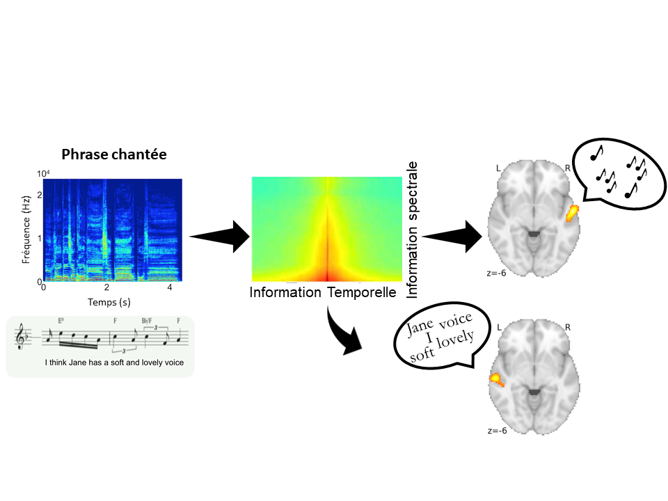
The research team found that for both languages, when the temporal information was distorted, participants had trouble distinguishing the speech content, but not the melody. Conversely, when the spectral information was distorted, they had trouble distinguishing the melody, but not the speech.

Functional magnetic resonance imaging (fMRI) of the participants’ neural activity showed that in the left auditory cortex the activity varied according to the sentence presented but remained relatively stable from one melody to another, whereas in the right auditory cortex the activity varied according to the melody presented but remained relatively stable from one sentence to another.

What is more, they found that degradation of the temporal information affected only the neural activity in the left auditory cortex, whereas degradation of the spectral information only affected neural activity in the right auditory cortex. Finally, the participants’ performance in the recognition task could be predicted simply by observing the neural activity of these two areas.

Original a capella extract (bottom left) and its spectrogram (above, in blue) broken down according to the amount of spectral and temporal information (center). The right and left cerebral auditory cortexes (right) decode melody and speech respectively.

“These findings indicate that in each brain hemisphere, neural activity depends on the type of sound information, specifies Morillon. While temporal information is secondary for recognizing a melody, it is essential for the correct recognition of speech. Conversely, while spectral information is secondary for recognizing speech, it is essential for recognizing a melody. “

The neurons in the left auditory cortex are therefore considered to be primarily receptive to speech thanks to their superior temporal information processing capacity, whereas those in the right auditory cortex are considered to be receptive to music thanks to their superior spectral information processing capacity. “Hemispheric specialization could be the nervous system’s way of optimizing the respective processing of the two communication sound signals that are speech and music,” concludes Morillon.

This research was supported by a Banting fellowship to Philippe Albouy, grants from the Canadian Institutes of Health Research and the Canadian Institute for Advanced Research to Robert J. Zatorre, ANR-16-CONV-0002 (ILCB) and ANR-11-LABX-0036 (BLRI) funding and the Excellence Initiative of Aix-Marseille Université (A*MIDEX).”

Egypt, Algeria and Republic of South Africa, main gates for coronavirus importation in Africa

©chuttersnap

Egypt, Algeria and Republic of South Africa are the African countries most at risk for coronavirus COVID-19 importation in the continent, due to high air traffic with the contaminated Chinese provinces. But these countries are also among the best equipped on the continent to quickly detect and deal with new cases. In other African countries, the risk of importation is lower, but health organization deficiencies raise concerns about rapid spread. This modeling work carried out by Vittoria Colizza, Inserm research director, and her team from Unit 1136 Pierre Louis Institute of epidemiology and public health (Inserm / Sorbonne University), in collaboration with the Université libre de Bruxelles, the Oxford Martin Programme on Pandemic Genomics and the University of California Los Angeles, is published in The Lancet.

The COVID-19 coronavirus continues to spread in China and cases have been reported in more than 25 countries. The African continent was spared for a long time until a first case was recently reported in Egypt. Vittoria Colizza, research director at Inserm (French Institute for Health and Medical Research), and her team from Unit 1136 Pierre Louis Institute of Epidemiology and Public Health (Inserm / Sorbonne University), in collaboration with the Université libre de Bruxelles, the Oxford Martin Programme on Pandemic Genomics and the University of California Los Angeles, assessed the risk of importing the virus into Africa, country by country, and the capacities of each of them to detect and deal with it.

The researchers evaluated the risk of the virus importation according to the number of cases declared by each chinese province and according to air traffic between the three main airports of each of these provinces (except Hubei due to flights suspension) and each African country. Moreover, they analyzed the potential of each country to face the risk of the spread of a contagious disease using WHO data and official data.

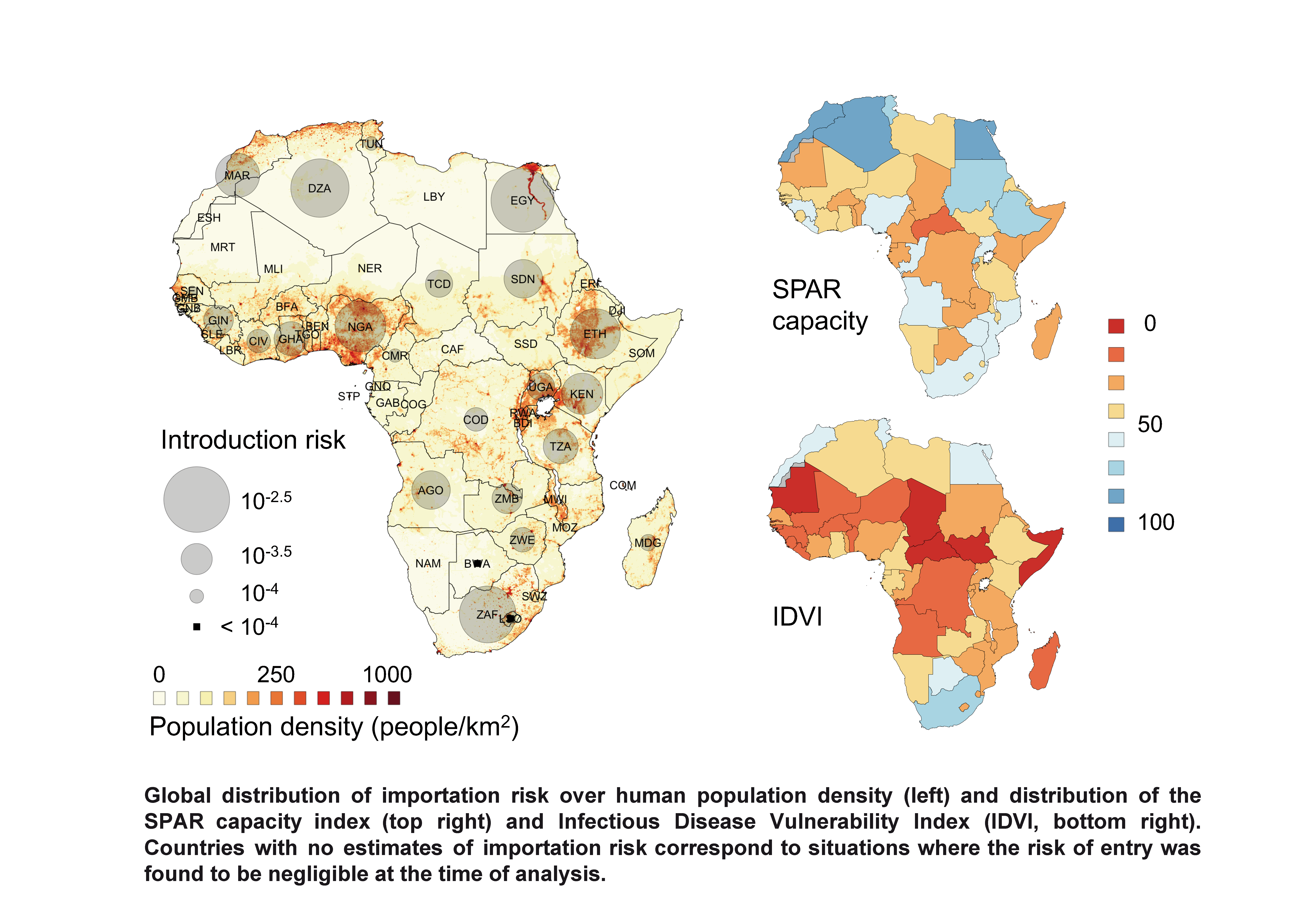
Each country makes a mandatory annual declaration to the WHO of its resources to deal with an epidemic (State Parties self-assessment Annual Reporting SPAR). It includes twenty-four items weighted into an overall score between 0 and 100, 100 showing a strong preparedness to face an epidemic. These indicators are legislation, adherence to WHO standards, laboratory skills, medical staff, emergency organization, food safety, level of equipment in healthcare centers and public communication.

The researchers also took into account the IDVI score (for Infectious Disease Vulnerability Index), also noted out of 100, 0 corresponding to an extreme vulnerability and 100 to the lowest vulnerability. The IDVI takes into account factors not directly linked to the health system but which can influence the response to an epidemic: the size of the population, the socio-economic level or even political stability. Thus, high IDVI and SPAR scores are predictive to an efficient response in case of virus importation.

The results show that Egypt, Algeria and Republic of South Africa are the countries most at risk of importing the virus to Africa due to high trade exchanges with China. On the other hand, their SPAR and IDVI scores are among the best on the continent, letting expect effective detection and containment of the virus. Other countries as Nigeria, Ethiopia, Sudan, Angola, Tanzania, Ghana and Kenya, are at lower risk of virus importation but their SPAR and IDVI scores are lower, raising fears of the non-detection of possible imported cases and of local or even national spread.

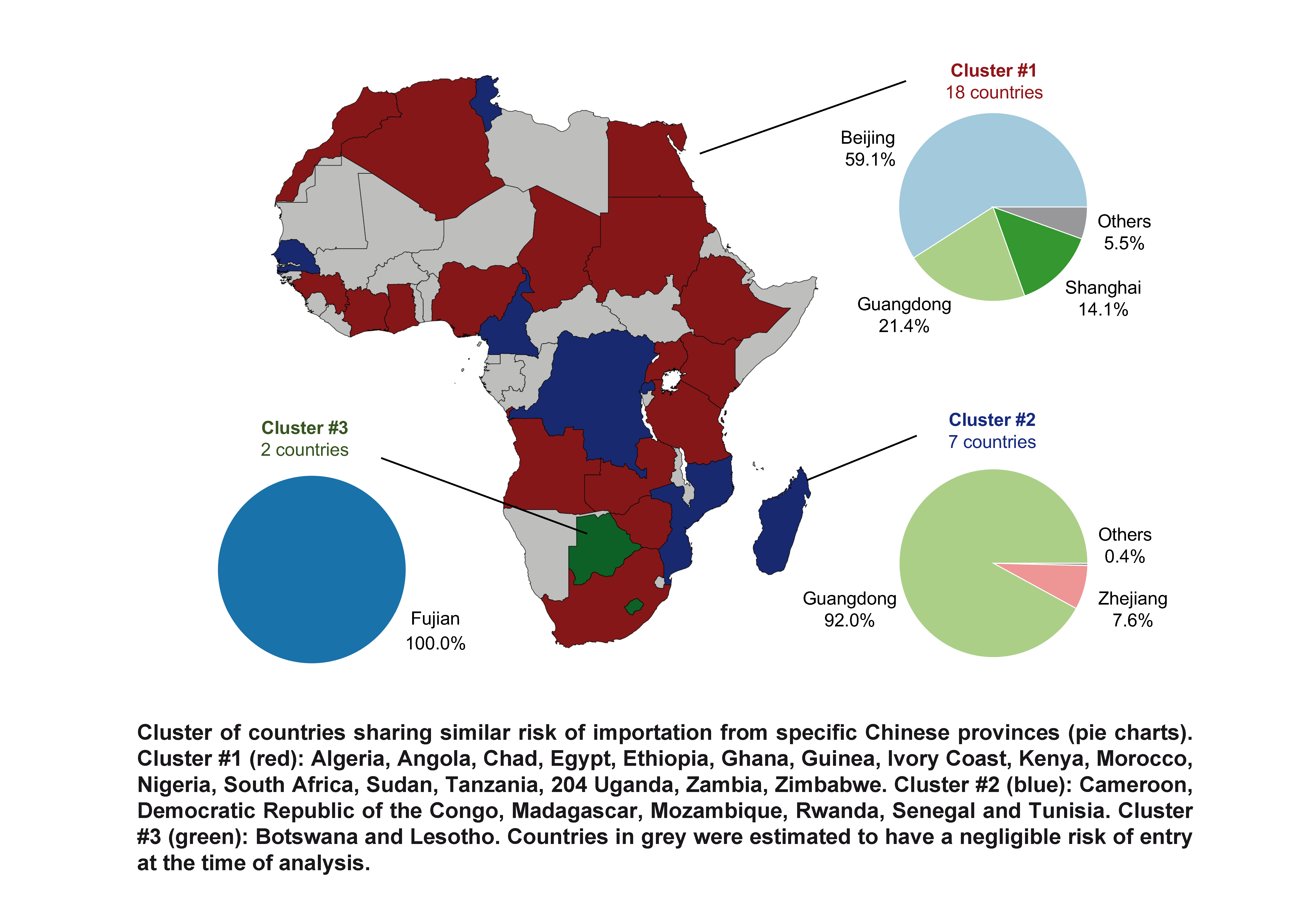
Finally, the researchers clustered the African countries at risk into three groups according to the influence of the Chinese provinces in these countries. Thus, a first group including 18 countries will be more vulnerable in the event of a major epidemic in the province of Beijing, a second comprising 7 countries will be more exposed in the event of a strong growth of the epidemic in the province of Guangdong and a third group of two countries is risking virus importation only from Fujian province.

“This work will allow the international community to make projections and plans according to the evolution in China. It also alerts the countries most exposed to the need of being prepared for the possible introduction of the virus. We can see how hard it is to quickly detect imported cases, as even well prepared developed countries missed some of them. For several poorly equipped African countries, the risks are significant of not having sufficient organization and infrastructure for detection, containment and urgent care, raising fears of a risk of epidemic on the continent”, concludes Vittoria Colizza.

Diabetes: the Gut Flora Yields Its Secrets for the Development of New Treatments


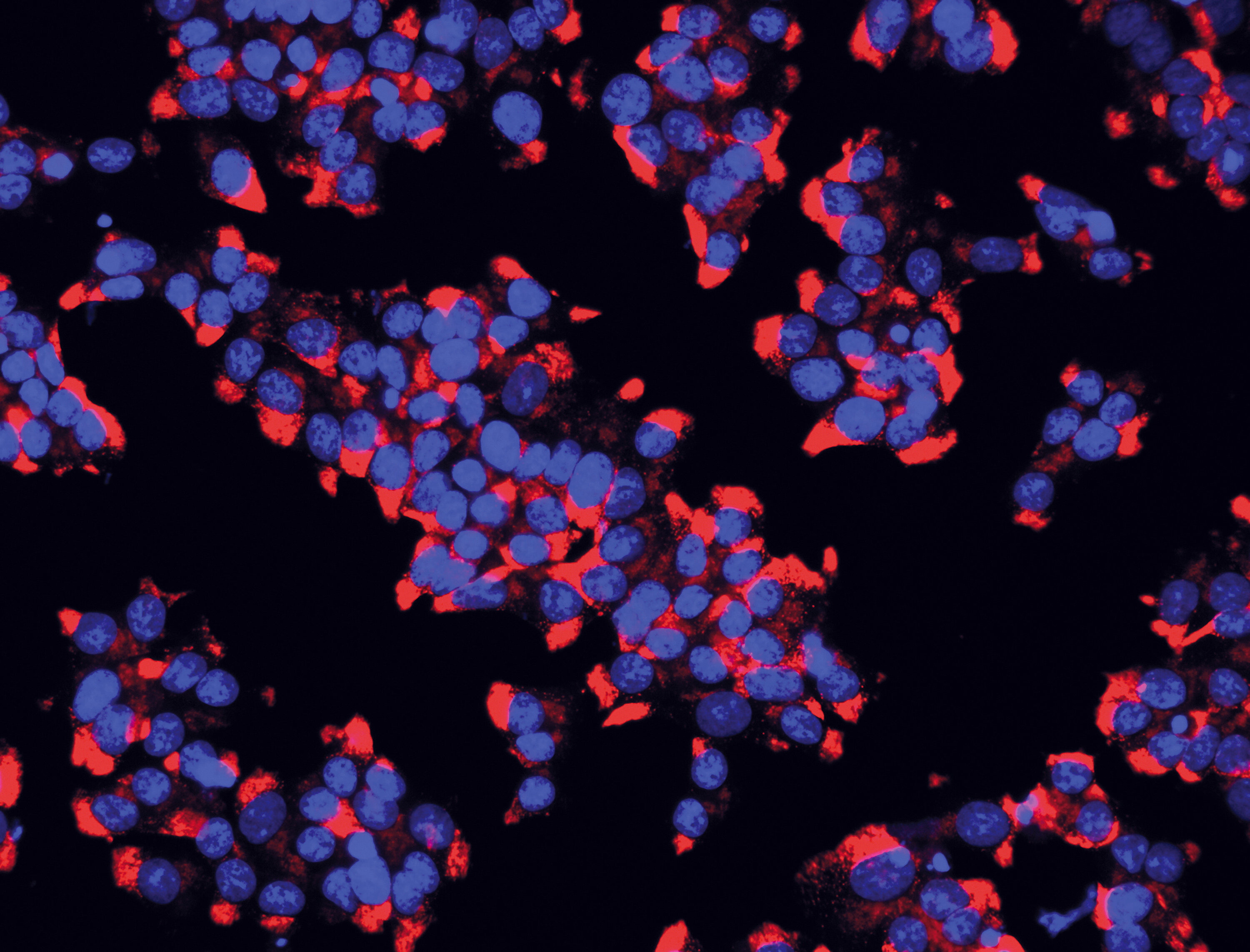
Insulin is produced by the beta cells of the pancreatic islets of Langerhans. In blue: the nuclei of the beta cells. In red: the insulin they contain. © Inserm/U845/JRUS975/EndoCells SARL

An organic compound produced by the gut flora – the metabolite 4-Cresol – is considered to have protective effects against both type 1 and 2 diabetes, notably by stimulating the growth of the insulin-producing pancreatic beta cells. This is according to a new study led by Inserm researcher Dominique Gauguier at the Environmental Toxicology, Therapeutic Targets, Cell Signaling and Biomarkers laboratory (Inserm/Université de Paris), published in Cell Reports. These findings open up new therapeutic avenues which could improve the situation of millions of patients.

Over three million people in France have some form of diabetes. This disease – whose prevalence continues to grow – is linked to an increased risk of developing cardiovascular diseases, making it a major public health concern. Developing and improving treatments for these patients is therefore crucial.

Recent studies have shown that the common forms of diabetes are caused by a mutation of several genes and by factors associated with the environment and certain gut flora compositions.

A study by Inserm researcher Dominique Gauguier at the Environmental Toxicology, Therapeutic Targets, Cell Signaling and Biomarkers laboratory (Inserm/Université de Paris), in collaboration with colleagues from Kyoto University (Japan) and McGill University (Canada), backs up these findings. It reveals a link between diabetes and the metabolite 4-cresol – an organic compound produced by the gut flora and which is also present in some foodstuffs.

The researchers began by performing a metabolic profiling study to identify the various types of metabolites present in the body, based on blood samples from 148 adults – some of whom diabetic. The idea was to identify markers that could be linked to the development of the disease. “4-Cresol really stood out. A product of the gut flora metabolism, this compound appears to be a marker of resistance to diabetes. We found lower levels of it in the serum of diabetic patients compared with non-diabetics”, explains François Brial, Inserm researcher and first author of the study.

Using rat and mouse models of obesity and diabetes, the researchers then tested the effects of 4-Cresol on the clinical signs of diabetes and on the functioning of the pancreatic beta cells, which secrete the hormone insulin whose role is to regulate blood glucose. As the disease progresses, beta cells burn out.

Treatment avenues

The researchers showed that chronic treatment with low concentrations of 4-Cresol led to an improvement in the diabetes. They observed reduced obesity and reduced fat accumulation in the liver, as well as an increase in pancreatic mass, the stimulation of insulin secretion and the proliferation of the pancreatic beta cells.

“At present, we lack therapies to stimulate the proliferation of pancreatic beta cells and improve their function, so these findings are particularly encouraging. They also confirm the impact of the gut flora on human health, demonstrating the beneficial role of a metabolite produced by intestinal bacteria, and open up new therapeutic avenues in diabetes, obesity and fatty liver disease”, emphasizes Gauguier.

The immediate objective of the researchers is now to study the possibilities for modulating the gut flora to restore 4-Cresol production in diabetic patients. To do that they will first try to identify the bacteria that naturally produce this metabolite, and then define what could prove to be potential safe and effective treatments in syndromes of insulin deficiency.

On the basis of recent studies, new treatment options can already be envisaged for diabetic patients. For example, gut flora transplants – even if their mechanisms of action are not yet fully understood, or bariatric surgery – although these are major and invasive operations. “Our aim is to find therapeutic avenues that enable refined modulation of the gut flora, favoring the proliferation of ‘good’ bacteria whose functioning is better understood, and the production of 4-Cresol at therapeutic doses”, concludes Gauguier.

PTSD: Resilience after trauma: the role of memory suppression

A study published in Science provides new insights into understanding post-traumatic stress disorder © Inserm

The terrorist attacks committed in Paris and Saint-Denis on November 13, 2015 have left lasting marks, not only on the survivors and their loved ones, but also on French society as a whole. A vast transdisciplinary research program, the 13-Novembre project is codirected by Francis Eustache, neuropsychologist and director of the Inserm Neuropsychology and Imaging of Human Memory laboratory (Inserm/Université de Caen Normandie/École pratique des hautes études/Caen university hospital/Cyceron imaging platform) and Denis Peschanski, historian and CNRS Research Director[1]. The program seeks to understand the ongoing construction and evolution of the individual and collective memory of these traumatic events and improve our understanding of the factors that protect people against the development of post-traumatic stress disorder (PTSD).

Part of this program is a brain imaging study called Remember, which is focused on the cerebral networks implicated in PTSD. The findings will be published in the journal Science on February 14, 2020. This study, which is sponsored by Inserm and led by Inserm researcher Pierre Gagnepain, shows that the untimely resurgence of intrusive images and thoughts in PTSD patients – a phenomenon long attributed to a deficiency of memory – is linked to a dysfunction of the brain networks that control memory. The researchers expect that these findings will lead to the identification of new treatment options for PTSD sufferers.

  1. Context: the 13-Novembre program

Unprecedented in their scale and violence, the terrorist attacks of November 13, 2015 sent shockwaves throughout French society. In their aftermath, the scientific community decided to act, to improve our understanding of the consequences of such a trauma and to improve the treatment options available to victims and their loved ones. A few days after the attacks, Alain Fuchs, then President of the CNRS, addressed the academic world and called upon researchers to come up with projects to tackle these challenges. He asked for all interested research teams to submit “proposals on any subjects capable of addressing the societal questions raised by the terrorist attacks and their consequences, and which open up avenues for new solutions – social, technical, and digital”.

The 13-Novembre transdisciplinary program was then launched by Inserm, CNRS and héSam Université[2].  One of its components “Étude 1000”, in which 1,000 volunteers are followed up over 12 years. Volunteers include people who were directly exposed to the violence – survivors and people close to the victims, members of the emergency response teams, residents of the neighborhoods targeted and of the Paris suburbs, as well as inhabitants of other French cities, in order to understand the construction and evolution of the memory of the attacks (see box).

The Remember project: understanding PTSD

One of the components of 13-Novembre, the Inserm-sponsored project Remember makes it possible to go much further in the understanding of human memory. With this brain imaging study conducted in Caen on a subgroup of 175 participants, the researchers are exploring the effects of a traumatic event on brain structure and function, identifying neurobiological markers of both PTSD and resilience to trauma. They hope that one day this research will open up new avenues for treatment, to complement those already available.

As such, Remember is tackling a major question that has intrigued neuroscientists for years: why does PTSD affect some trauma victims but not others? One of the objectives of this study, published in the journal Science, is to determine whether there is a link between the control mechanisms of our memory and the individual capacity for resilience.

“We focused this research program on the protective factors and brain markers associated with resilience to trauma. This is what makes our work original compared with previous studies that focused more generally on the impact of trauma on memory and its dysfunction”, emphasizes Pierre Gagnepain, scientific leader of Remember.

Étude 1000″ 

During four campaigns of filmed interviews initiated with the support of France’s Audiovisual Institute (INA) and Defense audiovisual communication and production unit (ECPAD), which are being conducted over a 10-year period (2016, 2018, 2021, and 2026), the participants are asked to share their accounts and evoke their personal memories of the attacks on the basis of an identical interview guide.

These individual accounts will then be analyzed in detail and put into perspective with the collective memory as it is constructed over the years, particularly within the media spaces (television and radio news, press articles, social networks, images of commemorations, etc.).

An approach inspired by that of William Hirst, Professor of Psychology at The New School (New York, USA), following September 11. Along with his teams, he collected some 3,000 written questionnaires of people affected by these attacks.  

13-Novembre takes this methodology further by using video recordings, by seeking to follow the same people over the 10-year period, and by adopting a transdisciplinary approach. Seen like this, it is a world first. “With 13-Novembre, the idea was to take things even further thanks to a very rich collaboration among multiple disciplines and the implementation of the biomedical study Remember. A study that gives us the unique scientific opportunity to observe the process of individual memory construction and how it interacts with collective memory”, emphasize Francis Eustache and Denis Peschanski, codirectors of 13-Novembre.


  1. PTSD

PTSD can develop in people who have been confronted to shocking, dangerous or terrifying events. First identified and studied by scientists in servicemen and women returning from the front, it can affect anyone – children and adults alike. PTSD can develop following any type of trauma, such as a natural catastrophe, sudden death of a loved one or terrorist attack, such as those of November 13, 2015. Studies performed in the USA and Canada estimate a 6 to 9% prevalence of the disorder in the general population.

Intrusive memories a core component

PTSD is a complex condition characterized by symptoms that can vary from one person to another. It can occur just after the trauma or years later. It can remain silent for relatively long periods of time. One of the most characteristic symptoms is the frequent intrusion of memories of the images, smells and sensations linked to the trauma.

These intrusions, which severely impact everyday life, cause great distress as well as other intense emotions, such as fear, guilt, and anger. These can be accompanied by physical symptoms triggered by the recollection of the event, such as muscle tension or increased heart rate.

In order to minimize the distress caused by intrusive memories, sufferers of PTSD also have a tendency to develop avoidance behaviors in the face of any circumstances that could evoke memories of the trauma. These can involve refusing to think or talk about the event, or gradually withdrawing from society and even their loved ones.


  1. Understanding more about the cause of intrusive memories

According to the traditional models of PTSD, the persistence of painful intrusive memories is caused by memory dysfunction – a bit like a scratched record playing the same fragments of our memories over and over again. From the anatomical point of view, such dysfunctions are particularly visible in the hippocampus – a key region for the formation of memory.

In addition, patients’ attempts to suppress their traumatic memories have long been considered an ineffective mechanism. Instead of confronting these painful images in order to leave them in the past, the way they were trying to repress or drive them out was seen more as a negative strategy, intensifying the intrusions and worsening the situation of those with PTSD.

The brain imaging study published in the journal Science challenges some of these ideas, hypothesizing that the untimely resurgence of intrusive images and thoughts could also be linked to a dysfunction of the brain networks implicated in controlling memory (going back to the previous metaphor of the record player, the turntable arm is not working correctly). “These control mechanisms act like a regulator of our memory and are engaged in halting or suppressing the activity of the regions associated with memories, such as the hippocampus”, states Gagnepain.
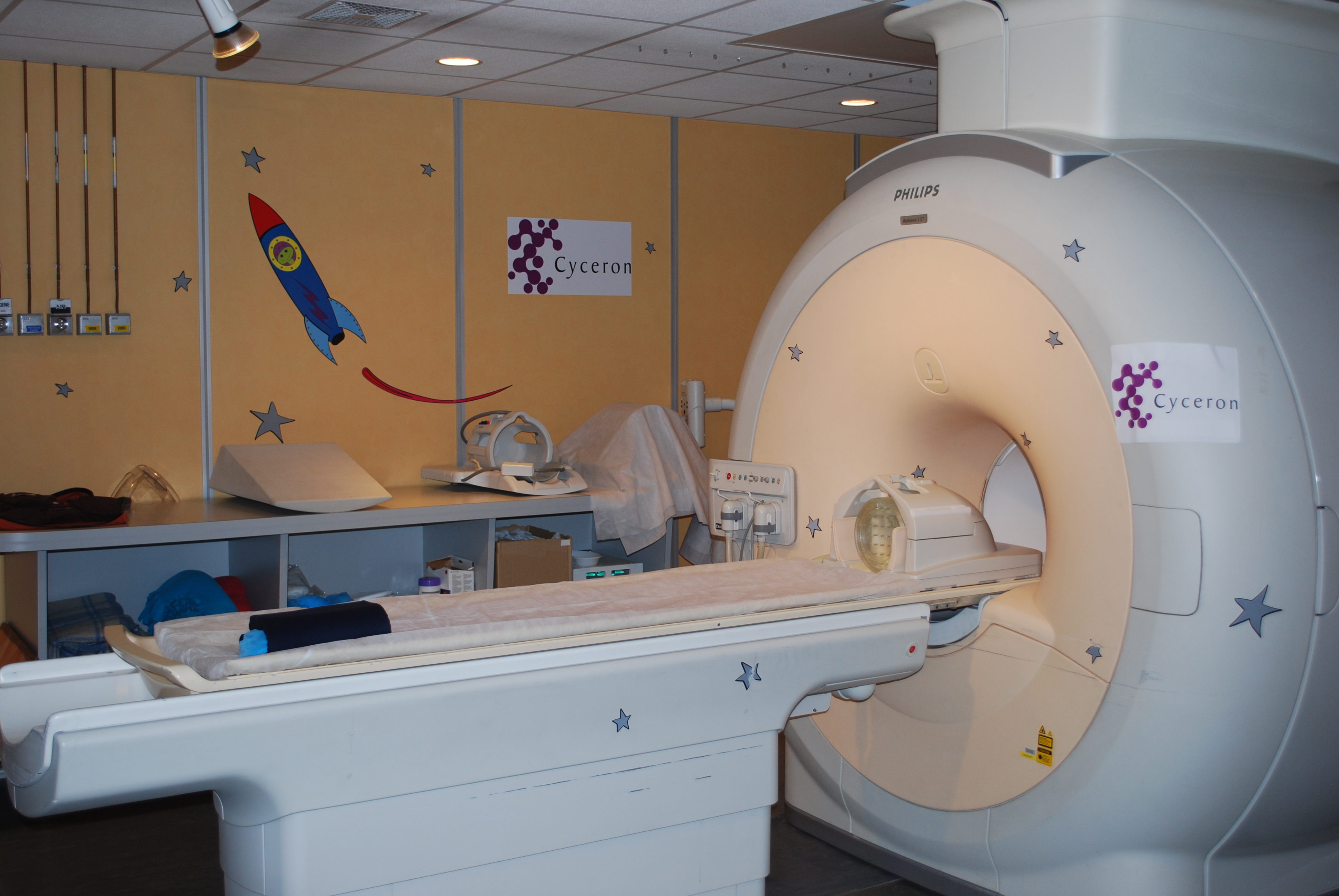
The participants had to perform the Think/No-Think task in the MRI machine.


Methods and results

Together with his colleagues, Gagnepain worked with 102 survivors of the Paris attacks, 55 of whom had PTSD. Also involved in the study were 73 people who had not been exposed to the attacks.

In order to model the resurgence of intrusive memories observed in the PTSD of these volunteers without putting them through the ordeal of viewing the shocking images of the attacks, the scientists opted for a brain imaging research protocol based on the Think/No-Think method (see box).

The aim of this method is to create associations between a cue word and an unrelated everyday object (for example, the word chair with the image of a ball), in order to reproduce the presence of an intrusion when confronted with the cue word. “Then we can study the capacity of the participants to drive and suppress from their mind the intrusive image that emerges against their will when confronted with the cue word”, says Alison Mary, researcher and co-author of the article.

The Think/No-Think method is used to obtain a model of memory intrusion (see box). Credits: Pierre Gagnepain


The Think/No-Think method

For the researchers, it was important not to expose survivors to new traumatizing images that could plunge them back into a state of distress. The Think/No-think paradigm models the situation of patients with PTSD, confronted with images and memories that frequently intrude on them, but without using potentially traumatic stimuli.

During the learning phase, the participants learn pairs of stimuli by heart (for example, the word chair associated with the image of a ball). The objective? When the word chair is subsequently presented to the participants, the image of the ball is automatically reactivated. The word chair acts as a cue for mental intrusion, triggering the associated memory of the ball. The emergence of this memory is spontaneous, simulating certain characteristics of the genuine intrusive memories of PTSD.

In the phase that follows, the brain activity of the participants is measured using functional MRI, according to two conditions:

·         Think: one of the words is presented written in green and the subject has to visualize the associated image precisely.

·         No-Think: one of the words is presented in red and the subject has to clear their mind and prevent the image from emerging, whilst maintaining their attention on the word. Here, researchers distinguish the situations in which the image does not emerge and those that lead to intrusion, even if the intrusion is brief. This means that they can observe the differences in brain activity in both cases, in order to closely study the memory control mechanisms used to repress an intrusive image.

Intrusive memory control and resilience

The researchers looked at the brain connections between the control regions located in the frontal cortex (at the front of the brain), and the memory regions, such as the hippocampus. They hoped to identify any differences among the three groups of participants (the first not exposed to the attacks, the second exposed but without PTSD, and the third also exposed but with PTSD).

The results show that the participants with PTSD present a deficiency of the mechanisms that suppress and regulate the activity of the memory regions during an intrusion (particularly the activity of the hippocampus).  

Conversely, the functioning of these mechanisms is to a large extent preserved in the individuals without PTSD, who are able to fight the intrusive memories. “In our study, we suggest that the memory suppression mechanism is neither intrinsically poor nor responsible for the intrusions, as was previously believed. However, its dysfunction is. If we use the analogy of a car’s brakes, it is not the act of applying them – or in our case the act of suppressing the memories – that poses a problem, but the fact that the braking system is faulty, which leads to their overuse”, explains Gagnepain.

  1. Implications of this research

These findings will make it possible to challenge traditional thinking on PTSD and devise new avenues for treatment.

Scientific implications

Firstly, the study highlights that the persistence of the traumatic memory is probably not solely linked to a dysfunction of memory but also to a dysfunction of the memory control mechanisms.

Secondly, although the memory suppression processes in victims of PTSD have long been considered problematic and ineffective (because they enable the traumatic memories to return in an even more violent way), the study shows that the problem is not this mechanism as such, but its poor implementation by the cerebral networks.

Patients with PTSD are actually considered to be in an ongoing state of memory “suppression”, even when there is no memory intrusion, in order to compensate this deficient memory control system. It remains to be determined whether these control difficulties arise after the trauma or were present before, rendering the individual more vulnerable.

Therapeutic implications

Many of the current PTSD therapies are aimed at recontextualizing the problematic memories, at making the patients aware that these memories belong in the past, and at reducing the feeling of fear that they generate.

Designing new interventions which are disconnected from the traumatic events and which stimulate the control mechanisms identified in this study could be a useful addition in training patients how to implement more effective mechanisms of suppression. “The current treatments all involve confronting the trauma, which is not always easy for the patients. It could be imagined that this type of task stimulates the mechanisms of suppression, thereby facilitating the processing of the traumatic memory in the traditional therapies”, the researchers point out.


Findings in interaction with the other components of 13-Novembre

The study also makes it possible to improve research into the brain function of the “resilient” survivors who have not developed PTSD. “This study goes further than all the others, which traditionally focus on military personnel exposed to traumatic situations. However, their exposure has rarely been to the same extent, to the same situations, or at the same frequency. So it is often difficult to study in parallel PTSD sufferers and resilient individuals who have been exposed to the same horrifying situation. We have that possibility here and I hope the progress made will have major positive impacts on trauma victims worldwide”, emphasizes Gagnepain.

Several studies are now emerging in order to supplement these findings. The researchers will in particular use the data obtained from the brain imaging sessions to take a closer look at specific alterations of the hippocampus, a key structure in the expression of intrusive memories. Since these participants have already been studied on two occasions separated by a two-year interval, the researchers will also be able to study their individual evolution and try to identify biomarkers to predict it. “It will be interesting to compare the data from 13-Novembre, which studies the collective and individual evolution of traumatic memories, with those of Remember, which studies the brain mechanisms used to fight this trauma. This research is original and unprecedented on the global scale”, concludes Gagnepain.


It is this ability to bring so many disciplines into dialogue and explore the entire complexity of human memory that makes the 13-Novembre program so rich.

Participant accounts*

Alice’s story

For Alice and the father of her children, November 13, 2015 was meant to be a special day – their first romantic meal out since the birth of their second son. Living in the XI arrondissement, they had chosen a local restaurant. Just after their meal was cut short by the babysitter because their youngest child was sick, and just after they decided that Alice would go and see to him, there was an explosion in the Comptoir Voltaire café across the road. Assuming a gas leak in one of the nearby buildings, Alice had no idea what was actually happening.

It was only when she was climbing the stairs to her apartment, whilst the news of the attacks was making the tour of the globe, that Alice began to realize what she had experienced. For her, the rest of the night was associated with a floating, anxious sensation while her husband, gone to join colleagues for a wrap party, found himself confined at the party location. This sensation was followed by incomprehension in the face of the horror throughout the weekend that followed.

The week following the attacks was hard. Alice had difficulty realizing what had happened, she cried a lot. Every day, she feared for the lives of her children, who were attending schools in the neighborhood. She was rapidly diagnosed with PTSD.

Participation in the studies

One year after the attacks, Alice heard of an enrolment campaign conducted at the INA, which was looking for volunteers to take part in the Inserm and CNRS 13-Novembre program. Encouraged by her psychologist, by a desire to better understand the events of that evening and by a wish to share her account with the scientific community, she went along. Everything then fell into place, with Alice also accepting to participate in the program’s brain imaging study sponsored by Inserm, Remember.

For Alice, taking part in a biomedical study has been a very intense but enriching human experience. “In Caen, the team welcomed me with a great deal of kindness. A team of doctors and psychologists was there to ask questions about my health and to explain the protocol and its objectives. I was exhausted when I left: learning the word list was not easy and doing the Think/No-Think task in the MRI machine was difficult because of the noise. But I was well looked after and happy to know that my participation has helped to improve knowledge of brain functioning in PTSD”, she explains.

Alice has continued to make progress with the support of a psychologist whom she met at a crisis unit at the XI arrondissement city hall. She is happy to have been able to participate in Remember and hopes that its findings will serve to improve the treatment of people living with PTSD, and to make the disorder less alarming.

Dominique’s story

It was at a meeting of an association for the victims of the attacks that Dominique first heard about the 13-Novembre program codirected by Francis Eustache and Denis Peschanski. A survivor of the Parisian café attacks, Dominique has always had a strong interest in science, particularly in the major challenge that represents exploring the human brain to elucidate its mysteries. The transdisciplinary aspect of the program – at the crossroads of social and neurosciences – as well as the researchers’ desire to study the construction of memory, aroused his curiosity.

As a survivor of the attacks, you feel a kind of guilt. I wanted to do something, I wanted to help move things forward after these events. Participating in a scientific study with such important themes as the collective memory of a country, the treatment of victims with PTSD, and coming together as a society after the attacks were important to me. And what is more, I felt capable of doing it”, he explains.

After participating in the 13-Novembre program filmed INA sessions, Dominique then accepted to go to Caen twice as a volunteer in Remember. An experience that was both difficult and highly enriching. “The discussions with the various researchers were fascinating, I had lots of questions about their work, about what they were looking for in the brain. However, the MRI part was a real challenge. While I am not claustrophobic, it is still a narrow space with little light, and the machine is very noisy, producing sounds that can evoke gunshots. It is very hard when you are trying to fight your intrusive memories. But I was well looked after by the team”, he specifies.

In order to conquer the MRI step during his second session in Caen, Dominique had practiced being in confined spaces by spending time under his bed at home. However, to his great surprise, the second session went without incident. He is ready to return to Caen next year for the third and final series of experiments in order to collect additional data for studying the longer-term evolution of the study participants, and to build on the study published in Science.

* Names were changed at the request of participants

For more information about the 13-Novembre program: Paris Attacks: What Will We Remember? (an interview with Francis Eustache and Denis Peschanski).


[1] Currently at the European Centre of Sociology and Political Science of the Sorbonne (CNRS/Université Paris 1 Panthéon-Sorbonne/EHESS)

[2] With the scientific component driven by the CNRS and Inserm and the administrative component by héSam Université, 13-Novembre is funded by the French Secretary General for Investment via the French Research Agency (ANR) as part of the Investments for the Future program (PIA). It has 31 partners and the support of 26 entities. It is a component of the facility of excellence, MATRICE. For more information: http://www.memoire13novembre.fr/partenaires-et-soutiens

Resilience after trauma: the role of memory suppression

Human Textiles to Repair Blood Vessels

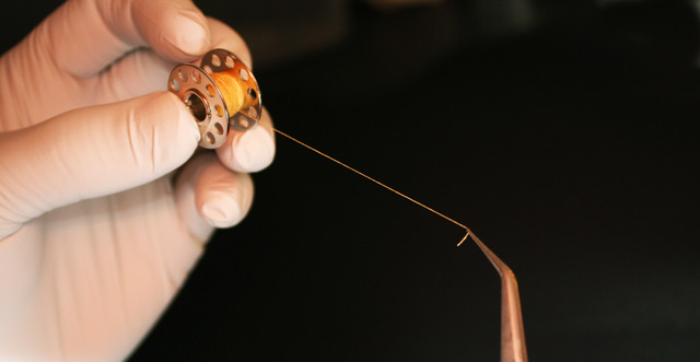
The researchers use extracellular matrix sheets to make yarn – a bit like that used to make clothing fabric. ©Nicolas L’Heureux

The leading cause of mortality worldwide, cardiovascular diseases claim over 17 million lives each year, according to World Health Organization estimates. To open up new research avenues into this serious public health problem, Inserm researcher Nicolas L’Heureux and his team at the Tissue Bioengineering unit (Inserm/Université de Bordeaux) are developing “human textiles” from collagen in order to repair damaged blood vessels. An innovation described in the journal Acta Biomaterialia, which still has to pass through several stages before it can be tested in humans.

What if we could replace a patient’s damaged blood vessels with brand new ones produced in a laboratory? This is the challenge set by Inserm researcher Nicolas L’Heureux, who is working on the human extracellular matrix – the structural support of human tissues that is found around practically all of the body’s cells.

In a study published in Acta Biomaterialia, L’Heureux and his colleagues at the Tissue Bioengineering unit (Inserm/Université de Bordeaux) describe how they have cultivated human cells in the laboratory to obtain extracellular matrix deposits high in collagen – a structural protein that constitutes the mechanical scaffold of the human extracellular matrix. “We have obtained thin but highly robust extracellular matrix sheets that can be used as a construction material to replace blood vessels”, explains L’Heureux.


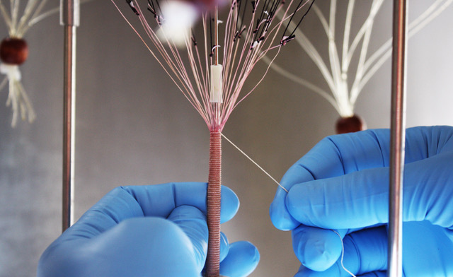
Made entirely from biological material, these blood vessels would have the advantage of being well tolerated by all patients.© Nicolas L’Heureux

The researchers then cut these sheets to form yarn – a bit like that used to make fabric for clothing. “The resulting yarn can be woven, knitted or braided into various forms. Our main objective is to use this yarn to make assemblies which can replace the damaged blood vessels”, adds L’Heureux.

Made entirely from biological material, these blood vessels would also have the advantage of being well-tolerated by all patients. Given that collagen does not vary from individual to individual, it is not expected that the body will consider these vessels as foreign bodies to be rejected.

The researchers would now like to refine their techniques used to produce these “human textiles” before moving on to animal testing, in order to validate this last hypothesis. If these are conclusive, this could lead to clinical trials.

Malaria: Vaccine clinical trial for Pregnant Women yields promising results

©Benoît Gamain. Gestational malaria is associated with low birth weight for the baby and an over-risk of neonatal mortality.

Malaria infection during pregnancy represents a major public health problem in the regions endemic for the disease, substantially increasing the risks to mothers and their unborn children. For newborns, malaria is linked to low birth weight and an excess risk of mortality. To protect this population, a team of researchers from Inserm and Université de Paris led by CNRS Research Director Benoît Gamain is developing a vaccine at the French National Institute of Blood Transfusion (INTS). Called PRIMVAC, the vaccine has undergone a clinical trial to study its safety and collect preliminary data on its ability to induce an immune response. The results of this clinical trial sponsored by Inserm[1] have been published in the prestigious journal Lancet Infectious Diseases.

According to the World Health Organization, malaria is responsible for over 400,000 deaths each year. Despite the progress made in fighting the disease in recent decades, some populations remain particularly vulnerable. One such population is pregnant women.

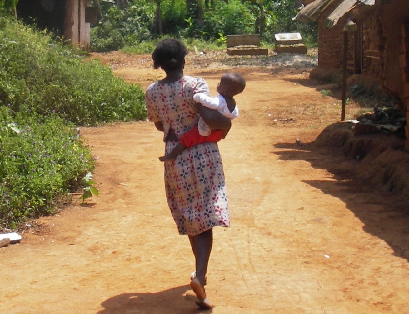
In the areas of the world where malaria is endemic, people acquire immunity throughout their childhood, meaning that they are generally protected against its most severe outcomes once they reach adulthood. However, pregnant women are an exception because the red blood cells infected with the Plasmodium falciparum parasite responsible for malaria accumulate in the placenta, promoting anemia and gestational hypertension. The disease is also linked to a higher risk of spontaneous abortion, premature birth and intrauterine growth delays which lead to low birth weight and a high rate of neonatal mortality. In Sub-Saharan Africa, 11 million pregnant women were infected with malaria in 2018, with around 900,000 of their babies born underweight.

To tackle this public health problem, a team of researchers from Inserm and Université de Paris led by CNRS Research Director Benoît Gamain has spent the past two decades developing a vaccine for gestational malaria. The goal is to prevent the deaths of up to 10,000 mothers and 200,000 babies each year. “Developing an effective vaccine for young women before their first pregnancy is a priority if we are to reduce malaria-related mortality. An effective strategy could focus on a population similar to that targeted by HPV vaccination, for example, before the women become sexually active”, emphasizes Benoît Gamain.

A safe and effective vaccine

Called PRIMVAC, the vaccine had recently been produced in large quantities in accordance with current regulations. In a clinical trial published in Lancet Infectious Diseases, the researchers provide data on its safety and ability to induce an appropriate immune response, up to 15 months after the initial vaccination.

The vaccine was evaluated in 68 non-pregnant women aged 18 to 35 at the Cochin Pasteur Clinical Investigation Center in Paris, then at the National Center for Research and Training on Malaria (CNRFP) in Ouagadougou, Burkina Faso. The participants were randomly assigned to 4 cohorts, receiving the vaccine at various doses, on 3 occasions over a period of 3 months. These women were then monitored for 15 months in order to identify and treat any side effects and study the immune response induced by the vaccination.

Antibody (green) of a vaccinated volunteer binding to the surface of a human red blood cell infected with the Plasmodium falciparum parasite (blue). Credits: Inserm/Chêne, Arnaud et Semblat, Jean-Philippe

The results of this study show that PRIMVAC is well tolerated. In addition, the researchers have shown that vaccine can produce an immune response, with the production of antibodies in 100% of women vaccinated after only two injections. The antibodies produced are capable of both recognizing the parasitic antigen on the surface of the infected red blood cells and inhibiting their adhesive capacity, which is responsible for their accumulation in the placenta.

“We were able to show that the vaccine is well tolerated, at all the tested doses. The side effects observed were mainly pain at the injection site. We also revealed that the quantity of antibodies generated by the vaccine increases after each vaccination and that they persist for several months. It therefore appears that the vaccine has the capacity to trigger a lasting and potentially protective immune response”, underlines Gamain.

Studying this immune response on the longer term will be the subject of future clinical trials. The researchers want to continue monitoring the 50 Burkinabe volunteers in order to evaluate whether the immune response induced by the vaccination is maintained until their first pregnancy.


[1] The trial was coordinated by the Cochin Pasteur Clinical Investigation Center in Paris and the EUCLID/F-CRIN clinical trials platform in Bordeaux in collaboration with the National Center for Research and Training on Malaria (CNRFP) in Ouagadougou, Burkina Faso, and the European Vaccine Initiative (EVI). Funding: Federal Ministry of Education and Research, through the development bank KfW, Germany; Inserm, and National Institute of Blood Transfusion (INTS), France; Irish Aid, Department of Foreign Affairs and Trade, Ireland.

fermer