Menu

Understanding Zebrafish Fin Regeneration Opens up Avenues in Regenerative Medicine

zebra fish _ poisson zèbre

The zebrafish (Danio rerio) is a tropical species commonly used as a model organism in research laboratories. © Adobe Stock


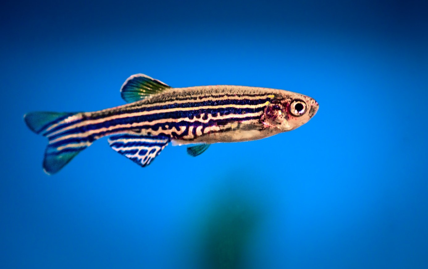
In the animal kingdom, several species share the extraordinary ability to regenerate their limbs or appendages following amputation. One of them is the zebrafish, which is particularly studied in research laboratories due to its ability to regenerate its caudal fin. This phenomenon is made possible by the formation of a blastema, a transient structure composed of undifferentiated cells, which initiates and controls the regeneration of the tissue. Improving our understanding of the cells that make up the blastema and deciphering their interactions opens up new avenues for improving our understanding of the regeneration processes, with the aim of developing clinical applications in the field of regenerative medicine. In a study published in Nature Communications, scientists from Inserm and Université de Montpellier have taken one step closer to this objective, by identifying within the blastema the cell population that coordinates the regeneration process in zebrafish.

The zebrafish, also known as Danio rerio, is a tropical species which has been commonly used as a model organism in research laboratories since the late 1990s. It offers many advantages to scientists, such as the transparency of the embryo and its external development, which is easier to observe than that of mammals. In addition, humans and zebrafish share 70% of the same genes. This genetic conservation with the other vertebrates makes Danio rerio a model of choice for deciphering several major biological processes and their conservation over the course of evolution.

Surprisingly, the zebrafish is also able to regenerate its caudal fin when amputated, thanks to the transient formation of a cell mass known as a “blastema”.

At the larval stage, this structure ensures regeneration of the sectioned appendage in only three days. This is enough to attract the interest of the scientific community, given that understanding the mechanisms associated with this process could pave the way for multiple regenerative medicine applications.

However, only a few cells of the blastema had been described until recently, with the underlying biological mechanisms poorly documented. In their previous work, Inserm Research Director Farida Djouad and her team had highlighted the unique role of macrophages – immune system cells – during the formation of zebrafish blastemas. The team had thus showed that macrophages coordinate the inflammatory processes necessary for the proliferation of the blastema cells and the regeneration of the caudal fin.


Identify the cell coordinating the regeneration

In their new study, these researchers went further in exploring the blastema and revealed the major involvement of a new cell population – cells derived from the neural crest[1]. These cells are found in all vertebrates, including in humans, and play a key role in embryonic development.

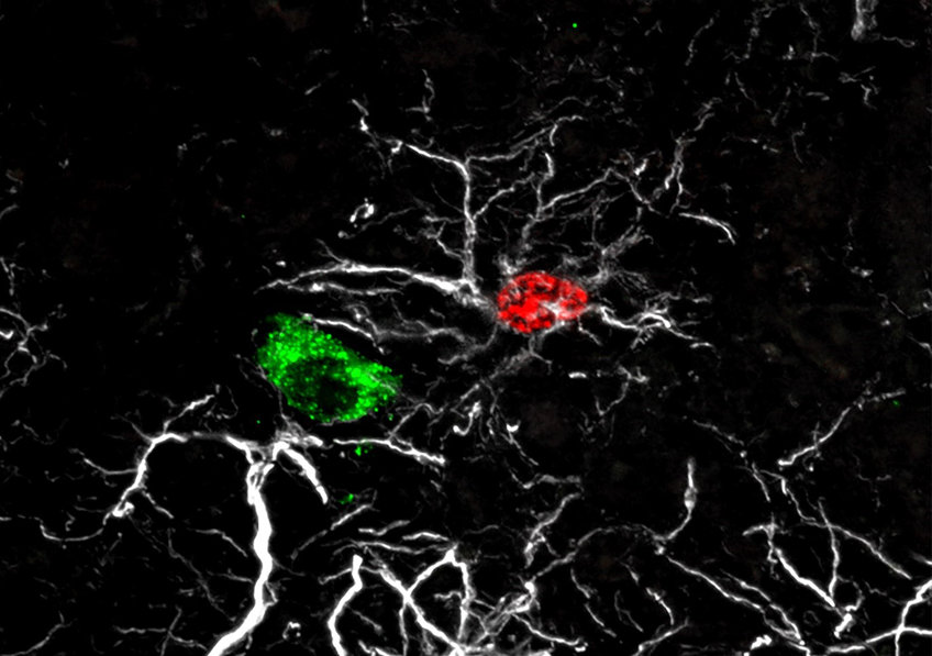
The scientists deployed several methodological approaches to observe and monitor the fate of the blastema cells. By combining the applications of real-time confocal imaging and single cell RNA-sequencing technology[2] on zebrafish larvae, the Montpellier-based team was able to demonstrate that cells derived from the neural crest coordinate the fin regeneration process, dialoguing with macrophages and other cells in the blastema in order to control and regulate their response. This dialog is conducted via a key factor called NRG1 (Neuregulin 1).

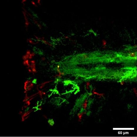
Interactions between the macrophages (in red) and neural crest cells (in green) during regeneration of the zebrafish larva caudal fin. © Farida Djouad

All of these data make it possible to go further in understanding the regeneration processes and their activation in zebrafish. Based on these findings, the next objective will be to understand why mammals, which also possess macrophages and cells derived from the neural crest, fail to regenerate their appendages.

We are continuing this work on other vertebrate models, including mice, in order to better understand when mammalian embryonic development loses this regeneration capacity, and why, whilst focusing on the role of cells derived from the neural crest,” says Djouad.

“The aim of this research on several animal models capable of regenerating is to identify ‘THE’ coordinating cell, which is common to all regeneration processes. A better understanding of its role, and especially of the factors it secretes, could open up new avenues to promote the regeneration of certain tissues in the treatment of degenerative diseases such as osteoarthritis.”


[1] The neural crest of vertebrates is a transient embryonic structure, involved in development, and capable of producing many tissues of the face and skull, particularly the cartilaginous and osteomembranous skeleton, the meninges, the vascular walls of the external and internal carotid system, the dermis… Source: French Academy of Medicine

[2] Single-cell sequencing is based on a set of molecular biology methods to analyze genetic information (DNA, RNA, epigenome…) on a single cell scale.

Creation of a Scientific Interest Group: A French Reference for All Questions Regarding the 3Rs

The use of animals for scientific purposes is based on the principles of the 3Rs (Replacement, Reduction and Refinement) to provide the best guidance to research and promote responsible and innovative methods. A group of research players comprising Inserm, CNRS, INRAE, Inria, CEA, Institut Pasteur de Paris, CPU, and the association Udice has announced the creation in France of FC3R (France Center 3R) to support the application of the rules resulting from these principles. Founded at the request of the country’s Ministry of Higher Education, Research and Innovation and taking the form of a Scientific Interest Group (GIS) endowed with substantial missions and resources, this structure aims to be recognized in France and Europe as a reference and contact point for all 3R-related questions, in both public and private research.


Structure and composition

Founded by major public research operators1, FC3R will promote synergy and collaboration between the existing entities in the use of animals for scientific purposes, mobilizing the various competences available in order to fulfil its missions. It will operate in close collaboration with the Ministry of Higher Education, Research and Innovation and the Ministry of Agriculture and Food, and will interact with partners in the private sphere.

FC3R will be structured around an initial team of five (director, secretary/administrator, webmaster, and individuals in charge of training and calls for projects, respectively) and it will have its own steering committee, scientific committee, and advisory board/think-tank. It will be attentive to the various stakeholders, especially the associations.

FC3R will be based at the National Veterinary School of Alfort, in Maisons-Alfort, where it will have at its disposal an operational structure and receive strong support from this major player in animal biology education for its training missions.


Main missions

Training in the 3Rs

    • Make all 3R training opportunities known thanks to a website that lists the existing opportunities in French and English,

    • Promote the emergence of training by encouraging its partners, whether private or public, to create courses in areas of insufficient provision,

    • Develop remote training tools (webinars, e-learning, MOOC, etc.),

    • Provide innovative content: create training modules on emerging approaches or that promote the harmonization of existing approaches,

    • Develop international openness: make its courses known and recognized in Europe, thereby contributing to the recognition of French training.


Engineering of 3R-compliant projects

  • Assist with project design: become a point of contact and advice,

  • Process unpublished data of relevance to the 3Rs: create a platform for the submission of negative results (on the FC3R website), for better dissemination of the knowledge obtained but not published by the authors.


Funding of 3R projects

  • Hold an annual call for projects to develop and validate research methods improving respect of the 3Rs,

  • Hold calls for projects on specific 3R-related issues in order to fund the best projects for the development and validation of innovative approaches, involving any public or private partners,

  • Fund 3R Prizes.


 Communication

  • Implement specific communication following the motto “For responsible and innovative research”,

  • Create a website and a social media communication strategy: disseminate relevant information on the correct application of the 3Rs and on FC3R-specific activities, primarily for the research sphere, but also civil society.


10-year objectives

  • Bring about a significant reduction in the number of animals used in experimentation, particularly through longitudinal multimodal approaches, ensure better replacement by invertebrate models or complementary approaches, particularly in vitro, and ensure refinement based on innovative practices,

  • Ensure training in rigorous and responsible practice, in accordance with the 3Rs, of all students and/or newcomers required to use animals for scientific purposes,

  • Position FC3R as a key player in France and Europe in the development of alternative methods to the use of animals and on other issues relating to the 3Rs.


1 The founding members of the GIS are Inserm, CNRS, INRAE, INRIA, CEA, Institut Pasteur de Paris, CPU and Udice.

The Impacts on Reproductive Function of Early Exposure to Endocrine Disruptors

Astrocyte né pendant la période infantile

Astrocyte born during the infancy period (red) (7 to 20 days after birth) adhering to a GnRH neural cell body (green). The astrocytes’ processes are shown in white. © Vincent Prévot, Inserm.


Researchers from Inserm, Lille University Hospital and Université de Lille, at the Lille Neuroscience and Cognition laboratory, have discovered one of the mechanisms by which endocrine disruptors can alter reproductive function development from birth. At the neural level, they saw in animals how exposure to low doses of bisphenol A – a known endocrine disruptor – a few days after birth disrupts the integration of GnRH neurons within their neural circuit and alters their reproductive function regulation activity. The findings of this study have been published in Nature Neuroscience.

In mammals, reproduction is regulated by the GnRH neurons, a population of neurons that, during embryonic development, appears in the nose and then migrates to the hypothalamus in the brain. Being well established in the brain at birth, these neurons go on to control the various processes associated with reproductive function: puberty, acquisition of secondary sexual characteristics, and fertility in adulthood.

To perform their functions, the GnRH neurons must be surrounded by another type of neural cell: the astrocytes. The adhesion of the astrocytes to the GnRH neurons plays a decisive role in their integration within the neural network. The encounter between these two cell types takes place during the so-called “mini-puberty” period that begins one week after birth in mammals, when the GnRH neurons are first activated, and which is when the first sex hormone secretions occur.

“Failure of GnRH neurons to integrate during mini-puberty may lead to a predisposition to developing puberty and/or fertility disorders, and also potentially affect brain development, thereby leading to learning disorders or metabolic disorders, such as being overweight,” explains Vincent Prévot, Inserm Research Director and last author of the study.

But how does this encounter between GnRH neurons and astrocytes take place? According to the findings of this research, the astrocytes do not get there by chance but respond to molecular signals emitted by the GnRH neurons, which recruit them as soon as they appear in the hypothalamus.

Early bisphenol A exposure prevents communication between GnRH neurons and astrocytes

Going further, the researchers wanted to understand the importance of this meeting between astrocytes and GnRH neurons in the development of mammalian reproductive functions during the mini-puberty period. With recent studies having shown that the GnRH neural network is particularly sensitive to endocrine disruptors and that there is a link between the latter and puberty disorders, the researchers investigated the impact of exposure to one of these endocrine disruptors, bisphenol A, in rats.

Bisphenol A is a compound used in the industrial manufacturing of plastics whose endocrine disruptive nature is now widely recognized. It is used for example in the production of food containers such as drinks bottles and baby feeding bottles. It is also found in the protective linings of drinks cans and tinned food, or on till receipts where it is used as a developer. In France, bisphenol A has been banned from use in baby feeding bottles and other  food containers since 2015. It has been replaced with substitutes such as bisphenol S (BPS) and bisphenol B (BPB) which also raise questions regarding their safety.

“Despite its ban, bisphenol A continues to remain present in our environment due to the slow degradation of plastic waste, and also because people are still using food containers they had purchased before 2015. With the recycling of waste, bisphenol A from plastics produced before 2015 has also found itself in new products,” explains Prévot.

During the 10 days following their birth, the female rats received low-dose injections of bisphenol A. Using an astrocyte labelling technique, the researchers saw that, under the effect of bisphenol A, the astrocytes were unable to permanently adhere to the GnRH neurons. The absence of such a phenomenon

occurring between these nerve cells then led to delayed puberty and the absence of estrous cycles in the adult female rats (equivalent to the menstrual cycle in women), suggesting that reproductive functions are affected.

Our findings suggest that early exposure to chemicals in contact with food, such as bisphenol A, can disrupt the onset of puberty and have a lasting impact on reproductive functions, by preventing GnRH neurons from building an appropriate and necessary environment in the hypothalamus for their role as fertility coordinator,” explains Ariane Sharif, lecturer at Université de Lille, who co-led the study.

Taking this research further, the scientists are now seeking to understand the exact mechanism by which bisphenol A prevents communication between GnRH neurons and astrocytes. One hypothesis is that bisphenol A acts directly on the astrocytes’ receptors, preventing them from adhering to the GnRH neurons. The research team is also interested in the action of bisphenol A on DNA and the traces it may leave.

Child Physical Abuse: Standardizing Guidelines to Optimize Diagnosis

In developed countries, 4% to 16% of children under 18 are physically abused. © Adobe Stock


Diagnosing physical abuse in infants can be difficult. To assist health care professionals, guidelines defining what to do in case of suspected physical abuse have been produced by learned societies, public authorities, and independent expert groups. Despite this reference framework, differences in management have been observed between practitioners, both in France and in other developed countries. To try to understand this phenomenon, researchers from Inserm and teacher-researchers from Université de Paris at the Center for Research in Epidemiology and Statistics (CRESS), the Paris hospitals group AP-HP and Nantes University Hospital analyzed and compared different clinical guidelines published in 15 countries. They have discovered substantial differences between countries and even within countries where several reference documents sometimes coexist. The findings of this systematic review highlight the need for an international consensus process in order to produce clear and standardized guidelines to optimize the practices of health care professionals. These findings have been published in JAMA Network Open.

Physical abuse affects 4% to 16% of children under the age of 18 in developed countries and around one third of diagnoses of such abuse are made late. Infants under two years of age are the most affected, which adds to the difficulties of rapid diagnosis based on a combination of clinical and social assessments, imaging examinations, and laboratory tests.

Several previous studies revealed discrepancies in the practices of health care professionals when physical abuse is suspected. A French study published in 20151 showed, for example, that in the theoretical case of a 9-month-old infant presenting with femoral fracture, multiple bruising, and a head injury, only 28% of the pediatricians surveyed would have requested a skull MRI, despite it being recommended following a CT scan within this context of potential inflicted injury.

Faced with this observation, and in an attempt to understand these differences between practices, the study looked at guidelines intended to assist health care professionals in diagnosing child physical abuse. These guidelines, accessible online, were produced by learned societies, health agencies, and pediatric expert groups, and include documents written by the French National Authority for Health (HAS), the UK Royal College of Paediatrics and Child Health, and the American Academy of Pediatrics.

The scientists extracted the data from 20 reference documents published between 2010 and 2020, by 15 of the 24 most developed countries according to the United Nations. They compared the diagnostic examination guidelines in the event of suspected child abuse, determining for each examination whether it was routinely recommended, conditional (on a case-by-case basis depending on the clinical context), not recommended, or not even mentioned.


Discrepancies between reference documents that create gray areas

According to the study findings, the international guidelines vary widely, which could explain some of the differences between the clinical practices observed in the diagnosis of child physical abuse.

While some examinations, such as skeletal X-rays, are recommended across the board, others are recommended on a case-by-case basis or not at all, depending on the country. For example, a bone scan – an imaging examination used to look for fractures in addition to skeletal X-rays – is recommended by the HAS and the American Academy of Pediatrics but not by the Royal College of Radiologists. Within the UK, the Royal College of Radiologists and the Royal College of Paediatrics and Child Health differ in their recommendation for the use of this scan.

The researchers also saw significant differences in the definition of “sentinel injuries,” – injuries found in infants who have not acquired the ability to move on their own and which require assessment for physical abuse. While all the documents analyzed propose a description of these injuries, the content differs: six give a brief description and focus solely on skin lesions, while ten also refer to intra-oral, intracranial and abdominal injuries, and fractures.

Finally, the researchers found that some of the elements frequently mentioned in the guidelines did not appear in all of them. For the researchers, even investigations that are not recommended should be mentioned in all of the documents, if only to explain to the practitioner why they are not recommended.

“Our objective was to systematically examine the completeness, clarity and consistency of the clinical guidelines for the early detection and diagnostic work-up of child physical abuse. We felt that, in the developed countries, the authors of guidelines theoretically all have access to the same knowledge and that all practitioners have the possibility to prescribe the same examinations. As such, there should be little variation between the examinations recommended for suspected child physical abuse and those actually carried out. We did not expect to identify such great disparities between the guidelines. We believe that these differences may partially explain the absence of the standardization of practices,” explains Flora Blangis, first author of the study and doctoral student at Inserm.

“Our objective was to systematically examine the completeness, clarity and consistency of the clinical guidelines for the early detection and diagnostic work-up of child physical abuse. We felt that, in the developed countries, the authors of guidelines theoretically all have access to the same knowledge and that all practitioners have the possibility to prescribe the same examinations. As such, there should be little variation between the examinations recommended for suspected child physical abuse and those actually carried out. We did not expect to identify such great disparities between the guidelines. We believe that these differences may partially explain the absence of the standardization of practices,” explains Flora Blangis, first author of the study and doctoral student at Inserm.

According to the researchers behind this systematic review, the findings highlight the need for an international consensus process in order to produce clear and standardized guidelines to optimize the practices of health care professionals in the diagnosis of child physical abuse. Such an approach has been carried out in many other areas of medicine, particularly for the screening of hypertension.

Primary care physicians and pediatricians are key players in the early detection and diagnosis of child physical abuse. They should be able to base their decisions on guidelines that are clear, comprehensive, and consistent, as is the case with other diseases,” concludes Blangis.

Standardizing practices2 is an issue that is all the more important given the serious consequences that incorrect diagnosis can have on a child’s health. In particular, false negative results expose infants to a risk of recurrence of abuse estimated as being between 35% and 50%.


1 Ledoyen A, Bresson V, Dubus J-C, et al. Explorations complémentaires face à une situation d’enfant en danger : état des lieux des pratiques en France en 2015. Arch Ped. 2016

2 Deployment throughout France of Pediatric Reception Units for Children at Risk (UAPED) should help to standardize practices in the diagnosis of abuse and the care pathway for abused minors. This is a strong focus of the plan to combat violence against children decided upon by the Ministry of Solidarity and Health and supported by the French Society for Forensic Pediatrics.

Lancement du Groupement d’intérêt scientifique Obépine sur les eaux usées

Myeloid Leukemia: Understanding Treatment Resistance to Move Towards Personalized Medicine

mitochondries © Adobe Stock

The patients who best respond to the dual therapy in this study have a “Mitoscore signature” associated with strong mitochondrial activity. © Adobe Stock


While the care and treatment of acute myeloid leukemia (AML) have greatly improved in recent years, overall survival remains low. Resistance to the various treatments continues to present a major clinical challenge. Using animal models, and also by working with patients themselves, scientists from Inserm, CNRS and Université Toulouse III – Paul Sabatier at the Cancer Research Center of Toulouse have identified a new biomarker predictive of response to dual therapy (chemotherapy plus targeted therapy) used in AML, as well as resistance mechanisms behind relapses. The findings of this research have been published in Nature Cancer.

Leukemia groups several types of blood cancer that affect nearly 10,000 people each year in France. These include acute myeloid leukemia (AML), which affects the hematopoietic cells[1] in the bone marrow.

For a long time, intensive chemotherapy has been the treatment of choice for patients. Although the majority respond favorably and go into remission, overall survival in the longer term remains low, with certain resistant cancer cells persisting in the body following chemotherapy and leading to relapse.

In recent years, the development of targeted therapies has improved the treatment and response of patients, prolonging survival a little – particularly in elderly people ineligible for chemotherapy. However, even with these therapies, relapse remains a major issue. Understanding the mechanisms underlying resistance to leukemia treatments and finding a way to resolve them are a central focus of the work of Inserm researcher Jean-Emmanuel Sarry and his team at the Cancer Research Center of Toulouse (Inserm/CNRS/Université de Toulouse III – Paul Sabatier).

While most scientists working on the subject are more interested in the genetic mechanisms associated with resistance, Sarry’s team is studying the non-genetic mechanisms in order to understand why some patients are more likely to relapse.

Identification of a “Mitoscore signature”

In their new study, the researchers looked at a recently approved dual therapy (conventional chemotherapy combined with a new targeted therapy), which is increasingly used in the treatment of AML.

Using patient transcriptomes (i.e. all messenger RNA derived from genome expression), they show that people who respond best to the dual therapy and who have prolonged survival have a specific biomarker – a “Mitoscore signature” – that is associated with a high level of mitochondrial activity[2]. “In other words, this strong Mitoscore signature, which reflects a high level of mitochondrial activity, is predictive of an improved response to these treatments,” explains Sarry.

Finally, thanks to single-cell sequencing[3]of residual disease[4] following this dual therapy, the researchers observed a particular remodeling of mitochondrial function allowing cancer cells to adapt to therapies and induce the patient’s relapse. In mice, the team also showed that treatment based on a molecule that inhibits the action of the mitochondria makes it possible to block this mitochondrial function remodeling, prevent relapse, and prolong the animals’ survival.

“The objective is now to test this Mitoscore signature on very large cohorts in order to validate its utility. Ultimately, the idea would be to use this biomarker to improve patient follow-up and offer more personalized therapies – by giving dual therapy, possibly also with the mitochondrial inhibitor, for those likely to benefit from it. This research could therefore have a real clinical impact in the years to come,” explains Sarry.


1 Hematopoietic stem cells are made by the bone marrow and develop into the various blood cells: red cells, white cells, and platelets. Source INCa

2 Mitochondria are intracellular organelles whose role is to provide the cells with the energy they need. They therefore play a central role in cellular energy metabolism.

3 Single-cell sequencing is a set of molecular biology techniques used to analyze genetic information at single-cell level, using next generation sequencing technologies.

4 Residual disease is the persistence in tissue of malignant cells below the detection limit of conventional techniques.

Using Mechanical Tools Improves Our Language Skills


aires cérébrales liées au langage

The brain regions associated with language have increased during periods of technological boom, when the use of tools became more widespread. © Adobe stock


Our ability to understand the syntax of complex sentences is one of the most difficult language skills to acquire. In 2019, research had revealed a correlation between being particularly proficient in tool use and having good syntactic ability. A new study, by researchers from Inserm, CNRS, Université Claude Bernard Lyon 1 and Université Lumière Lyon 2 in collaboration with Karolinska Institutet in Sweden, has now shown that both skills rely on the same neurological resources, which are located in the same brain region. Furthermore, motor training using a tool improves our ability to understand the syntax of complex sentences and – vice-versa – syntactic training improves our proficiency in using tools. These findings could be applied clinically to support the rehabilitation of patients having lost some of their language skills. This study is published in November 2021 in the journal Science.

Language has long been considered a very complex skill, mobilizing specific brain networks. However, in recent years, scientists have revisited this idea.

Research suggests that brain areas, which control certain linguistic functions, such as the processing of word meanings, are also involved in controlling fine motor skills. However, brain imaging had not provided evidence of such links between language and the use of tools. Paleo-neurobiology[1] has also shown that the brain regions associated with language had increased in our ancestors during periods of technological boom, when the use of tools became more widespread.

When considering this data, research teams couldn’t help wondering: what if the use of certain tools, which involves complex movements, relies on the same brain resources as those mobilized in complex linguistic functions such as syntax?


Syntax exercises and use of tongs

In 2019, Inserm researcher Claudio Brozzoli in collaboration with CNRS researcher Alice C. Roy and their team had shown that individuals who are particularly proficient in the use of tools were also generally better at handling the finer points of Swedish syntax.

In order to explore the subject in greater depth, the same team, in collaboration with CNRS researcher Véronique Boulenger[1], developed a series of experiments that relied on brain imaging techniques (functional magnetic resonance imaging or MRI) and behavioral measurements. The participants were asked to complete several tests consisting of motor training using 30 cm-long pliers and syntax exercises in French. This enabled the scientists to identify the brain networks specific to each task, but also common to both tasks.

They discovered for the first time that the handling of the tool and the syntax exercises produced brain activations in common areas, with the same spatial distribution, in a region called the “basal ganglia”.


ganglions de la base

The handling of the tongs and the syntax exercises proposed to the participants produced activations in a region called “basal ganglia”. © Claudio Brozzoli


Cognitive training

Given that these two skill types use the same brain resources, is it possible to train one in order to improve the other? Does motor training with the mechanical tongs improve the understanding of complex phrases? In the second part of their study, the scientists looked at these issues and showed that this is indeed the case.

This time, the participants were asked to perform a syntactic comprehension task before and after 30 minutes of motor training with the pliers (see box for details of the experiment). With this, the researchers demonstrated that motor training with the tool leads to improved performance in syntactic comprehension exercises.

In addition, the findings show that the reverse is also true: training of language faculties, with exercises to understand sentences with complex structure, improved motor performance with the tool.

Motor training and syntax exercises

The motor training involved using the pliers to insert small pegs into holes that matched their shape but with differing orientations.

The syntax exercises which were completed before and after this training consisted of reading sentences with a simple syntax, such as “The scientist who admires the poet writes an article” or with a more complex syntax, such as “The scientist whom the poet admires writes an article.” Then the participants had to decide whether statements such as “The poet admires the scientist” were true or false. Sentences with the French object relative pronoun “que” are more difficult to process and therefore performance was generally poorer.

These experiments show that after motor training, the participants did better with the sentences that were considered to be more difficult. The control groups, which performed the same linguistic task but after motor training using their bare hands or no training at all, did not show such an improvement.

The scientists are now thinking about how to best apply these findings in the clinical setting. “We are currently devising protocols that could be put in place to support the rehabilitation and recovery of language skills of patients with relatively preserved motor faculties, such as young people with developmental language disorders. Beyond these innovative applications, these findings also give us an insight into how language has evolved throughout history. When our ancestors began to develop and use tools, this proficiency profoundly changed the brain and imposed cognitive demands that may have led to the emergence of certain functions such as syntax,” concludes Brozzoli.


[1] A field in which scientists study the evolution of our ancestors’ brain anatomy.

[2] Involved in these findings are the Lyon Neuroscience Research Center (Inserm/CNRS/Université Claude Bernard Lyon 1) and the Dynamics of Language laboratory (CNRS/Université Lumière Lyon 2).

Covid-19 : Le lourd impact de l’épidémie sur la santé mentale des étudiants, notamment pendant les périodes de confinements

Cohort study identifies genetic cause for rare form of diet-induced Cushing syndrome

Coupe de rein humain grossie 400 fois par un microscope à immunofluorescence polychromatique

Human kidney section magnified 400 times by a polychromatic immunofluorescence microscope. © Inserm/Oriol, Rafael


The team made up of researchers from the endocrinology and reproductive diseases department of Bicêtre AP-HP hospital, Inserm and Paris-Saclay University, carried out work, coordinated by Professor Peter Kamenický, to study the genetic cause of bilateral macronodular adrenal hyperplasia with diet-induced Cushing’s syndrome. This rare disease affects the two adrenal glands located above the kidneys and causes an overproduction of cortisol, a steroid hormone whose excess has harmful consequences for the body . Researchers were able to determine the molecular explanation for the occurrence of this disease 30 years after its initial description. This work was published on October 13, 2021 in the journal The Lancet Diabetes & Endocrinology .

This rare form of adrenal Cushing syndrome, studied by these researchers, is due to the abnormal expression of the GIP receptor (Glucose-dependent insulinotropic peptide) in both adrenal glands of patients. GIP is a hormone produced by the small intestine in response to the ingestion of food. In patients with this particular form of Cushing’s syndrome, cortisol levels increase abnormally after each food intake. Patients with this disease develop the typical clinical signs of Cushing’s syndrome such as weight gain associated with muscle atrophy, high blood pressure, diabetes mellitus, osteoporosis, and depression. The pathology is associated with an increase in mortality, especially cardiovascular causes.

In this international study involving researchers from six countries, and based in particular on close Franco-Quebec collaboration, the team reports that GIP-dependent macronodular hyperplasia of the adrenals, in both familial and sporadic forms, is a genetic disease caused by germline mutations of Lysine Demethylase 1A (KDM1A) with secondary loss of the second KDM1A locus, comprising the second copy of the gene, in adrenal tissue. KDM1A acts mainly as a transcriptional repressor (ie a regulator which prevents a gene from being expressed), the loss of its function results in deregulation of the expression of various genes in the adrenal tissue, including the GIP receptor but also of other receptors coupled to G proteins.

This discovery will make it possible to offer genetic counseling and earlier detection of this rare disease to patients and their relatives. Rare diseases are generally underdiagnosed. This is all the more important as the pathogenic variations of KDM1A also predispose to myeloma and other types of cancer.

In addition, this new role of KDM1A as an epigenetic regulator of the expression of the GIP receptor and other receptors coupled to G proteins could have pharmacological implications.

COVID-19: Artificial Intelligence Identifies Gene Signature Specific to Patients Suffering from Critical Forms

SARS-CoV-2

Covid-19: Intracellular observation of reconstituted human respiratory epithelium MucilAir™ infected with SARS-CoV-2. © Manuel Rosa-Calatrava, Inserm ; Olivier Terrier, CNRS ; Andrés Pizzorno, Signia Therapeutics ; Elisabeth Errazuriz-Cerda  UCBL1 CIQLE. VirPath (Centre International de Recherche en Infectiologie U1111 Inserm – UMR 5308 CNRS – ENS Lyon – UCBL1). Colorisé par Noa Rosa C.


What are the molecular and genetic characteristics that distinguish patients with critical forms of COVID-19 – and particularly acute respiratory distress syndrome (ARDS)? To answer this question, researchers from Inserm and Université de Strasbourg at Unit U1109 Molecular Immunology and Rheumatology, in collaboration with clinician-researchers at the Strasbourg University Hospitals, have investigated the biological and genomic data of a targeted cohort of young patients. Patients hospitalized in intensive care with ARDS were compared with COVID-19 patients hospitalized in a non-critical care ward.

As part of a Franco-US collaboration with researchers from the company Genuity Science in Boston and the University of Southern California in Los Angeles and using the most advanced artificial intelligence techniques to interpret these data, the scientists have succeeded in identifying a gene signature that differentiates these critical patients from their non-critical counterparts. Some of the genes included in this signature could ultimately become therapeutic targets for severe forms of COVID-19 or ARDS. The findings from this research have been published in Science Translational Medicine.

COVID-19 varies broadly from one patient to another. While some are asymptomatic, others develop flu-like symptoms. Then there are others who progress towards severe forms of the disease, in some cases developing acute respiratory distress syndrome (ARDS) that requires mechanical ventilation in an intensive care unit. Although this group of patients accounts for only a small proportion of those infected with the virus, its mortality rate is high – reaching around 25%.

While age and comorbidities such as diabetes and cardiovascular diseases are the main risk factors for developing these severe, potentially fatal, forms of COVID-19, scientists do not yet know why some younger and previously healthy patients also develop these forms. In molecular and genetic terms, what is it that distinguishes these patients with severe respiratory symptoms from the others?

There has been research into the subject since the start of the pandemic and some avenues have been identified, but so far each study addressed the question from a single methodological angle, generally focusing only on one aspect – genetic and metabolic factors, immune response parameters, etc.

Young patients with no comorbidities

The scientists from Inserm and Université de Strasbourg, in conjunction with the Strasbourg University Hospitals, were in this case interested in a patient cohort with restrictive and strict inclusion criteria. The patients had been hospitalized during the first wave of the pandemic, were under 50 years of age and had no major comorbidities. A total of 72 patients were recruited into two groups, one consisting of intensive care patients with ARDS and the other of less severe COVID-19 patients hospitalized in a non-critical ward. A “control” group of 22 healthy individuals was also studied.

“We chose to focus on a restricted but very well-defined patient cohort, excluding confounding factors such as age and certain diseases so that we could really study the molecular and genetic mechanisms directly associated with the severe forms, which are exclusively linked to viral infection and not to other pre-existing risk factors,” emphasizes Seiamak Bahram [1], last author of the study.

The scientists collected various samples in order to perform a multi-omics analysis –that is say retrieve and analyze the various genomic, proteomic, transcriptomic (investigation of all messenger RNA) data and other virological, immunological, and serological data from these patients. This allowed them to confirm that ARDS is associated with a major inflammatory state and an immune system surge (the so-called “cytokine storm”).

Using artificial intelligence

However, given the considerable mass of data generated as part of this multi-omics analysis, it was impossible to take the interpretation further without the help of artificial intelligence (AI). Thus, in collaboration with the AI Research Institute of Genuity Science [2] , a biotech in Boston (USA), the team was able to identify a network of 600 genes involved in the progression towards the critical forms of COVID-19, thanks to the cross-application of several AI algorithms (including one having run on the quantum computer made available by the University of Southern California in Los Angeles).

As part of this transatlantic collaboration, these large quantities of data were modelled and analyzed with the help of AI, making it possible to more accurately identify five genes that are overexpressed in these patients.

One of them, ADAM9, is a particularly interesting “driver gene,” with previous studies having shown that it interacts with SARS-CoV-2 proteins. The findings obtained here are consistent with that, suggesting that ADAM9 overexpression would “drive” some patients towards severe forms of COVID-19 and ARDS.

The researchers then conducted in vitro experiments which showed that blocking ADAM9 in cell lines is associated with a reduction in the quantities of SARS-CoV-2 in these cells, as well as reduced replication of the virus, thereby confirming not just its importance in critical disease but also its potential as a therapeutic target.

Of course, further studies will have to be carried out to confirm this last point, but the scientists believe that these findings have opened up an interesting therapeutic avenue, especially given the current clinical trials in oncology that are testing monoclonal antibodies which inhibit ADAM9. Therapeutic repurposing strategies could therefore be considered in the longer term.


[1] Professor Seiamak Bahram, a university professor and hospital practitioner, is the director of Inserm Unit 1109, head of the Strasbourg Precision Medicine Interdisciplinary Thematic Institute, and head of the Biological Immunology Department at Strasbourg University Hospitals.

[2] It has since become the company HiberCell

fermer