Researcher Contact
Stéphane Marinesco
Chercheur Inserm
Unité 1028 / UMR5292 "Centre de recherche en neuroscience de Lyon"
Equipe : "Recherche translationnelle et intégrative en épilepsie "
04 78 78 56 81
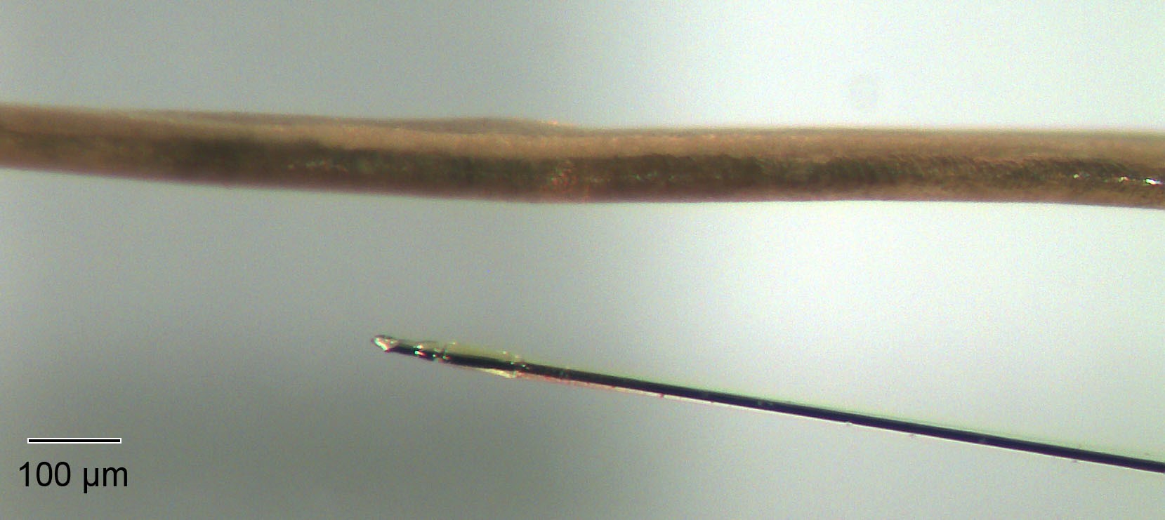
©Stéphane Marinesco / Inserm, Photograph of an implantable chemical sensor (bottom right) made with platinized carbon fiber and coated with a recognition enzyme, placed next to a human hair (top).
A team of Inserm and CNRS researchers has succeeded in developing new-generation chemical sensors to monitor the brain’s metabolism, particularly during stroke, trauma or epileptic seizure. Measuring less than 15 µm in diameter, these minimally-invasive tools monitor what is happening in the brain in order to obtain data that are much more reliable and representative of the neurochemical exchanges. This research has been published in ACS Central Science.
Analyzing the interstitial fluid of the brain can reveal important chemical information about the state of the latter. In the clinic or in laboratory animals, the ability to detect, over time, the levels of metabolites characteristic of brain energy (such as glucose) can help detect the onset of brain lesions, enabling doctors to act before it is too late. In addition, the activation of neuronal networks leading to a release of neurotransmitters can be detected in interstitial fluid. However, up until now the size of the probes and the local injury caused by their implantation were parameters which disrupted the quality of the measurements obtained. In particular, the rupture of small cerebral blood vessels during implantation represents a major trigger for inflammation. Within the first hour after implantation, local chemical brain tissue composition can be affected.
Marinesco clarifies that: “This minimally invasive device represents a major advance in our ability to analyze the brain interstitial fluid, paving the way for the measurement of new physiological parameters and multiple applications. This novel tool could be used to test the effect of certain medicinal products on the brain. Finally, in the longer term, monitoring the human brain could provide invaluable information to doctors in order to better understand how a patient with brain lesions recovers after a head injury or stroke. This device could also help them to take the best therapeutic decisions depending on the patient’s condition”.
Stéphane Marinesco
Chercheur Inserm
Unité 1028 / UMR5292 "Centre de recherche en neuroscience de Lyon"
Equipe : "Recherche translationnelle et intégrative en épilepsie "
04 78 78 56 81
Minimally Invasive Microelectrode Biosensors Based on Platinized Carbon Fibers for In Vivo Brain Monitoring
Charles Chatard a, b, e, f, Andrei Sabac c, d, e, Laura Moreno-Velasquez a, f, Anne Meiller b, f, and Stephane Marinesco a, b, f*
a INSERM U1028, CNRS UMR5292; Lyon Neuroscience Research Center - CRNL, Team TIGER, and b AniRA-Neurochem Technological Platform, Lyon, France
c CNRS UMR5270, Lyon Nanotechnologies Institute – INL, Villeurbanne, France
d CNRS UMR5005, Ampère Laboratory, Villeurbanne, France
e INSA de Lyon, Villeurbanne, France
f Université Claude Bernard Lyon 1, Lyon, France
ACS Central Science. doi: https://pubs.acs.org/doi/abs/10.1021/acscentsci.8b00797