Menu
Press releases

Diabetes: Study Of Satiety Mechanism Yields New Knowledge

Diabète

In purple, the tanycytes that form the brain’s cellular gateway to the hormone leptin; in yellow, the appetite-inducing neurons and, in blue, the appetite-suppressing neurons. Leptin targets both neuron types, inhibiting the former and using its appetite-suppressant signal to activate the latter. © Vincent Prévot


Diabetes, a disease in which blood sugar levels remain too high for too long, can lead to health complications in the long term. Type 2 diabetes (T2D) accounts for 90% of cases. Patients are usually obese or overweight, with risk factors that include sedentary lifestyle and unbalanced diet. To increase their understanding of the disease, a team of researchers from Inserm, Université de Lille, and Lille University Hospital in the Lille Neuroscience and Cognition laboratory[1] has for several years studied the role of leptin, a hormone involved in appetite control that sends satiety signals to the brain. In a new study published in the journal Nature Metabolism, in addition to furthering scientific knowledge of the mechanism of satiety, the scientists developed a new mouse model of diabetes that will be useful for and relevant to future research in this area.

Leptin, the satiety or appetite-suppressant hormone, is secreted by the adipose tissue at levels proportional to the body’s fat reserves and regulates appetite by controlling the feeling of fullness.  It is transported to the brain by tanycytes – cells which it enters by attaching to the LepR receptors. Tanycytes are therefore leptin’s gateway to the brain, helping it to cross the blood-brain barrier and deliver satiety information to the neurons.

Previous research has revealed that such transport is impaired in subjects who are obese or overweight. This goes some way to explaining their dysfunctional appetite regulation given that it is more difficult for the information on satiety to reach the brain. In their new study, the researchers took a closer look at this transport mechanism, and more precisely the role played by the LepR receptors.


The key role of satiety hormone receptors in glucose management

In mouse models, the researchers removed the LepR receptor that is located on the surface of the tanycytes. After three months, the mice experienced a marked increase in their fat mass (which doubled over the period) as well as a loss of muscle mass (reduced by more than half). The total amount of weight gained was only fairly moderate. The scientists also regularly measured the animals’ blood sugar levels following the injection of glucose.

They found that in order to maintain blood sugar at normal levels (between 0.70 and 1.10 g/L), the mice secreted more insulin during the first four weeks of the experiment. Three months after removing the receptor, their ability to secrete insulin from the pancreas appeared to be exhausted.

Removing the LepR receptors and impairing leptin transport to the brain therefore led the mice to initially develop a pre-diabetic state. This occurs when the body releases more insulin than usual in order to control blood sugar. Then, in the longer term, the mice became unable to secrete insulin and as such unable to control their blood sugar levels. These data therefore suggest that impaired leptin transport to the brain via the LepR receptors plays a role in the development of type 2 diabetes.

In a healthy animal or person, blood sugar levels rise slightly after the ingestion of glucose and then decrease rapidly. In order to bring blood sugar back to within normal limits, the pancreas secretes insulin that helps the glucose to penetrate the body’s cells. 

In animals deprived of the LepR receptor where leptin enters the brain, blood sugar levels are abnormally high in the fasting state and even more so after ingesting glucose. The pancreas becomes unable to secrete the insulin needed for the body to absorb the glucose. The brain’s “deafness” to the information conveyed by leptin thereby renders the pancreas ineffective.

In the last part of their research, the scientists reintroduced leptin to the brain and observed the immediate resumption of its pancreatic function-promoting action – particularly the ability of the pancreas to secrete insulin to regulate blood sugar. The mice quickly regained a healthy metabolism.

This study therefore elucidates the brain’s role in type 2 diabetes and also helps to further research into a disease that until then had not been considered to involve the central nervous system.

“We show that the brain’s perception of leptin is essential for the management of energy homeostasis[2] and blood sugar. We also show that blocking the transport of leptin to the brain impairs the functioning of the neurons that control pancreatic insulin secretion,” concludes Vincent Prévot, research director at Inserm and last author of the study.

Another interesting finding of this study: by removing the LepR receptor where leptin enters the brain, the animal model obtained exhibits the characteristics of so-called East Asian Diabetes, still little studied by researchers. This diabetes phenotype mainly affects the populations of Korea and Japan.

While so-called Western Diabetes is mainly associated with people who are markedly overweight (BMI >25) or morbidly obese (BMI >30), this other type 2 diabetes phenotype is often associated with people who are slightly overweight, have increased levels of abdominal fat, and insulin insufficiency related to deficient insulin secretion by the pancreas.

According to the scientists, the development of this new animal model will make it possible to further research into this disease that affects millions of people.


The research team started by describing the mechanism by which leptin passes through the cell gate: tanycytes (Figure opposite: cells in yellow). These cells capture circulating leptin from the blood vessels which at that location have the particularity of letting it through (step 1). Whilst in the tanycyte, the leptin captured by LepR activates the EGF receptor (or EGFR) which itself activates an ERK signaling pathway (step 2), triggering its release into the cerebrospinal fluid (step 3). The leptin then activates the brain regions that convey its anorectic (appetite suppressant) action, as well as control of pancreatic function (step 4).


[1] This research was performed in collaboration with two laboratories at Institut Cochin and Université de Strasbourg as part of a project funded by the French National Research Agency (ANR) and two European laboratories, one at Lübeck University in Germany and the other at the University of Santiago de Compostela in Spain, within the framework of European Community funding. In addition, the Lille Neuroscience and Cognition laboratory is a member of LabEx EGID (European Genomic Institute for Diabetes) and DISTALZ (Development of Innovative Strategies for a Transdisciplinary approach to ALZheimer’s disease).

[2] Stabilization, regulation in living organisms, of certain physiological characteristics (food intake, energy expenditure, etc.).

Medias
Researcher Contact

Vincent Prévot

Directeur de Recherche Inserm

Responsable de l’équipe « Développement et plasticité du cerveau neuroendocrine »

Unité U1172 – Lille Neuroscience & Cognition – Lille 

E-mail : rf.mresni@toverp.tnecniv

Téléphone sur demande

Press Contact

rf.mresni@esserp

Sources

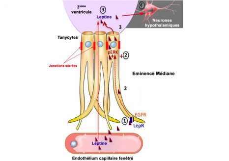
Leptin brain entry via a tanycytic LepR:EGFR shuttle controls lipid metabolism and pancreas function

Manon Duquenne1, Cintia Folgueira2#, Cyril Bourouh3#, Marion Millet4,#, Anisia Silva5#, Jérôme Clasadonte1#, Monica Imbernon1, Daniela Fernandois1, Ines Martinez-Corral1, Soumya Kusumakshi6, Emilie Caron1, S. Rasika1, Eleonora Deliglia1, Nathalie Jouy1,12, Asturo Oishi5, Massimiliano Mazzone7, Eric Trinquet8, Jan Tavernier9, Young-Bum Kim11, Stéphane Ory4, Ralf Jockers5, Markus Schwaninger10, Ulrich Boehm6, Ruben Nogueiras2, Jean-Sébastien Annicotte3, Stéphane Gasman4&, Julie Dam5&, Vincent Prévot1&*

1      Univ. Lille, Inserm, CHU Lille, Laboratory of Development and Plasticity of the Neuroendocrine Brain, Lille Neuroscience & Cognition, UMR-S1172, EGID, DISTALZ, F-59000 Lille, France

2    CIMUS, Universidade de Santiago de Compostela-Instituto de Investigación Sanitaria, Santiago de Compostela, 15782, Spain- CIBER Fisiopatología de la Obesidad y Nutrición (CIBERobn), 15706, Spain

3    Univ. Lille, Inserm, CHU Lille, Institut Pasteur de Lille, CNRS, U1283 – UMR 8199 – EGID, F-59000 Lille, France

4    Centre National de la Recherche Scientifique, Université de Strasbourg, Institut des Neurosciences Cellulaires et Intégratives, F-67000 Strasbourg, France.

5    Institut Cochin, Inserm U1016, CNRS UMR 8104, University Paris Descartes, Sorbonne Paris Cité, Paris, France

6      Experimental Pharmacology, Center for Molecular Signaling (PZMS), Saarland University School of Medicine, 66421, Homburg, Germany

7    Laboratory of Tumor Inflammation and Angiogenesis, Center for Cancer Biology, VIB, Department of Oncology, KU Leuven, Leuven, B3000, Belgium

8    Cisbio Bioassays, Parc Technologique Marcel Boiteux, BP84175, F-30200 Codolet, France

9    VIB-UGent Center for Medical Biotechnology, Gent, Belgium.

10  Institute for Experimental and Clinical Pharmacology and Toxicology, University of Lübeck, Lübeck, Germany

11    Division of Endocrinology, Diabetes, and Metabolism, Beth Israel Deaconess Medical Center and Harvard Medical School, Boston, MA, USA.

12    Flow core Facility, BioImaging Center of Lille, campus HU, UMS2014-US41, F-59000 Lille, France


# These authors contributed equally

& These authors jointly supervised this work

Nature Metabolism 2021

https://doi.org/10.1038/s42255-021-00432-5

fermer