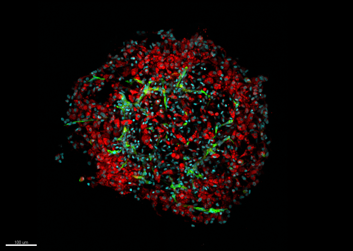
©STROMALab
Can human adipose tissue be reproduced in a laboratory? It can now, thanks to a research team with members from Inserm, CNRS, Université Toulouse III-Paul-Sabatier, the French Blood Establishment (EFS) and the National Veterinary School of Toulouse (ENVT) working together at STROMALab. This team used 3D culture to develop adipose tissue organoids (or adipospheres) – small cellular units that mimic the characteristics and organization of adipose tissue as it presents in vivo. In their article, published in Scientific Reports, the researchers describe the various stages of the experimental conditions needed to obtain these adipospheres from human cells. An innovation that could make it possible not just to study diseases related to the impaired functioning of this tissue, such as obesity and type 2 diabetes, but also to develop new drugs to treat them.
Human adipose tissue, highly vascularized by a network of capillaries, is made up of fat cells known as adipocytes. Until now, laboratory researchers used 2D models that did not take into account the 3D architecture of this tissue as found in the human body.
Mini organs, which are known as organoids and capable of reproducing the cellular organization of a specific organ, have already been developed for some tissues, such as that of the intestine. However, none had been able to reproduce in 3D the cellular and vascular organization of the adipose tissue in a laboratory setting.
However, this is now possible thanks to researchers from Inserm, CNRS, Université Toulouse III-Paul-Sabatier, the French Blood Establishment (EFS) and the National Veterinary School of Toulouse (ENVT) working together at STROMALab. Thanks to the advent of the new 3D cell culture methods, the control of the selection and characterization of adipose tissue stromal cells (support cells), the team was able to develop organoids of this tissue, called adipospheres.
Generating organoids in 3D
From these stromal cells of the human adipose tissue, the researchers developed new 2D – followed by 3D – culture conditions, making it possible to obtain both adipocytes and endothelial cells from this tissue. The adipospheres obtained contained an intact vascular network organized around adipocytes in the same way as in actual human tissue. Better still, the adipocytes obtained were capable of differentiating into those of brown or white tissue (the two types of human adipose tissue) in the same way as those encountered in the human body.
Transplantation in mice
The research team then transplanted these adipospheres into mice in order to verify the functionality of their vascular network. They observed that not only was this network maintained in the body but also that it extended itself by establishing connections with the host’s circulatory system.
The researchers also observed so-called chimeric vessels, constituted of both mouse and human cells. “These are all signs that the host tolerates the transplanted organoids well, explain Isabelle Ader, Inserm researcher, and Frédéric Deschaseaux, from the French Blood Establishment (EFS), authors of the study. From this we can conclude that not only are these small structures faithful to the organization of human tissue, but also that they are capable of staying alive by establishing connections with the host circulatory system that provides them with the necessary oxygen and nutrients.“
According to the researchers, this innovation will enable continued study of the functioning and properties of human adipose tissue. By working directly on this tissue, the use of animals will be reduced.
“This innovation will also make it possible to test various drugs that could be used to treat certain diseases related to a pathology of adipose tissue, such as obesity or type 2 diabetes“, conclude Isabelle Ader and Frédéric Deschaseaux.
These contents could be interesting :