
Researcher Contact
Soraya Taleb
Inserm Research Director
Unit 970 Inserm/Université Paris Cité – Paris-Center for Cardiovascular Research (Parcc)
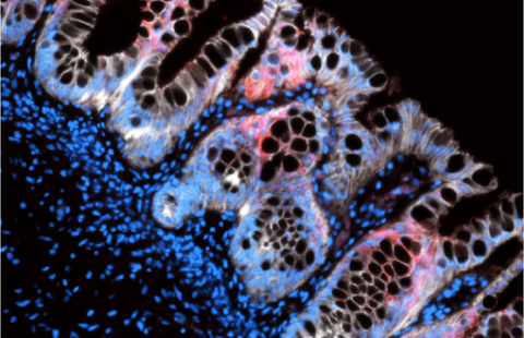
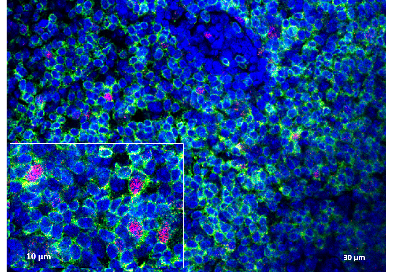 Visualization of immune cell (lymphocyte) proliferation in the mesenteric lymph nodes, under the influence of a microbiota modulated by a high-fat diet. © Soraya Taleb/PARCC
Although a high-fat, low-fiber diet is recognized as promoting cardiovascular diseases such as atherosclerosis, the mechanisms involved have not yet been fully identified. Researchers from Inserm and Université Paris Cité have studied the role of the gut microbiota in the development of atherosclerosis. Their work in mice reveals that the low fiber content of the high-fat diet leads to an imbalance in the gut microbiota, which itself causes systemic inflammation, worsening the development of atherosclerotic plaques in the arteries. These findings, published in Cell Reports, provide further evidence of the importance of fiber in the diet, both for good bowel function and for preventing the onset of cardiovascular diseases.
Cardiovascular diseases constitute one of the leading causes of death worldwide. Among them, atherosclerosis is characterized by the formation of an atherosclerotic plaque (atheroma), composed mainly of lipids, on the walls of the arteries. Over time, these plaques can cause damage to the arterial wall, obstruct the vessel, or rupture – often with serious consequences. Among the major risk factors for atherosclerosis is obesity, particularly when caused by a diet that is too high in fat and low in fiber. As such, it is not just diet but also its impact on the gut microbiota that are now avenues of interest for research into cardiovascular diseases.
A team led by Soraya Taleb, Inserm research director at the Paris-Center for Cardiovascular Research (Inserm/Université Paris Cité), looked at the influence of a high-fat, low-fiber diet on the gut microbiota of mice and how this could contribute to the development of atherosclerosis.
The researchers used a mouse model of diet-induced atherosclerosis to compare the effects of several diets on the metabolism, microbiota and development of atherosclerosis.
Unsurprisingly, in the mice fed a high-fat, low-fiber diet, their findings show an increase in metabolic risk factors linked to cardiovascular diseases (significant weight gain, hyperglycemia, insulin resistance, increased weight of the liver and its triglyceride content, etc.).
However, these are not the only effects observed of this diet, which also appears to be associated with an overall imbalance in the microbiota – in its composition and immune response –, reflected in the altered production of metabolic derivatives by its component bacteria. In particular, short-chain fatty acids, derived from the fermentation of fiber and recognized for their positive impact on health, are produced in smaller quantities.
However, this imbalance itself appears to be associated not only with metabolic risk factors but also with a worsening of the manifestations of atherosclerosis at vascular level, with an increase in atheromatous plaque size in the aorta as well as a systemic inflammatory phenomenon which results in an increase in the number of immune cells in these plaques. However, supplementation with fiber made it possible to counteract these effects.
“These findings show, in mice fed a high-fat diet, that a pathological gut microbiota accelerates the development of atherosclerosis,” comments Taleb. Our observations also show that, more than its high fat content, it is the small amount of fiber in this diet that causes the microbiotal imbalance and as such the worsening of the atherosclerosis. This further supports the idea of the essential role of fiber in structuring a healthy microbiota and in preventing systemic inflammatory diseases such as cardiovascular diseases”, she continues.
But how can we explain the surprising link that appears between the composition of the microbiota and the accumulation of immune cells in atheromatous plaques? In mice grafted with a gut microbiota initially modulated by a high-fat diet, the research team observed an increased proliferation of immune cells in the mesenteric lymph nodes[1], the site of their activation in the gastrointestinal tract.
Techniques used to track migration of the immune cells confirmed that it was indeed cells from the mesenteric lymph nodes that, after passing from the gut into the bloodstream, accumulated in atheromatous plaques, thereby contributing to the development of atherosclerosis.
“The fact that we have seen that immune cells are capable of migrating from the gut to the periphery and thereby generate systemic inflammation that worsens the atheromatous plaques adds a new dimension to our understanding of the link between diet, gut, microbiota and atherosclerosis,” explains Taleb. Additional work is needed in order to identify which of the bacteria in the microbiota are involved in this mechanism, in order to envisage targeted therapeutic approaches and study these mechanisms in humans”, concludes the researcher.
[1] The mesenteric lymph nodes are located in the mesentery, a fold of the peritoneum (the membrane lining the abdominal cavity and covering the abdominal organs) that suspends the small intestine from the posterior abdominal wall.
Soraya Taleb
Inserm Research Director
Unit 970 Inserm/Université Paris Cité – Paris-Center for Cardiovascular Research (Parcc)
An obesogenic diet increases atherosclerosis through promoting microbiota dysbiosis-induced gut lymphocyte trafficking into the periphery
Cell Reports, 31 octobre 2023
https://doi.org/10.1016/j.celrep.2023.113350
Ludivine Laurans,1 Nirmala Mouttoulingam,1,14 Mouna Chajadine,1,14 Aonghus Lavelle,2,3,14 Marc Diedisheim,4,5 Emilie Bacquer,1 Laura Creusot,2,3 Nadine Suffee,1,6 Bruno Esposito,1 Nada Joe Melhem,1 Wilfried Le Goff,6 Yacine Haddad,1,7,8 Jean-Louis Paul,9 Dominique Rainteau,3,10 Alain Tedgui,1 Hafid Ait-Oufella,1 Laurence Zitvogel,7,8,11,12 HarrySokol,2,3,13 and SorayaTaleb 1,15,*
1 Inserm, Université Paris Cité, Paris Cardiovascular Research Center, 75015 Paris, France
2 Sorbonne Université, Inserm, Centre de Recherche Saint-Antoine, CRSA, AP-HP, Saint-Antoine Hospital, Gastroenterology Department, 75012 Paris, France
3 Paris Centre for Microbiome Medicine (PaCeMM) FHU, Paris, France
4 Clinique Saint Gatien Alliance (NCT+), 37540 Saint-Cyr-sur-Loire, France
5 Institut Necker-Enfants Malades (INEM), Université Paris Cité, Inserm UMR-S1151, CNRS UMR-S8253, 75015 Paris, France
6 Inserm UMRS1166, ICAN-Institute of Cardiometabolism and Nutrition, Sorbonne Université, 75013 Paris, France
7 Gustave Roussy, Villejuif, France
8 Inserm, Gustave Roussy, UMR1015, Villejuif, France
9 Université Paris-Sud, Equipe d’Accueil 4529, UFR de Pharmacie, Chatenay-Malabry, France and Assistance Publique Hôpitaux de Paris, Hôpital Européen Georges Pompidou, Paris, France
10 Sorbonne Université, Inserm, Centre de Recherche Saint-Antoine, CRSA, AP-HP, Saint Antoine Hospital, Clinical Metabolomics Department, 75012 Paris, France
11 Université Paris-Saclay, Faculté de Médecine, Le Kremlin Bicêtre, France
12 Center of Clinical Investigations BIOTHERIS, Inserm CIC1428, Gustave Roussy, Villejuif, France
13 INRAE, Micalis & Agro Paris Tech, Jouy-en-Josas, France
14 These authors contributed equally
15 Lead contact
*Corresponding author