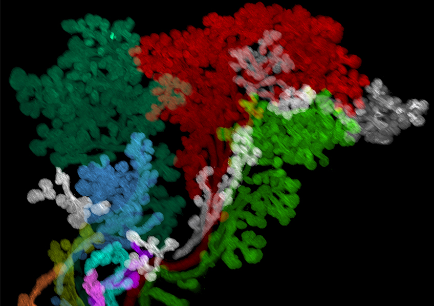
3D light-sheet microscope image of a lacrimal gland of a tissue-cleared 12-week-old human embryo. The different elements of the gland were coloured using virtual reality software. © Raphael Blain/Alain Chédotal, Vision Institute (Inserm/CNRS/Sorbonne Université)
Improving our knowledge of the development of the complex structures of the human head to shed new light on the congenital abnormalities that cause malformations: this is the challenge that a team of researchers from Inserm, CNRS and Sorbonne Université at the Vision Institute, Université Claude Bernard Lyon 1 and Hospices civils de Lyon is well on its way to fulfilling. Thanks to an innovative technique in which the skull structures are made transparent and 3D photos are taken of their component cells, this team has been able to establish the very first 3D atlas of the embryonic human head. These findings, to be published in Cell, have already provided deeper insights into how certain complex structures of the head are formed, such as the lacrimal and salivary glands or the arteries of the head and neck. They pave the way for new tools to study embryonic development.
The head is the most complex structure in the human body. In addition to the muscles and skin that protect it, and the brain encased within the skull, it contains blood vessels, nerves, endocrine glands – which secrete hormones directly into the bloodstream – such as the pituitary glands, and exocrine glands – which secrete substances to the outside environment – such as the salivary glands, which produce saliva, or the lacrimal glands, which secrete tears.
Our current knowledge about the development of the human head and its complex structures is rudimentary and comes from studies mostly carried out in the first half of the XX century, using simple histological sections. As such, despite head malformations occurring in around one third of newborns with congenital defects, the mechanisms that control the development of the human head remain poorly understood.
A team led by Alain Chédotal, Inserm research director at the Vision Institute (Inserm/CNRS/Sorbonne Université) and professor at the MéLiS laboratory of Mechanisms in Integrated Life Sciences (Inserm/CNRS/Université Claude Bernard Lyon 1/Hospices civils de Lyon), and Yorick Gitton, CNRS staff scientist also at the Vision Institute, used an innovative microscopy method to shed new light on the development of the human head.
The team had previously used the same technology in the embryo to study the development of other human organs[1]. This technology is called ’tissue clearing’ because it makes organs transparent to light. The cleared sample is then imaged in 3D using a special microscope that scans with a fine sheet of laser light. This makes it possible to locate in situ the cells that make up the embryonic tissues.
The researchers were able to apply this technique to embryos at different stages of development, obtained from the human tissue biobank created as part of the Human Developmental Cell Atlas (HuDeCA) programme coordinated by Inserm[2]. Thanks to the images obtained, they established the first 3D map of the embryonic human head[3].
Next, the team used virtual reality to analyse the 3D images and thus ‘navigate’ within the embryos.
‘This enabled us to discover previously unknown characteristics of the development of the cranial muscles, nerves, blood vessels and exocrine glands, states Chédotal. For example, it had never been possible to study the very early stages of development of the human salivary and lacrimal glands. Our research has enabled us to begin to visualise and better understand the mechanisms behind the establishment of these anatomically extremely complex structures’, he adds.
 3D light-sheet microscope image of a 12-week-old tissue-cleared human embryo eye. The 6 oculomotor muscles responsible for eye movement and the 3 motor nerves (in white, green and red) were coloured using virtual reality software. ©Raphael Blain/Alain Chédotal, Vision Institute (Inserm/CNRS/Sorbonne Université)
The scientists have also set up a web interface (hudeca.com) to access not only the images obtained in this research, but also models for 3D printing and interactive 3D reconstructions of human embryos. This platform provides valuable resources that can also contribute to the training of medical students.
In future research, the team will attempt to map the various cells of certain organs, such as the retina.
‘At this stage, it is kind of as if we have mapped the continents and countries but still have to position the cities and their inhabitants’, explains Chédotal, whose team will also collaborate with physicians to apply the technology to pathological samples.
‘The new knowledge of human embryology provided by this research, as well as the new tools developed, has major implications for understanding craniofacial malformations and neurological disorders, as well as for improving diagnostic and therapeutic strategies’, concludes the researcher.
[1] See our press release of 23 March 2017: https://presse.inserm.fr/en/the-human-embryo-as-you-have-never-seen-it/57363/
[2] Launched in 2019, the objective of the cross-cutting HuDeCa programme coordinated by Inserm is to build the first atlas of human embryonic and foetal cells. It also aims to structure human embryology research at French level and develop databases. In the longer term, HuDeCa is expected to serve as a basis for understanding the origin of chronic diseases or congenital malformations.
[3] With the specific exception of the brain, a structure that was not covered by this research.
These contents could be interesting :