Menu

A “Nano-Robot” Built Entirely from DNA to Explore Cell Processes

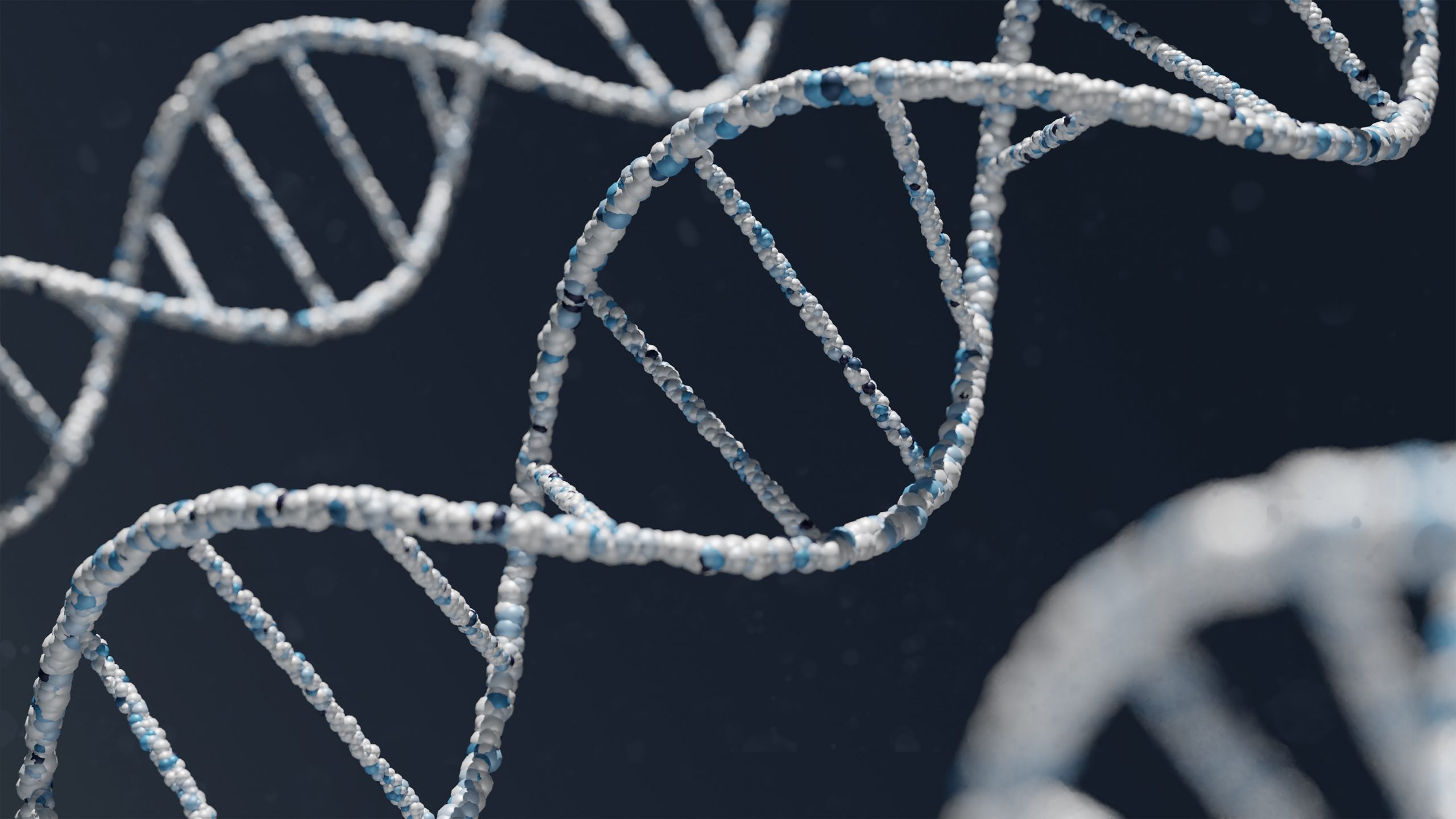
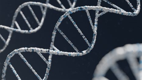
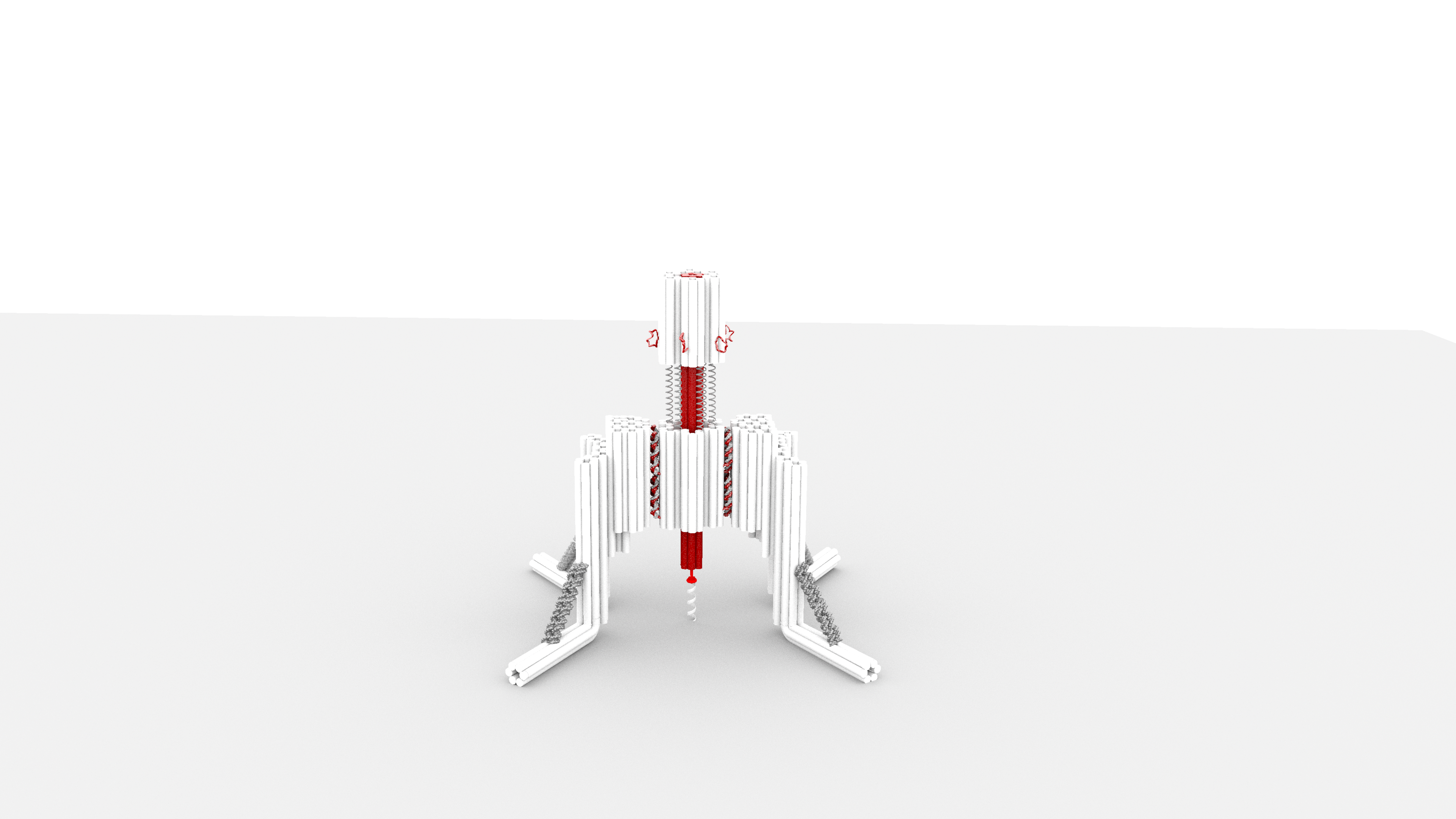
Scientists have designed a “nano-robot” made up of three DNA origami structures. © Gaëtan Bellot/Inserm

Constructing a tiny robot from DNA and using it to study cell processes invisible to the naked eye… You would be forgiven for thinking it is science fiction, but it is in fact the subject of serious research by scientists from Inserm, CNRS and Université de Montpellier at the Structural Biology Center in Montpellier[1]. This highly innovative “nano-robot” should enable closer study of the mechanical forces applied at microscopic levels, which are crucial for many biological and pathological processes. It is described in a new study published in Nature Communications.

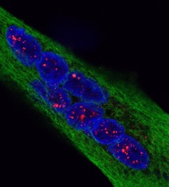
Our cells are subject to mechanical forces exerted on a microscopic scale, triggering biological signals essential to many cell processes involved in the normal functioning of our body or in the development of diseases.

For example, the feeling of touch is partly conditional on the application of mechanical forces on specific cell receptors (the discovery of which was this year rewarded by the Nobel Prize in Physiology or Medicine).

In addition to touch, these receptors that are sensitive to mechanical forces (known as mechanoreceptors) enable the regulation of other key biological processes such as blood vessel constriction, pain perception, breathing or even the detection of sound waves in the ear, etc.

The dysfunction of this cellular mechanosensitivity is involved in many diseases – for example, cancer: cancer cells migrate within the body by sounding and constantly adapting to the mechanical properties of their microenvironment. Such adaptation is only possible because specific forces are detected by mechanoreceptors that transmit the information to the cell cytoskeleton.

At present, our knowledge of these molecular mechanisms involved in cell mechanosensitivity is still very limited. Several technologies are already available to apply controlled forces and study these mechanisms, but they have a number of limitations. In particular, they are very costly and do not allow us to study several cell receptors at a time, which makes their use very time-consuming if we want to collect a lot of data.

DNA origami structures

In order to propose an alternative, the research team led by Inserm researcher Gaëtan Bellot at the Structural Biology Center (Inserm/CNRS/Université de Montpellier) decided to use the DNA origami method. This enables the self-assembly of 3D nanostructures in a pre-defined form using the DNA molecule as construction material. Over the last ten years, the technique has allowed major advances in the field of nanotechnology.

This enabled the researchers to design a “nano-robot” composed of three DNA origami structures. Of nanometric size, it is therefore compatible with the size of a human cell. It makes it possible for the first time to apply and control a force with a resolution of 1 piconewton, namely one trillionth of a Newton – with 1 Newton corresponding to the force of a finger clicking on a pen. This is the first time that a human-made, self-assembled DNA-based object can apply force with this accuracy.


The team began by coupling the robot with a molecule that recognizes a mechanoreceptor. This made it possible to direct the robot to some of our cells and specifically apply forces to targeted mechanoreceptors localized on the surface of the cells in order to activate them.

Such a tool is very valuable for basic research, as it could be used to better understand the molecular mechanisms involved in cell mechanosensitivity and discover new cell receptors sensitive to mechanical forces. Thanks to the robot, the scientists will also be able to study more precisely at what moment, when applying force, key signaling pathways for many biological and pathological processes are activated at cell level.

“The design of a robot enabling the in vitro and in vivo application of piconewton forces meets a growing demand in the scientific community and represents a major technological advance. However, the biocompatibility of the robot can be considered both an advantage for in vivo applications but may also represent a weakness with sensitivity to enzymes that can degrade DNA. So our next step will be to study how we can modify the surface of the robot so that it is less sensitive to the action of enzymes. We will also try to find other modes of activation of our robot using, for example, a magnetic field,” emphasizes Bellot.


[1] Also contributed to this research: the Institute of Functional Genomics (CNRS/Inserm/Université de Montpellier), the Max Mousseron Biomolecules Institute (CNRS/Université de Montpellier/ENSCM), the Paul Pascal Research Center (CNRS/Université de Bordeaux) and the Physiology and Experimental Medicine: Heart-Muscles laboratory (CNRS/Inserm/Université de Montpellier).

Lancement de la campagne de recherche participative Science à la Pelle : trouver des médicaments sous nos pieds

Significant Increase in Infant Mortality in France

In France, for the first time in peacetime, the infant mortality rate has risen significantly in the last ten years. ©Adobe Stock

The infant mortality rate (IMR) is a key indicator of population health. In the absence of updated data on the statistical trends of this indicator in France, researchers from Inserm, Université de Paris, the Paris public hospitals group (AP-HP) and Nantes University Hospital, in collaboration with teams from the University of California, analyzed civil registry data from the French National Institute of Statistics and Economic Studies (INSEE) from 2001 to 2019. They identified a significant increase in the IMR since 2012, thereby setting France apart from other high-income countries. The findings, published in The Lancet Regional Health – Europe, reflect the importance of more in-depth research into the precise causes of these 1200 excess deaths observed each year in France before one year of age.

The United Nations have made one of its priority objectives the elimination of preventable deaths in children by 2030. Given that the vast majority of child deaths occur during the first year of life, the infant mortality rate (IMR)1 is used to track progress towards this goal.

IMR serves as a key indicator of population health, given its strong relationship with the socio-economic development and quality of preventive and curative care in the country. In some high-income countries, such as Finland and Sweden, the IMR has been continuously decreasing since World War II. In other countries, such as France, this decrease appears to be slowing down.

Scientists from Inserm, Université de Paris, the Paris public hospitals group (AP-HP), Nantes University Hospital and the University of California wanted to go further in the statistical analyses of the evolution of the French IMR, and more specifically over the 2001 to 2019 period.

During this study period, the deaths of 53,077 infants were recorded for 14,622,096 live births, giving an average IMR of 3.63/1,000 (4.00 for boys, 3.25 for girls). Around one quarter of the deaths (24.4%) occurred during the first day of life and half (47.8%) in the early neonatal period – the first week following birth.

An in-depth statistical analysis identified two inflexion points, in 2005 and 2012 (see figure above). The IMR saw a sharp decrease from 2001 to 2005, and then a slower decrease from 2005 to 2012. From 2012, a significant 7% increase in the IMR was observed. This meant that infant mortality rose from 3.32 in 2012 to 3.56 deaths per 1,000 live births in 2019. Sensitivity analyses2 showed this trend to be unrelated to changes in registering practices or changes in medical practices for the management of newborns with serious conditions. Subgroup analyses showed this increase to be mainly due to an increased IMR in the early neonatal period.

Thanks to in-depth statistical analyses, we have identified a significant increase in the infant mortality rate in France since 2012. When comparing the data against other European countries with similar economies, such as Sweden and Finland, we observe that every year in France there is an excess of around 1,200 deaths of children under one year of age,” explains Prof. Martin Chalumeau, last author of the study. “It is essential to be able to explore in detail the causes of this increase by having, for example, systematic information on the specific medical and social circumstances of these deaths and by making this population, which is the most vulnerable, a real research and public health priority, which is not the case at present,” the researcher concludes.

1 Infant mortality rate (IMR) is defined as the number of deaths of children under one year of age (D0-D364) per 1,000 live births over a given period

2 Additional analyses to support the robustness of the main analyses

Consumption of ultra-processed food and risk of cardiovascular disease

©Photo Christopher Flowers / Unsplash

In an article published May 30, 2019 in the British Medical Journal, researchers from Inserm, Inra, Université Paris 13 and Cnam in the Nutritional Epidemiology Research Team (EREN) report an increased risk of cardiovascular disease in consumers of ultra-processed foods in the NutriNet-Santé cohort.

In recent decades, dietary habits have shifted towards an increased consumption of ultra-processed foods (see boxed text below), which currently account for over half of the total daily energy intake in many western countries. Such foods are often characterized by lower nutritional quality, in addition to the presence of additives, neoformed compounds and substances from packaging and other contact materials.

Recent studies have shown links between the consumption of ultra-processed foods and an increased risk of dyslipidemia, overweight, obesity, and hypertension.

While the Nutritional Epidemiology Research (EREN) team researchers have already observed links between the consumption of such foods and the risk of cancer, mortality, depression symptoms and functional gastrointestinal disorders, no epidemiological studies had up until now investigated the risk of cardiovascular disease. However, this has changed, thanks to the NutriNet-Santé cohort study by the EREN team – and more specifically by epidemiologist and PhD candidate Dr. Bernard Srour, led by Inserm Research Director Dr. Mathilde Touvier, in collaboration with the University of São Paulo in Brazil.

Over 100,000 participants from the French NutriNet-Santé cohort (followed up between 2009 and 2018) were included. On entry into the study, dietary intakes were collected using repeated 24-hour dietary records (on average, 6 per participant), designed to register their usual consumption of 3,300 different foods and drinks. These were categorized by degree of processing using the NOVA classification (see boxed text below).

During the follow-up period, ultra-processed food intake was found to be linked to a higher risk of cardiovascular disease (n = 1409 cases out of the 105,159 participants), particularly coronary heart disease (n = 665 cases), as well as cerebrovascular disease (n = 829 cases).

An absolute increase of 10% in the proportion of ultra-processed foods in the diet (for example, when comparing two individuals with diets consisting of 15% and 25% of ultra-processed foods, respectively) was linked to a 12% increase in the risk of overall cardiovascular disease (13% for coronary heart disease and 11% for cerebrovascular disease).

This observational study in itself does not enable a causal relationship to be established. However, in addition to the prospective design of the study, the results take into account a large number of sociodemographic and lifestyle factors, including age, sex, smoking status, alcohol consumption, educational level, physical activity and weight, metabolic comorbidities and family history. The results obtained also show that the lower overall nutritional quality of ultra-processed foods may not be the only factor involved.

The nutritional guidelines published recently by the French Public Health Agency (2019) recommend limiting the consumption of ultra-processed foods and opting for unprocessed or minimally processed foods. This is in line with the High Committee for Public Health objective of reducing by 20% the consumption of ultra-processed foods in France by 2022.

Definition and examples of ultra-processed foods

Food and drinks are assigned to one of the four groups in the NOVA classification, based on their degree of processing (unprocessed or minimally processed foods, processed culinary ingredients, processed foods, ultra-processed foods). This study focused on the “ultra-processed foods” group, which includes, for example, sugary and artificially-sweetened soft drinks, vegetables preserved with the addition of sauces containing food additives, vegetable nuggets reconstituted with the addition of additives, confectionery and any processed products with the addition of preservatives other than salt (for example, nitrites), as well as food products made mostly or entirely from sugar, oils and fats and other substances not used in culinary preparations, such as hydrogenated oils and modified starches. Industrial processes notably include hydrogenation, hydrolysis, extruding, and pre-processing by frying. Colors, emulsifiers, texturizing agents, non-sugar sweeteners and other additives are often added to these products.

Examples:

– Salted red or white meats are considered “processed foods”, whereas smoked meats and/or with added nitrites and preservatives, such as sausages and ham, are considered “ultra-processed foods”.

– Liquid soups in cartons prepared using just vegetables, herbs and spices are considered “processed foods” whereas dried soup mixes are considered “ultra-processed foods”.


NutriNet-Santé is a public health study coordinated by the Nutritional Epidemiology Research Team (EREN, Inserm U1153 / Inra U1125 / Cnam / Université Paris 13) which, thanks to the commitment and loyalty of over 160,000 participants (known as “Nutrinautes”), advances research into the links between nutrition (diet, physical activity, nutritional status) and health. Launched in 2009, the study has given rise to over 160 international scientific publications. To mark its 10-year anniversary, a call to enroll new participants is being launched so that together we can continue to further research into the relationship between nutrition and health.

By devoting a few minutes per month to answering various online questionnaires relating to diet, physical activity and health, participants contribute to furthering knowledge of the links between diet and health. With this civic gesture, we can each easily participate in research and, in just a few clicks, play a major role in improving the health of all and the wellbeing of future generations. These questionnaires can be found on the secure platform www.etude-nutrinet-sante.fr

Explaining Chronic and Relapsing Eczema

©AdobeStock

Why do eczema patches caused by skin contact with an allergen reappear in the same areas despite having had time to heal? This is what an International Center for Infectiology Research team with members from Inserm, Université Claude Bernard Lyon 1, Ecole Normale Supérieure de Lyon and CNRS were keen to find out. The researchers discovered that not only do the allergens persist in the skin for several weeks but also that they are not alone in doing so. Indeed, immune cells – known as tissue-resident memory T cells – proliferate at the lesion sites and remain there for long periods, reactivating the onset of eczema patches in the event of re-exposure to the allergen. This research, published in The Journal of Allergy and Clinical Immunology, opens up new perspectives when it comes to understanding the mechanism and treatment of allergic contact dermatitis.

Allergic contact dermatitis (ACD) (a type of eczema) is a skin reaction triggered by exposure to allergens. The resulting inflammation of the upper layers of the skin can last for several days, persists for as long as the area remains in contact with the allergen in question and can even become chronic. It manifests as localized skin rashes (eczema patches) accompanied by itching and burning, and reappears if the healed areas are re-exposed to the allergen.

Tissue-resident memory T (TRM) cells are immune cells that persist in peripheral tissues, such as the skin, over the long term. They contribute to the secondary immune response that – while especially rapid and effective against pathogens encountered previously – can cause the exacerbation of some inflammatory diseases, such as ACD. In the eczema patches caused by ACD, a build-up of TRMs is indeed observed.

A research team at the International Center for Infectiology Research (CIRI), with members from Inserm, Université Claude Bernard Lyon 1, Ecole Normale Supérieure de Lyon and CNRS, studied the contribution of TRMs to the severity and chronicity of ACD in mice. They observed that the TRMs proliferate locally in the areas of the skin in contact with the allergen.

When ACD-induced inflammation resorbs, TRMs gradually accumulate in the epidermis and persist there for several weeks.

If the eczema lesion is re-exposed to the allergen – even if it appears to be healed – these cells are then responsible for the appearance of eczema patches.

The team, in its desire to find out why TRMs persist in the skin, observed that the allergens can remain in the epidermis for much longer than was previously thought (at least one month).

This persistence of the allergens in the healed areas could explain the stimulation over several weeks of the proliferation of the TRMs that are specific to them, as well as their persistence in the eczema lesion.

Finally, the researchers observed that the reactivation of the TRMs responsible for the eczema patches was subject to a retro-control enabled by a specific set of inhibitory receptors carried by the TRMs. When re-exposed to a low dose of allergen, these receptors are activated and suppress the activity of the TRMs, thereby preventing an excessive immune reaction.

This research helps to elucidate the role played by the TRMs in the local reappearance of eczema patches. It also shows that the development of therapeutic strategies to prevent the local reactivation of the TRMs through their inhibitory receptors should open up new perspectives in the treatment of ACD.

Flu Shot: Cutaneous Administration Improves Efficacy

©Photo by Kelly Sikkema on Unsplash

Are there ways to improve the efficacy of flu vaccines? Are there markers that could, at the time of vaccination, predict the quality of the immune response several weeks down the line? Thanks to the work of Inserm Research Director Béhazine Combadière’s team at Unit 1135 “Center for Immunology and Infectious Diseases”, the answer to these two major questions is “yes”.

Their findings were published on April 8, 2019 in JCI.

While flu continues to claim lives every year[1], a vaccine exists to protect the populations. And while this vaccine is the best means of preventing the disease and reducing the risk of severe complications and death, it is not 100 % effective. This is due to the fact that its formulation is determined each year by the WHO several months before the epidemic peak and that it is based only on the probability that such and such a flu strain will be in circulation during the coming winter. Flu viruses are highly unpredictable, and the vaccine formulation must change from one year to the next. However, given that 5 to 6 months are needed to develop it, the vaccine does not always target all of the circulating strains.

The team of Béhazine Combadière, Inserm Research Director at Unit 1135 “Center for Immunology and Infectious Diseases”, has been working for years on the impact of vaccine administration routes on the quality of immune responses. The vaccines are usually administered by the muscular route and are effective in inducing humoral responses (production of antibodies), whereas the other immune response component, the cytotoxic response (production of T cells that directly destroy the infected cells) is poorly-induced by this route of administration.

The team studied the utility of administration via the skin – either by intradermal injection or transcutaneous application (via the hair follicles) – in inducing cytotoxic responses during flu vaccination. This involved conducting a phase I/II clinical trial on 60 people between 18 and 45 years of age in collaboration with the Vaccinology CIC led by Dr. Odile Launay. The study, published in JCI, demonstrates that in some subjects the cutaneous routes induce a cytotoxic response following flu vaccination. “This finding argues in favor of considering this vaccine injection route given that it triggers an immune reaction additional to that obtained with a standard vaccination. These cytotoxic responses would be particularly protective in elderly people following flu vaccination. ” explains Combadière.

In addition to these findings, the team brings new elements to the table concerning the specific imprints left by these injection routes in the body. For this, the researchers studied the gene signature of innate immunity, i.e. the expression of the messenger RNA of the genes in the blood the day after vaccination for each administration route. “Since previous findings showed that each administration route had its own innate response, we expected to have three signatures of innate immunity corresponding to the three administration routes, yet our findings only show two. These two signatures are correlated with the immune response of the individual: those that respond to the vaccine by increasing their humoral response and those that respond by inducing a cytotoxic response. “

Among these signatures, a certain number of biomarkers expressed the day after vaccination are thought to be predictive of the quality of immune response three weeks later. “However, these latest findings require other studies to validate the utility of these biomarkers and their future use”, conclude the researchers.

[1] For the winter of 2018-2019, over 2,000 deaths were attributed to the flu according to French Public Health Agency data.

Nanoblades: shuttles for genome surgery


©Adobestock

Researchers are now able to edit the genome with precision using the “gene editing scissors” of CRISPR-Cas9, which is a highly promising tool for gene therapy. The technical challenge now is to get this tool into the genome of certain cells. With this in mind, a joint team from Inserm, the CNRS, the Université Claude Bernard Lyon 1, and the École Normale Supérieure de Lyon, working within the International Center for Infectiology Research (CIRI), have developed capsules that allow CRISPR-Cas9 to reach the target DNA: Nanoblades. Described in a recent article in Nature Communications, they open up avenues of research for genome editing in human stem cells.

Since 2012, the scientific community has had access to a revolutionary method for highly precise genome “surgery”: the CRISPR-Cas9 system. These molecular scissors are able to cut DNA at a precise place in a wide variety of cell types. The technique therefore offers significant prospects for research and human health. However, getting these “gene editing scissors” to their target—including the genome of certain stem cells—remains technically challenging.

Tackling this problem has been the focus for research teams from Inserm, the CNRS, the Université Claude Bernard Lyon 1, and the École Normale Supérieure de Lyon, who have developed Nanoblades,[1] particles that enable CRISPR-Cas9 to be delivered into numerous different cells, including human cells.

The scientists had the idea of encapsulating the CRISPR-Cas9 system in structures that strongly resemble viruses as a way to deliver it into target cells, by fusing with the target cell membrane.

In developing Nanoblades, researchers exploited the properties of the retroviral Gag protein, which is able to produce viral particles that have no genome and are therefore non-infectious. The research team fused the Gag protein from a mouse retrovirus with the Cas9 protein—the scissor component of the CRISPR system. This new “fusion” protein is what makes Nanoblades original.

As a result, and unlike classic genome modification techniques, Nanoblades encapsulate a CRISPR/Cas9 complex that is immediately functional rather than delivering a nucleic acid coding for the CRISPR-Cas9 system in the treated cells. “The action of CRISPR-Cas9 on the cells is therefore temporary. It is also more precise and preserves the non-target regions of the genome, which is a particularly important feature in the context of therapeutic applications”, explain the authors.

Legend:

Représentation schématique d’une particule Nanoblades livrant CRISPR CAS9

Schematic diagram of a Nanoblades particle delivering CRISPR-Cas9

La protéine GAG tapissant l’intérieur des particules rétrovirales

The Gag protein internally lining the retroviral particles

La protéine CAS9, ciseau effecteur du système CRISPR, pouvant cliver l’ADN

The Cas9 protein, the scissor component of the CRISPR system, is able to cleave DNA

L’ARN guide, qui va placer CAS9 sur la région ADN cible. Il a une affinité naturelle pour CAS9

The RNA guides Cas9, then positions it at the target DNA region. It has a natural affinity for Cas9

Les deux enveloppes virales conférant un tropisme large aux particules

The two viral envelopes give the particles a broad tropism

La bicouche lipidique qui entoure la particule

The lipid bilayer surrounding the particle

Finally, researchers used an original combination of two viral envelope proteins on the surface of Nanoblades to enable them to enter a wide range of target cells.

The scientists have demonstrated the efficacy of Nanoblades in vivo, in mouse embryos, for a broad range of applications and in a broad panel of target cells for which other methods have had limited success. “Nanoblades have turned out to be particularly effective for editing the genome of human stem cells. These cells are of major therapeutic interest (particularly in tissue regeneration), but remain difficult to manipulate using standard methods”, explain the study authors.

[1] Nanoblades have been tested in mice and were patented by Inserm Transfert in 2016.

fermer