Two studies, one theoretical and the other biological and clinical, conducted by Nicolas Foray, radiobiologist at the Combined Research Unit 1052 ‘Cancer Research Centre of Lyon’ (Inserm/CNRS/Centre Léon-Bérard/Lyon I University), have just been published in the International Journal of Radiation Biology and the International Journal of Radiation Oncology. These two studies allow a better understanding of the adverse side effects of radiotherapy. They propose a new theory about the cellular response to ionising radiation. This theory is based on having revealed a protein called ATM passing from the cytoplasm to the nucleus of irradiated cells. Once inside the nucleus, the ATM protein initiates repair of breaks in the DNA: the longer this transit is delayed, the greater the radio-sensitivity of the cells and the more marked the adverse side effects of the radiotherapy.
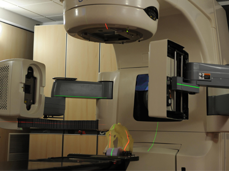
(c) Inserm/Institut Curie/ Guénet, François
These two studies were possible by virtue of the COPERNIC collection (started in 2003) of more than a hundred cell lines from radiosensitive patients.
- The theoretical study resolves an enigma in radiotherapy dating back 50 years, by giving a biological interpretation to an empirical formula. This formula links the survival of cells to the radiation dose.
- The biological and clinical study validates the theoretical study. It combines 67 co-authors, including 50 French radiotherapists, representing thirty cancer research centres or hospitals. It now enables us to develop predictive tests of radiosensitivity for the adverse side effects of radiotherapy.
What innovation have these two studies provided?
The ATM protein was discovered in 1995. This protein is known because its mutations cause the syndrome associated with the strongest form of human radiosensitivity, ataxia telangiectasia. Up until now, the ATM protein has always been considered as cell nucleus protein and no study had probed its presence in the cytoplasm. The radiobiology group of Inserm Unit 1052 analysed more than a hundred cell lines from patients suffering from adverse tissue reactions following radiotherapy (dermatitis, proctitis, etc.). These analyses showed that the ATM protein is instead found in the cytoplasm of cells and transits into the nucleus after irradiation.
How was the theoretical study conducted? What was its scope?
Larry Bodgi and Nicolas Foray mathematically modelled all the steps leading the ATM protein from the cytoplasm to the nucleus. Firstly, the oxidation produced by irradiation leads to a change in the shape of the ATM protein. This change facilitates its passage into the nucleus. An ATM then activates recognition of double-stranded breaks (DSB) in the DNA, triggering their repair. The result of this modelling was that the final mathematical formula linking cell survival to the radiation dose proved to be identical to that from the empirical model (called linear-quadratic), proposed in the 1970s. As a result of this theoretical study, we can now better understand at cellular level why a cell is radiosensitive and the consequences arising from a recognition or repair defect for DSB after irradiation. It remained to confirm it in the patient by means of a biological and clinical study, to predict the adverse side effects of radiotherapy.
How was the biological and clinical study conducted? What was its scope?
Out of 380,000 cancer case per year [in France], half of patients are treated using radiotherapy. From 5 to 20% of these patients can suffer adverse tissue reactions that extend from simple redness to even radio-induced burn, and in exceptional cases death. From the COPERNIC collection, the cells of each patient were amplified in the laboratory then irradiated under the exact conditions of a radiotherapy session (a dose of 2 Gy[1]). Researchers in the radiobiology group of Inserm Unit 1052 examined the DSB repair rate by using 3 immunofluorescence biomarkers (a technique used to visualise certain proteins on the actual site of DNA breaks) applied under many irradiation conditions. In parallel, 50 radiotherapists, co-authors of the study, supplied patient-specific clinical information, particularly the severity score[2] of tissue reactions (grade 0, no reaction; grade 5, death of patient), reflecting their tissue radiosensitivity. A significant correlation was obtained between the transit speed of ATM protein and the severity score of radio-induced tissue reactions. Such a correlation remains valid irrespective of the type of cancer treated or how early or late the adverse tissue reaction occurs after radiotherapy. From this correlation, human radiosensitivity is classified into 3 groups:
- Group I: Rapid ATM transit: radioresistance
- Group II: Delayed ATM transit: moderate radiosensitivity.
- Group III: Massive defect in recognition or repair of DSB: hyper radiosensitivity
This classification now makes it possible to anticipate care strategies for radiotherapy treatments, because this approach is also valid for tumours. Given the impact and applications of this work, patents applications have been submitted jointly with the Neolys Diagnostics start-up created in 2014, with the support of Inserm Transfer and Lyon SATT. The aim of Neolys Diagnostics is to industrialise radiosensitivity tests arising from these 2 studies, to give radiotherapy a new tool to improve personalisation of anti-cancer radiotherapy treatment.
Financial support
These two studies are part of the Investissements d’Avenir [Future Investments] (INDIRA project, Radioprotection and Nuclear Safety) directed by the Commissariat Général à l’Investissement [French General Investment Commission]. Other [French] financial support was received from: Association for research on Ataxia Telangiectasia (APRAT), AVIESAN- Cancer Plan calls to tender (Physicancer and Health and Environment programmes), the EDF Radioprotection Committee, National Centre for Space Studies (CNES), Rhône-Alpes Auvergne CLARA cancer centre (Oncostarter and Proof of Concept programmes). All experiments were performed at the Léon-Bérard Cancer Centre in Lyon.
[1] The radiation dosage unit is the Gray (Gy).
[2] Agreed international scales now allow tissue reactions to be classified irrespective of the location and organs affected (e.g. CTCAE and RTOG scales).
These contents could be interesting :

Medias
Sources
The nucleo-shuttling of the ATM protein as a basis for a novel theory of radiation response: resolution of the linear-quadratic modelBodgi, L.and Foray, N.International Journal of Radiation Biology Influence of the nucleo-shuttling of the ATM protein in the healthy tissues response to radiotherapy: towards a molecular classification of human radiosensitivity[1] Granzotto, A. …65 co-auteurs… and Foray, N.International Journal of Radiation Oncology