
Researcher Contact
Fabian Docagne
Unité Inserm 1237 ” Physiopathologie et imagerie des maladies neurologiques”
Multiple sclerosis (MS) is an autoimmune inflammatory disease of the central nervous system. It generally affects young people, in whom it is the leading cause of non-traumatic motor disability. This disability develops either progressively or in the form of relapses interspersed with periods of remission.
At present, magnetic resonance imaging (MRI) is widely used to diagnose and follow up patients with MS. However, no imaging tools exist to predict the onset of relapse.
MS is caused, at least partially, by inflammatory cells (notably lymphocytes) migrating into the brain and spinal cord through the walls of blood vessels. The cells migrate by binding to adhesion molecules that are present on the surface of these vessels.
In this study, published in the journal PNAS, the authors demonstrate that this technique is able to visualize the migration of the inflammatory cells, thereby making it possible to predict the onset of relapse in asymptomatic mice, and remission in sick mice.
This technique could in the future be adapted for use in humans to improve prognosis and follow-up in patients with MS.
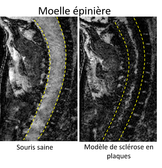
In the right-hand image, the iron beads (in black) have entered the spinal cord, thus revealing inflammation
Fabian Docagne
Unité Inserm 1237 ” Physiopathologie et imagerie des maladies neurologiques”
Prediction of disease activity in models of multiple sclerosis by molecular magnetic resonance imaging of P-selectin