Menu

Discover the new Inserm newsroom

The Inserm press room is updating its design, and continues to adapt to your needs and evolving practices. Now more ergonomic, its navigation has been reworked to make reading easier on mobile devices such as tablets and smartphones. Presentation of content has been revised to make it easier to read, and to quickly find the contact and sources of information to save you time. The search engine on the homepage has also been optimised to find information quickly by offering you suggestions.


Since its launch in 2012, the French National Institute of Health and Medical Research (Inserm) press room provides real-time information to journalists and Internet users on the latest health/research stories and discoveries from its laboratories. Inserm’s press releases are official news items disseminated by Inserm in France and internationally.

En 2014, the Inserm press room received an Honorable Mention at the Digital PR Awards, a competition organised by the American group PR News, which “salutes the most innovative digital initiatives in the field of digital public relations.”


If you have any comments, and wish to share your ideas in order to continue to improve the newsroom, do not hesitate to write to us: rf.mresni@bew-esserp

Final results of JIKI trial

The final conclusions of the JIKI clinical trial, testing the efficacy of favipiravir in reducing mortality in individuals infected by Ebola virus in Guinea, are published in PLoS Medicine this week. This work was carried out under the leadership of Prof. Denis Malvy, Inserm, with the help of a large team of international researchers. Although the conclusions are qualified, they have nonetheless made it possible to collect data and benchmarks that will enable researchers to base their future trials on robust hypotheses and criteria.


Ebola fever is an extremely lethal disease, for which there is no proven effective treatment. In September 2014, at the height of the epidemic, the World Health Organisation released a shortlist of drugs suitable for studies on Ebola virus (EBOV) disease, including favipiravir, an antiviral drug developed for the treatment of severe forms of influenza. An international team under the auspices of Inserm carried out a study called JIKI (meaning “hope” in the Malinke language) in Guinea. The goal of the study was to test the feasibility and acceptability of an emergency trial carried out during a large epidemic of Ebola virus disease, and to collect preliminary data on the safety and efficacy of favipiravir in reducing mortality and viral load in patients with Ebola virus disease. Because of the exceptional circumstances of the recent epidemic of Ebola fever, the study was a pilot, proof-of-concept, non-randomised, multicentre trial, with a so-called historical comparator group, in which 126 participants received favipiravir in addition to standard basic care.


According to the results of the study, it is unlikely that favipiravir monotherapy is effective in patients presenting with very high viraemia, but the drug deserves continued evaluation in patients presenting with intermediate to high viraemia. This conclusion is based on two results—the mortality rates observed, and the dynamics of the EBOV RNA load during treatment.

In patients presenting with very high viraemia, mortality was 7% higher than the pre-trial value, and the viral load did not decrease. This suggests that a future trial is unlikely to demonstrate any benefit of favipiravir in these patients

In patients with lower viraemia, mortality was 33% lower than the pre-trial value, and the viral load decreased rapidly with treatment. The trial was non-randomised, and the 95% confidence interval included the pre-trial value. As a result, the present findings do not prove that favipiravir was effective in these patients, but they do suggest that the question remains open, and provide an indication on how to better address it.

The authors conclude, “In the middle of a health crisis such as an epidemic of Ebola fever, researchers can be faced with a situation in which random assignment of patients to groups receiving standard care or standard care plus an experimental treatment is not acceptable from an ethical point of view. In these rare circumstances, it may be decided not to conduct a trial, but to wait for more favourable circumstances, or to conduct a non-randomised trial. In this pilot experiment, we chose this second option. Our conclusions are qualified. On the one hand, we cannot draw conclusions on the efficacy of the drug, and our conclusions regarding tolerance, although encouraging, cannot be as definite as they would have been with randomisation. On the other hand, we have learned a great deal on how to define and conduct a trial under such unusual circumstances, working closely with the community and non-government organisations. We incorporated research work on care, so that care can be improved; we rapidly generated interim data useful in designing studies on Ebola fever, and shared them with the scientific community; and we collected evidence enabling researchers to base their future trials on solid preliminary hypotheses and criteria.

Treating depressive symptoms from their roots

A wide range of compounds is on the market to ameliorate depressive symptoms, however their efficiency is achieved only after long periods of treatment and not in 100% of patients. Inserm researchers identified early cellular changes in the brain for the emergence of depressive symptoms, and a novel promising drug target.

These results are published in the journal Nature Medicine on Janaury 25th, 2016.

The aim of Manuel Mameli, Inserm researcher and Dr. Salvatore Lecca in his team, at Inserm Unit 839 the “Institut du Fer a Moulin (IFM)” (Inserm/UPMC), was to understand the initial cellular modifications occurring after a stressful aversive experience. Protracted stress and aversive experiences are indeed a trigger to engage depressive behaviors in animals and humans.

Using electrophysiological, viral-based and pharmacological approaches researchers found that the activity of neurons located in the lateral habenula – a cerebral nucleus for aversion and disappointment – increased after a stressful experience due to a reduced function of two proteins controlling neuronal function (GABAB and GIRK).

Inserm scientists designed a rescue strategy that reversed the cellular modifications and ameliorated depressive symptoms after aversive experience by targeting a specific phosphatase (PP2A). By employing a rodent model of mood disorders (Learned Helplessness), that recapitulates a number of behavioral phenotypes typical of human depression, they have shown that the inhibition of PP2A was efficient to rapidly ameliorate the behavioral phenotype of mice.

“Our study unravels unknown early cellular mechanisms able to trigger complex behavioral responses. Our study further highlights the role of the lateral habenula in the aetiology of depression. Our results provide insights on a novel potential pharmacological target that could be studied for a therapy of mood disorders”

explain Manuel Mameli, Inserm researcher.

Looking at the brain to prevent depression in adolescents

Depression is a real public health issue with 8% of adolescents affected by it, according to the French National Authority for Health (HAS). Adolescence is a time of transition during which young people are prone to episodes of depression. This often complicates the diagnosis of this disease.

According to some studies, adolescents with severe depression appear to have deviations in the areas of the brain associated with reward response. It may explain that lack of interest and moroseness are more common symptoms than sadness.

To better understand this phenomenon, researchers at Inserm Unit 1000 “Neuroimaging and Psychiatry”, led by Jean-Luc Martinot, in collaboration with a team from King’s College (London) have studied over 1,500 young people (at 14 years old and two years later) using magnetic resonance imaging (MRI) through the European study IMAGEN. Participants were split into three groups: one group with depression, a second group with isolated symptoms of depression with no actual diagnosis and finally, a group of healthy subjects

Each participant had to carry out a task where the brain’s reward response could be assessed (winning points in a game). The results of the simulated MRI confirms the scientists’ hypothesis—that adolescents with depression or occasional symptoms of depression have reduced activity in a specific area of the brain, the ventral striatum, which is involved in the reward circuit. It is significant that the response in this region is even weaker than the depressed person’s lack of interest.

The low activity in this region detected in healthy adolescents at 14 years old is correlated with the onset of depression or symptoms of depression at age 16”, explains Jean-Luc Martinot, Research Director at Inserm.

This study shows that impaired reward circuitry function is a factor that renders adolescents vulnerable to depression. The detection of symptoms involving loss of interest in adolescents and taking them into account in the early stages may help predict the onset of the disease or relapse and, as such, enable targeted early intervention well in advance.

Red blood cell fragments attack blood vessels

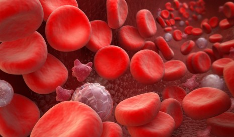
A new study published in the journal Blood shows that red blood cells can turn against blood vessels by releasing aggressive fragments that contribute to the development of cardiovascular diseases.Blood Cells : erythrocyte, thrombocyte, leukocyte

©Fotolia

There are approximately 5 million red blood cells in every microlitre of blood. Their stock is constantly renewed, and each one is discreetly removed every three months. But abnormal red blood cells persist in the bloodstream in some diseases. They ultimately rupture and release their contents, a process referred to as intravascular haemolysis.

Does the red blood cell disappear then? Not quite…

Researchers led by Olivier Blanc-Brude, a CNRS (French National Centre for Scientific Research) research fellow at the Inserm Paris Cardiovascular Center and Paris Descartes University, have just shown that at the exact moment of haemolysis, the red blood cells do not disappear: they actually release a large number of fragments, known as microparticles. And these fragments are not harmless. These observations were made using red blood cells from patients with sickle cell disease, the most common genetic disorder in France. Their rigidity leads to a considerable degree of haemolysis, which is therefore easier to study.

Red blood cells contain haemoglobin, the protein that carries blood gases and enables us to breathe. As a result, the red blood cell fragments also contain haemoglobin, but in a degraded and toxic form: the iron previously contained in the haemoglobin becomes exposed on the surface of these thousands of red blood cell microparticles.

It is this feature that makes the microparticles dangerous.

Effectively, the red blood cell particles act as carriers, and deposit the toxic substances on the cells lining the blood vessels. This transfer of haem and iron from the red blood cells to the vascular lining interferes with dilation, limiting the blood supply to the tissues that need it. This also leads to oxidative stress and promotes obstruction of the blood vessels.

In patients with sickle cell disease, red blood cell fragments may be partly responsible for very painful vaso-occlusive crises. These microparticles also probably contribute to the many lesions caused by chronic lack of oxygen, e.g. in the kidneys.

These innovative results make it possible to anticipate new therapeutic possibilities for preventing and treating vaso-occlusive crises, as well as for preventing some more common cardiovascular diseases.

These new therapies could target various components of the red blood cell microparticles, e.g. membrane components, such as phosphatidylserine, or free haem. Haemopexin is a molecule naturally found in the bloodstream, and which has the role of binding to free haem to eliminate it. It might represent a therapeutic opportunity, i.e. a haemopexin supplement might help to neutralise free haem carried by red blood cell microparticles.

These results are the subject of a patent filed by Inserm Transfert.

Seniors: Developing one’s balance and muscle strength is effective

Following a programme of physical exercise based on balance and building muscle strength reduces the risk of injury from falls by nearly 20% in women aged over 75 years. This study, conducted by Patricia Dargent, has just been published in the British Medical Journal (BMJ).Sport seniors activités physique

©fotolia

Falls are very common in older people, with approximately one in three people over the age of 65 experiencing at least one fall per year. The resulting injuries often require medical care, and lead to a significantly altered quality of life and accelerated functional decline that can in turn lead to a progressive loss of autonomy and admission to a healthcare facility.

In total, 706 women aged 75-85 years (mean age 80), living in their own homes and with reduced balance and walking ability, participated in the Ossébo trial in 20 study centres distributed throughout France. Participants were randomly assigned to 2 groups, one group that underwent the exercise programme and a “control” group with no intervention.

The exercise programme[1]  involved weekly sessions in small groups supervised by a facilitator, supplemented by exercises to do at home at least once a week, all for a period of 2 years.

For the 2 years of the study, women in the “intervention” group had significantly fewer traumatic falls (falls causing moderate to severe injuries, requiring medical care or impeding activities of daily living for at least 3 days) than women in the control group. They also scored better on clinical tests of balance and walking, and considered themselves to be in better physical health than women in the control group.

These results also show that subjects living in disadvantaged areas are more vulnerable to even brief episodes of atmospheric pollution. Most importantly, they indicate that this population, although chronically exposed to high levels of nitrogen dioxide (like other populations), is at a still higher risk during peaks of pollution.

“We are seeing populations being continuously compromised by the effects of chronic pollution. People thus compromised then ‘succumb’ to peaks of pollution, and the less privileged social categories are the main victims,” explains Denis Zmirou, co-author of the study.

[1] Designed and established by Group Associatif Siel Bleu

In Paris, inhabitants of disadvantaged areas are more vulnerable to the effects of atmospheric pollution

In a new study published in the journal Plos One, researchers from Inserm have analysed the causes of 79,107 deaths of Paris inhabitants aged over 35 years between 2004 and 2009. Their objective was to explore whether or not a combination of neighbourhood characteristics (socioeconomic profile and daily exposure to pollution) modified the risk of mortality during episodes of pollution such as those experienced in France during the heatwave of early summer 2015.

The choice of Paris as a city was not a random one, since the French capital is characterised by average pollution concentrations that vary enormously depending on the area, and by a variety of districts that are home to populations with diverse socioeconomic profiles.

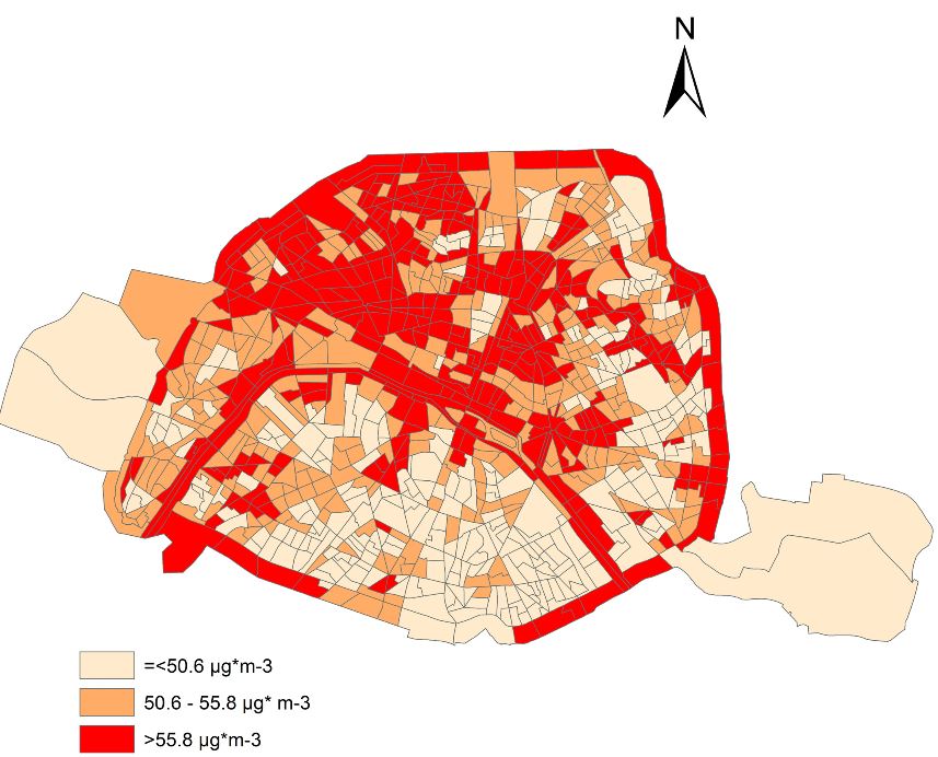
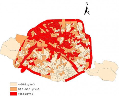
Two maps of the Paris districts were constructed. In the first, one can see the distribution of populations according to socioeconomic status of the residential districts (units known as IRIS). The second depicts the mean annual concentrations of nitrogen dioxide (NO2) over the study period. In Paris, NO2 in the outdoor air is mainly associated with heating buildings and car traffic.Carte de Paris catégories socio-pro

Dwellings housing the most disadvantaged people (category 3) are mainly located in the east and north of the city, whereas categories 1 and 2 are found in the centre and west part of Paris

Carte de Paris pollution

The most polluted regions are seen close to the main major traffic routes, along the Périphérique (ring road) and the Seine, and in northwest Paris.

The results of the study show that short-term variations in pollution and mortality are generally linked, and that there is a genuine risk of excess deaths during peaks of pollution.

These results also show that subjects living in disadvantaged areas are more vulnerable to even brief episodes of atmospheric pollution. Most importantly, they indicate that this population, although chronically exposed to high levels of nitrogen dioxide (like other populations), is at a still higher risk during peaks of pollution.

“We are seeing populations being continuously compromised by the effects of chronic pollution. People thus compromised then ‘succumb’ to peaks of pollution, and the less privileged social categories are the main victims,” explains Denis Zmirou, co-author of the study.

Discovery of a new mechanism of action of a protein that is toxic in Parkinson’s disease

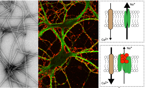
A team coordinated by Antoine Triller, Inserm Research Director, Director of the Institute of Biology at the École Normale Supérieure, and Ronald Melki, CNRS Research Director (Paris-Saclay Institute of Neuroscience), has just identified the target of an alpha-synuclein protein, which is pathogenic in Parkinson’s disease. This target is an ATP-dependent sodium/potassium pump. It may potentially be used in the development of symptomatic treatments for Parkinson’s disease. Details of this work are published in the 31 August 2015 issue of The EMBO Journal.Triller

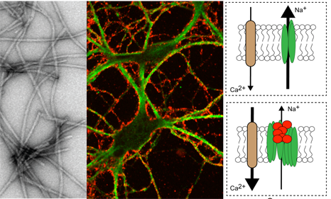
Alpha-synuclein forms fibrils (grey) that adhere (red) to the membrane of neurons (green). On the right side of the figure: the fibrils (red), on aggregating, disrupt the function of the pump (green) that maintains the sodium (Na+) gradient. This depolarises the neuron and increases the entry of calcium (Ca2+), which is toxic to the neuron. © Inserm/Antoine Triller

Alpha-synuclein is one of the pathogenic proteins (along with the tau and beta-amyloid proteins for Alzheimer’s disease, or prion protein for Kreutzfeldt-Jacob disease) that spread from cell to cell, and are associated with the physiopathological changes observed in neurodegenerative diseases.

Antoine Triller and his colleagues have shown that this protein aggregates on the neuronal membrane, and interacts with a protein on the surface of the neuron, the a3 subunit of the (Na+)/potassium (K+) ATPase pump. This pump controls the flow of sodium and potassium ions to and from the neurons, and hence the electrical activity of these neurons.

In humans, mutations in this pump are responsible for motor symptoms of early onset Parkinson’s disease, and alternating hemiplegia of childhood (AHC). The researchers have just demonstrated that alpha-synuclein, which diffuses between the cells, interacts with the Na+/K+ ATPase pump in the membrane. The pump, when bound to alpha-synuclein, is less well able to perform its pumping activity. Neuronal excitability is disrupted. Over time, the signals are no longer transmitted normally between neurons, and the symptoms of Parkinson’s disease or AHC appear.

This discovery was made possible through a combination of molecular biology and super-resolution microscopy techniques making it possible to follow individual molecules. In 2014, this latter approach was rewarded by the conferring of the Nobel Prize in Chemistry on Eric Betzig, Stephan W. Hell and William E. Moerner.

“This is a new mechanism that makes it possible to explain at cellular level the neuronal malfunctions in Parkinson’s disease,” explains Antoine Triller, Inserm Research Director. “This work sheds light on the fundamental and initial processes of the disease, and enables exploration of new therapeutic strategies to control its progression and symptoms,” he adds.

Observing the brain of moving animals

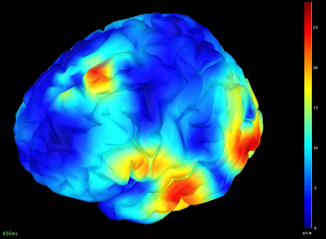
Such is the brain’s complexity that it is a particularly difficult organ to examine despite technological progress in this area. While electroencephalograms (EEG) and optical techniques can be used to record neurone activity in mobile animals, it is only possible to examine certain areas of the brain due to the size of the electrodes and light diffraction. Alternatively, functional ultrasound imaging (fUS) and magnetic resonance imaging (MRI) can be used to record variations in blood flow in the brain. Since active neurones need to be well-supplied with blood, blood flow in a given area reflects neurone activity in this area. However, these techniques require subjects to be completely immobile.Imagerie électro-encéphalographique

Electroencephalographic image. © Inserm/CRICM – Plateau MEG/EEG – Inserm U975

Two teams led by Ivan Cohen from Inserm Unit 1130 “Neuroscience Paris Seine” and Mickaël Tanter from Inserm Unit 979 “Wave physics for medicine” at the Langevin Institute (ESPCI/CNRS) have improved the fUS method to make it portable and usable with conscious and mobile rats at the same time as an EEG.

In order to demonstrate the benefits of this new method for pathological applications, the researchers looked at cerebral mechanisms in rats that reproduce epileptic seizures.

Thanks to this cutting-edge technology, we have precisely observed changes in blood flow which are precursors of epileptic seizure onset“, explains Inserm Research Fellow Ivan Cohen.

A study of cerebral mechanisms of mobile animals offers multiple opportunities for understanding behaviour and neurological conditions by mapping the regions of the brain associated with them. In particular, this technology will allow us to combine neurological and vascular data that play a key role in conditions such as dementia, strokes, epilepsy and Alzheimer’s disease.

Coffee colour…

Birthmarks, café au lait spots—although usually harmless (over 10% of the population has one or two), the presence of more than 5 of them in a child can conceal a hereditary disease, neurofibromatosis type 1 (NF1).

This is one of the most common genetic diseases (affecting 1 in 3,000 individuals). Until now, the molecular mechanisms associated with altered skin pigmentation were poorly understood, particularly since few appropriate models have been developed in the laboratory.

Inserm researchers at I-Stem (Institute for Stem Cell Therapy and Exploration of Monogenic diseases) have developed a model reproducing in vitro the hyperpigmentation associated with type 1 neurofibromatosis  from embryonic stem cells carrying the mutation responsible for this disease[1].

By establishing a protocol for differentiating these cells into melanocytes (the cells in the skin that can produce the melanin that gives it colour), the researchers were able to find the cause of generalised hyperpigmentation and formation of “café au lait” spots. The loss of neurofibromin expression in these NF1 melanocytes leads to a cascade of dysregulation. First, dysregulation of the AMPc and ERK signalling pathways, followed by dysregulation of the transcription factor MITF, and ultimately, increased expression of the enzymes involved in melanin production.

To correct this, the researchers used pharmacological inhibitors specific for these different signalling pathways, and were able to restore the cells to normal pigmentation levels.

This hyperpigmentation defect could be corrected by single small molecules (such as kojic acid, which is already contained in skin creams or lotions for lightening the skin), demonstrating the potential of pluripotent stem cells as a model for the study of pigmentation disorders.

The researchers now plan to identify and characterise the molecular mechanisms associated with other pigmentation-related conditions to find new therapeutic strategies, which are currently based on the use of mouse models, in which the melanocytes do not have the same location as in humans.

[1] These stem cells represent an unlimited source of cells that can theoretically differentiate into all the types of cells in the body, thus making it possible to demonstrate pathological phenotypes.
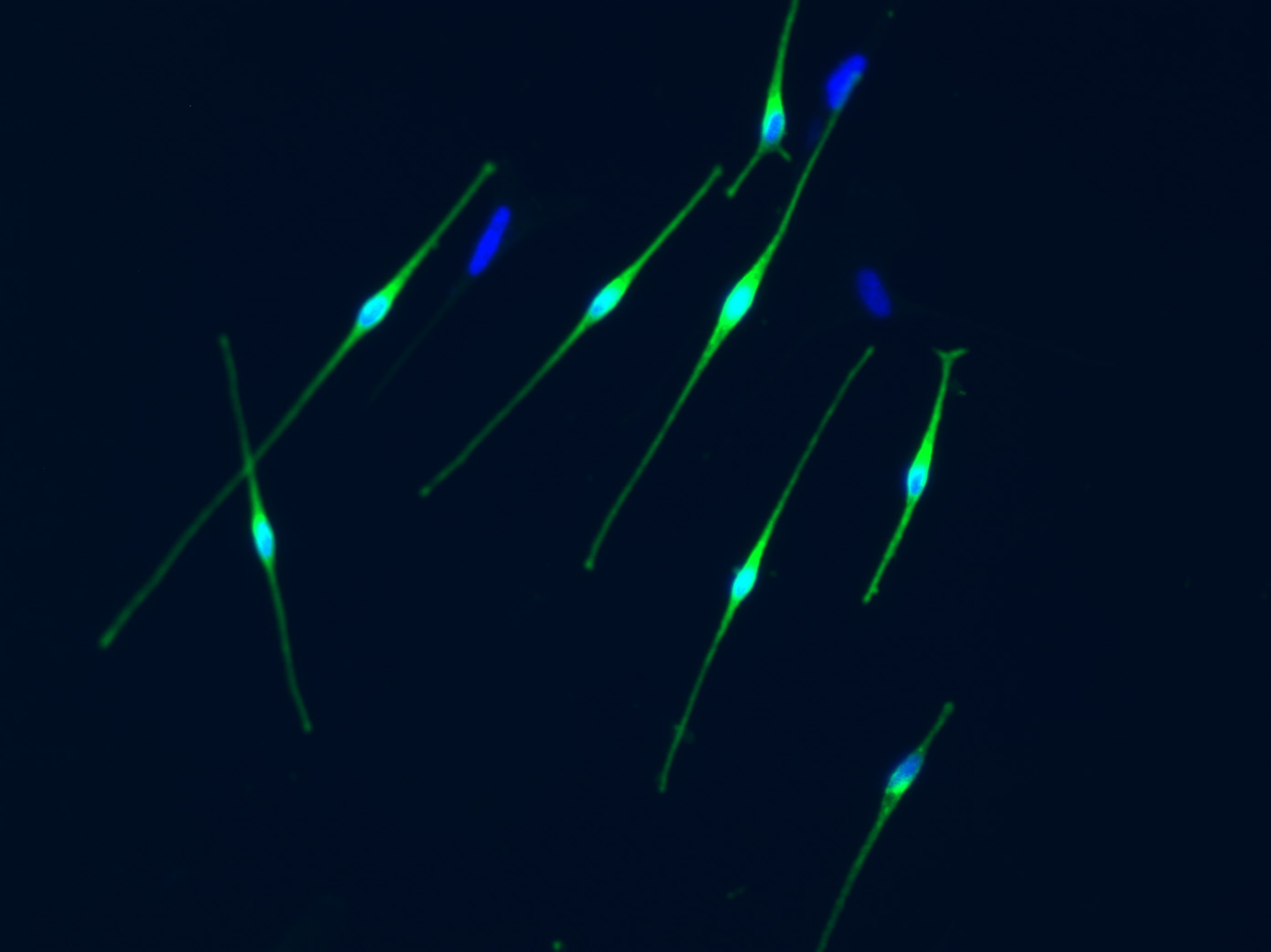
Mélanocytes

Melanocytes (cells producting melanin) © Inserm/Baldeschi, Christine

fermer