Menu

“Small” and “big” dreamers: what is happening inside our brains?

Dreams are still a field that is far from being understood by researchers. The mystery is particularly deep with respect to knowing whether people who claim to dream a lot are actually dreaming more than others or whether they are simply more able than others to remember their dreams.

It is currently impossible, using the resources available to researchers, to make this distinction. Yet the INSERM researchers of Unit 1028 “Centre de recherche en neuroscience de Lyon) have succeeded in developing a test that makes it possible to analyse brain activity in small and big dreamers and to draw a few conclusions. Their work has been published in the journal entitled Cerebral Cortex.

To do this, they used electrodes to record brain activity in two groups of volunteers, those who very often remembered their dreams and those who remembered them rarely. To obtain brain activity that was analysable by the researchers they subjected them to sounds while they were asleep and during the day.

An analysis of the electrical signals from the brain exhibited a significant difference in the responses produced by sounds in the small and big dreamers, not only during sleep but even when they were awake.

They suggest that the people who often remember their dreams have a special type of brain organisation in each state of awareness, whether during sleep or when they are awake, and this promotes either dreaming or the memory of a dream. These results do not support the hypothesis that has prevailed since the 1960s suggesting a strong link between dreaming and paradoxical sleep , with “different” brain function in small and big dreamers.

These observations appear to support a different hypothesis, namely that we cannot remember anything while dreaming. The memory of dreams is rather associated with phases of micro-awakening during sleep.

This theory is confirmed by the results obtained by researchers because “big dreamers” accumulate an average 15 minutes of wakening during the night, as opposed to five minutes for small dreamers. It remains to be discovered why certain people wake up for shorter or longer periods during the night.

Another of the researchers’ findings suggests that big dreamers wake up more often during the night because they are more sensitive to noise in the environment.

Their brains are more “reactive” to the environment or “distractible” than those of the people who dream little.


©Fotolia

What is at risk in the teenage drinker?

Alcohol addiction involves five million French people. It is currently very worrying to discover that more and more of the very young are drinking alcohol, and developing dangerous habits, such as that of binge drinking.

As part of the European AlcoBinge Project, coordinated by Mickaël Naassila’s team (INSERM Unit ERi 24), researchers used rats to explore the long-term effect of repeated intoxication during adolescence (the rat is considered to be an adolescent at 30 to 40 days from birth) and whether a predisposition and motivation to consume alcohol could lead to an addiction in adulthood.

The results published in the Neuropharmacology journal show that repeated intoxication during adolescence, before the brain has fully matured, produces a loss of control in alcohol consumption in the adult, resulting in long-term neurological changes.

From a behavioural point of view, the study headed by Mickaël Naassila shows that adult rats who were exposed to drunkenness early in their adolescence were more vulnerable to alcohol, and lost control of their consumption. They became desensitised to the negative properties specific to alcohol.

Again from the behavioural point of view, the INSERM researchers studied the incentive to consume alcohol in adult rats. The researchers showed for the first time and after performing numerous experiments involving rewards, that adult rats that had been exposed to alcohol earlier in their lives proved to have an excessive incentive to obtain alcohol.

From a neurological point of view, repeated intoxication during adolescence also produces changes inside the brain. The researchers showed that a very specific sub-region in the   nucleus accumbens, (the part of the brain that plays a leading role in addictive behaviour) becomes less reactive, in the long term, to re-exposure to alcohol. This could explain greater vulnerability when confronted with alcohol.

Although these are results produced from rats, they confirm that repeated intoxication in adolescence (reproducing the effect of binge drinking) renders adult subjects more vulnerable to alcohol and produce long-term neuro-adaptations.

©Fotolia

The carrot or the stick?

Should a child be punished, for example, to get him/her to produce good results at school or should the child be offered a reward for success?

Although they did not answer this question directly,  the INSERM researchers under Mathias Pessiglione at the Neuroscience Research Centre of the Pitié Salpêtrière Hospital recently showed that very specific regions are activated in the brain when faced with either situation. Certain regions of the brain (the anterior insula and the dorsal striatum) constitute a system dedicated to avoiding punishment, as opposed to the well-known system for obtaining rewards. In other words, it would appear that different regions of the brain are activated when confronted with the threat of punishment (one does all one can to avoid it) or, on the other hand, when faced with the promise of a reward (one does all one can to obtain it).

The general method adopted by the researchers in reaching this conclusion was to use an MRI scan to identify the regions of the brain that teach how to avoid punishment, and then to observe the behaviour of patients in whom these regions are affected by neurological disease such as a brain tumour (glioma) or Huntington’s Disease.

The results confirm the hypothesis of the existence of two different systems (punishment and reward) since the researchers showed that these patients remained capable of learning through reward (“the carrot”) but could not learn by punishment (“the stick”).

The computer modelling then performed by the researchers subsequently suggested complementary roles for these structures. The anterior insula is involved in the learning process, in other words it makes it possible to anticipate what sorts of behaviour are liable to be punished in a given context, while the striatum dorsal intervenes at the moment of decision-making, i.e. in choosing to avoid behaviour that is likely to be punished. The research was recently published in the journal entitled Neuron.

credit fotolia

The “still, small voice” in the brain has been found!

Joan of Arc is said to have heard voices, it’s a well-known fact. But we ourselves also hear voices, without even being mystics – and especially our own. We are constantly talking to ourselves. Where does this imaginary sound impression come from, this invented sound? Where does it resonate in our heads?

A collaborative effort between the INSERM researchers at the Lyon Centre de Recherche en Neurosciences and the Grenoble C.H.U. has just shown that our brains can react as if we were hearing someone talking to us even though no one else is in the room. The research has been published in The Journal of Neuroscience.

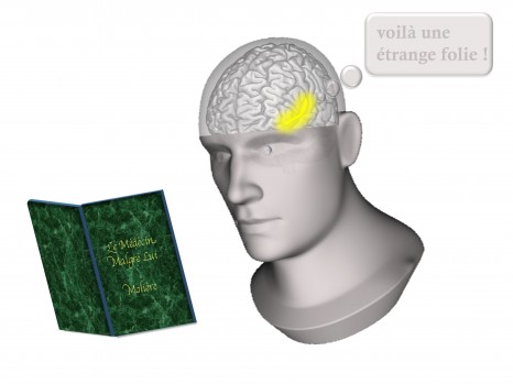
The team directed by Jean-Philippe Lachaux, INSERM’s Director of Research was interested in a situation particularly suited to hearing the “still, small voice”, that of reading to oneself. By directly recording the activity in the auditory regions of the brain that specialise in processing voices, the researchers were able to establish that they were active when reading to oneself, i.e. when the only auditory impression is indeed that of the still, small voice inside us.

Although this research only covers the reading situation, the researchers established the proof that it is possible to directly detect the moments in which someone is thinking, and even to know which thoughts are of a rather verbal nature.

But be aware that we are still a long way away from finding out what a person is thinking. In the long term, there are numerous potential applications, for example, to consider creating learning tools that would prevent a jumble of thoughts when they become too numerous, or in the case of depressing rumination (successive, interlinked negative thoughts that eventually preoccupy the whole attention of  those suffering from depression) or schizophrenia.

© Jean Philippe Lachaux/Inserm

Hearing the brain talking to itself. One often hears the sound of one’s own voice resonating in one’s head – when reading in the silence of a library, for example. The impression of hearing is due to spontaneous activation of the auditory parts of the cortex, those normally used to analyse the sounds that reach us. It is therefore possible to observe the brain talking to itself.

The origin of blindness identified for some types of hearing loss and visual impairment

Researchers from the Institut Pasteur, the Institut de la Vision, Inserm and the Université Pierre et Marie Curie have shed light on the origin of blindness that occurs in Usher I (the most common cause of deafness-blindness in humans). The scientists also demonstrated why the rat, the only animal model available today for this illness, does not suffer from the same blindness observed in humans. This work involves directing future research towards producing a primate animal model. The latter will then make it possible to progress towards a therapy-based approach for blindness in patients suffering from Usher I. This research was published on 8 October in the Journal of Cell Biology.

Usher syndrome is a genetic illness that causes congenital deafness and progressive visual impairment caused by retinitis pigmentosa. The prevalence rate of Usher syndrome is estimated at 1/30,000. Currently, a good level of care is provided for patients with hearing disorders. However, today, there is no treatment able to stop the end result of retinitis pigmentosa.

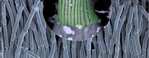
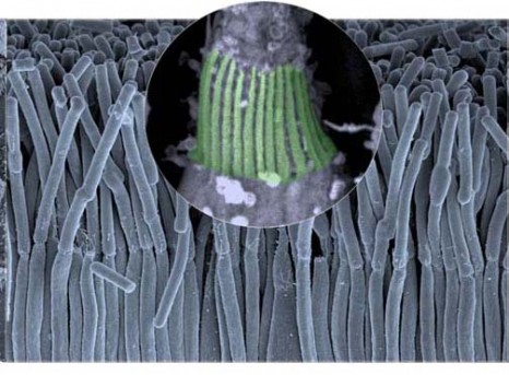
Research conducted by Professor Christine Petit, head of the “Genetics and physiology of hearing” research unit at the Institut Pasteur, in collaboration with Dr Aziz El-Amraoui (Institut Pasteur) and Professor José-Alain Sahel (Institut de la Vision), has provided fresh grounds for hope: researchers have just discovered the origin of retinitis pigmentosa in patients suffering from Ushers  I. It stems from a fault in the organization of cell structures essential to maintain vision, calyceal processes.  This failure is caused by the malfunction of one or several proteins; in this case, five were identified by the researchers that ensured cohesion in the calyceal processes. It was possible to observe the structure of the calyceal processes in high definition using electronic microscopic techniques (see photos).

For more information

©Copyright Institut Pasteur / Cataldo Schietroma et Vincent Michel

Hereditary neurological disorders: a gene identified in spinocerebellar ataxia

Efforts from four research teams in France (team led by Giovanni Stévanin, Inserm unit 975 “Centre of research into neuroscience at the Pitié-Salpêtrière”), theUS,Taiwanand theNetherlands, have identified the gene responsible for a hereditary neurological disorder affecting the cerebellum: type SCA22 spinocerebellar ataxia.

Almost ten years of work had previously not resulted in identifying the genes responsible for this illness.

The four research teams used a combination of genetic linkage analysis applied to the entire genome and new sequencing technology of the exome in four different patient groups. The four studies resulted in identifying three different mutations in the KCND3 gene that encodes a potassium channel. Electrophysiological studies demonstrated that the mutations led to disturbance in neuronal excitability.

For the researchers, “This study offers confirmation diagnosis for patients in these groups and can be used to target the research for treatments in these specific cases”.

This study also demonstrates the benefits of analyzing the exome, including in small groups, which have not been studied in any depth until now.

These results were published on line in the Annals of neurology review.

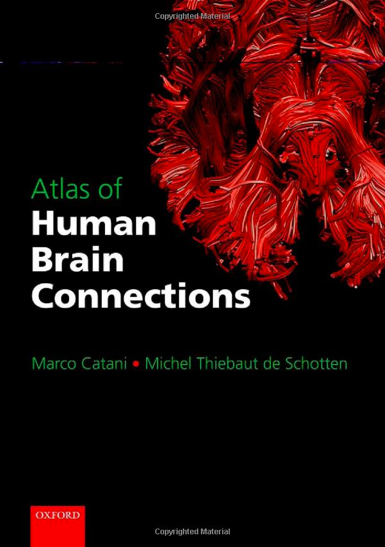
An atlas of human brain connections !

One of the major challenges of modern neuroscience is to define the complex pattern of neural connections that underlie cognition and behaviour. Brain connections have been investigated extensively in many animal species, including monkeys. Until recently, however, we have been unable to verify their existence in humans or identify possible tracts that are unique to human brain.

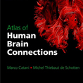
The Atlas of Human Brain Connections realized by Marco Catani and Michel Thiebaut de Schotten, researcher at Inserm, capitalizes on novel diffusion MRI tractography methods to provide a comprehensive overview of connections derived from virtual in vivo tractography dissections of the human brain.

It starts with an historical overview of the giant steps taken in neuroanatomy, from its birth more than 2000 years ago, to contemporary neuroimaging insights. Next, detailed descriptions of the major white matter connections, their function, and associated clinical syndromes are dealt with in detail. The composite maps of the atlas are an excellent anatomical resource for teaching, clinical, and research purposes. By reviewing both the basic principles of neuroanatomy, its historical roots, and its modern achievements in the field of DTI tractography, the book constitutes a valuable reference work for experienced clinicians and researchers working in the field of neurology, psychiatry, neurosurgery, and neuroradiology.

L’atlas offre un aperçu historique des grandes découvertes en neuroanatomie, depuis sa naissance il y a plus de 2000 ans, jusqu’aux récentes avancées scientifiques. Il décrit des connexions importantes de la substance blanche, leur fonction, et leurs syndromes cliniques associés. Les “cartes composites” de l’atlas sont une source d’information qui peut être utilisée pour l’enseignement anatomique, clinique et à des fins de recherche.

En examinant à la fois les principes de base de la neuroanatomie, ses racines historiques, et ses récentes découvertes dans le domaine de la tractographie des IRM DWI, ce livre est un ouvrage de référence pour les cliniciens expérimentés et les chercheurs travaillant dans le domaine de la neurologie, la psychiatrie, la neurochirurgie, la neuroradiologie et la neuropsychologie.

fermer