Researcher Contact
Stefan Catheline
Inserm researcher
Laboratory of Therapeutic Applications of Ultrasound (Inserm/Université Claude Bernard Lyon 1/Centre Léon Bérard)
E-mail : fgrsna.pnguryvar@vafrez.se
Telephone number provided upon request

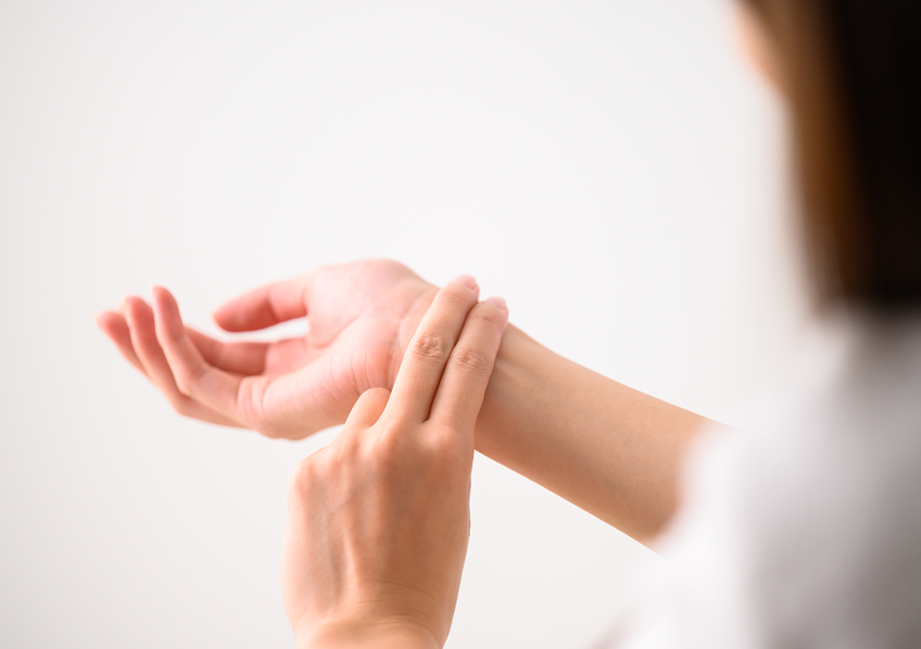
The pulse wave is used in everyday life to check heart rate. © Adobe Stock
We are all familiar with taking our pulse to check our heart rate. This signal is due to the propagation of a wave caused by the arteries dilating under the surge of blood from the heart. While we thought we knew the pulse well, the latest research by an international team led by Inserm researcher Stefan Catheline at the Laboratory of Therapeutic Applications of Ultrasound (Inserm/Université Claude Bernard Lyon 1/Centre Léon Bérard) shows that this was not the case. Their findings, published in Science Advances, show that the arteries not only dilate but also twist under the effect of the blood flow. This phenomenon generates a second “flexural” wave which propagates much more slowly. While it ultimately provides information on the same parameters – heart rate and arterial elasticity – the unprecedented measurement of this wave adds to our knowledge of the pulse.
Since 1820, pulse wave has been used in everyday life to check the heart rate of an athlete or inanimate person, or to assess arterial health, for example. It corresponds to the dilatation of the arterial wall following the surge of blood caused by the heart’s contractions, which propagates in an undulating manner along the arteries throughout the body.
An international research team led by Stefan Catheline, Inserm researcher at the Laboratory of Therapeutic Applications of Ultrasound (Inserm/Université Claude Bernard Lyon 1/Centre Léon Bérard), has just shown that in reality there is not one pulse wave but two. In addition to the principal wave, which is well-known and felt when touching the carotid artery or base of the wrist, there is a second one, which is more discreet but easily observable on ultrasound: the “flexural wave”, which had never been described until now.
A Chance Finding
It was somewhat by chance that Catheline’s team made this discovery. Specializing in waves and ultrasound therapies, it had been asked to test an innovative tool to analyze the retina: laser Doppler holography. This consists of photographing the organ at high speed and in very fine resolution to observe what is happening, and particularly to follow the arteries in motion. The researchers who developed this tool wanted to know if it could be used to calculate the speed of propagation of the pulse wave in the retina. Catheline’s team not only managed to measure this wave – which circulates at around one meter per second – but also detected a second wave signal nearly one thousand times slower.
The principles of fundamental physics on wave circulation in tubes are what enabled the scientists to better understand this phenomenon. Along the arteries, the two wave types actually propagate in two ways under the effect of the passage of the blood. The first is symmetrical to the central axis of the vessel and is when the arterial walls dilate and increase in diameter. The second is asymmetrical and results from the tube twisting in a so-called “sinusoidal” manner.
“Imagine a snake that swallows a prey which slides down the digestive tract – with the snake undulating away at the same time,” explains Catheline.
Following this discovery, the research team performed new ultrasound pulse measurements along the carotid artery of individuals and found both waves.
“It took us less than one afternoon to confirm the finding. This second wave, called a ‘flexural wave’, is present on all the recordings and is not difficult to observe. If it has never been described, it is simply because no-one had been looking for it,” explains Catheline.

The most well-known pulse wave (dilatation wave) is caused by the walls symmetrically separating outwards from the central arterial axis under the effect of the blood surge. And the newly-discovered flexural wave is caused by the artery twisting from side to side of this axis. © Stefan Catheline
And the Clinical Applications?
The principal pulse wave is widely used in medicine and reflects an individual’s cardiovascular health. Its speed of propagation depends on the condition of the artery walls: the younger and more supple they are, the slower the speed – and vice versa with age, with rigid arteries being a risk factor for cardiovascular events. However, given the high propagation speed of this wave, it is necessary to measure it over several centimeters to obtain a reliable value.
“With the flexural wave that we are describing here, whose slow speed ranges from one tenth to one thousandth of a meter per second depending on the arterial diameter, it is easier to study the signal on very short fragments and with other types of equipment than ultrasound, especially X-ray and MRI, explains Catheline. One millimeter is sufficient to obtain an accurate value, for example, to assess the state of the arteries in the retina,” he explains.
The researcher sees a second advantage in using this flexural wave in the clinic: by continuing to propagate in the veins there where the principal pulse wave is no longer detectable due to the distance from the heart, it would also provide information on the rigidity of the venous wall. He specifies, however, that in order to make it a clinical tool, research is needed in humans in order to correlate propagation speed and wall elasticity, as had been done previously for the principal dilatation wave.
Stefan Catheline
Inserm researcher
Laboratory of Therapeutic Applications of Ultrasound (Inserm/Université Claude Bernard Lyon 1/Centre Léon Bérard)
E-mail : fgrsna.pnguryvar@vafrez.se
Telephone number provided upon request
Observation of natural flexural pulse waves in retinal and carotid arteries for wall elasticity estimation
Gabrielle Laloy-Borgna1*, Léo Puyo2, Hidero Nishino3, Michael Atlan4, Stefan Catheline1*
1 LabTAU, Inserm, Centre Léon Bérard, Université Lyon 1, Univ Lyon, F-69003, LYON, France.
2 Institute of Biomedical Optics, University of Lübeck. Peter-Monnik-Weg 4, 23562 15 Lübeck, Germany.
3 Department Science and Technology, Tokushima University, 770-8506, Tokushima, 17 Japan.
4 Centre Hospitalier National d’Ophtalmologie des Quinze-Vingts, Inserm-DHOS CIC 1423, 28 rue de Charenton, 75012 Paris, France.
Science Advances, juin 2023