 L’alimentation serait responsable d’environ 30% des décès dus aux maladies cardiovasculaires. © Mathilde Touvier
Cardiovascular diseases are the leading cause of mortality in Western Europe, accounting for 1/3 of deaths in 2019. Diet is thought to be responsible for around 30% of such deaths. Nutrition-related prevention policies therefore constitute a major public health challenge for these diseases.
In an article to be published on 11 September 2024 in Lancet Regional Health – Europe, researchers from the Nutritional Epidemiology Research Team (CRESS-EREN), with members from Inserm, Inrae, Cnam, Université Sorbonne Paris Nord and Université Paris Cité, in collaboration with researchers from the International Agency for Research on Cancer (WHO-IARC), report an increased risk of cardiovascular diseases associated with the consumption of foods that rank less favourably on the Nutri-Score scale (new 2024 version) within the European cohort EPIC. A total of 345,533 participants from the cohort, spread across 7 European countries and followed for 12 years, were included in the analyses.
Officially adopted in France in 2017 (and in 6 other European countries since), the Nutri-Score aims to provide rapid information on the nutritional quality of foods and drinks to help and encourage consumers to compare them and choose those that offer a better nutritional quality. In parallel, it encourages manufacturers to improve the nutritional quality of their products.
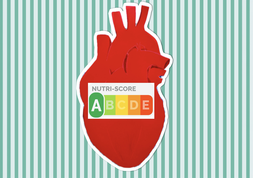
The Nutri-Score has 5 categories, ranging from A (dark green – higher nutritional quality) to E (dark orange – lower nutritional quality). An algorithm ranks each product according to its levels – per 100 g – of energy, sugars, saturated fatty acids and salt (to limit) and proteins, fruits, vegetables and pulses (to favour).
A number of studies published in international scientific journals have shown the validity of Nutri-Score in characterising the nutritional quality of foods and its efficacy in guiding consumers towards more nutritious choices (over 140 publications). In particular, links between the consumption of foods with a less favourable Nutri-Score (lower nutritional quality) and an increased risk of cardiovascular diseases have so far been observed in French studies (SU.VI.MAX and NutriNet-Santé cohorts). Studies in France, UK, Spain and Italy have also seen similar associations with an increased risk of various chronic diseases as well as higher mortality.
In this new study, the researchers focused on the latest version of the Nutri-Score algorithm (updated in 2024, see box), linked to the risk of cardiovascular diseases, in a large population spread across 7 European countries, with the aim of providing new scientific evidence for validating the Nutri-Score on a European scale. It follows two studies published in 2018 and 2020 in the same population on cancer risk and mortality.
A total of 345 533 participants from the EPIC (European Prospective Investigation into Cancer and Nutrition) cohort were included in the analyses. During the follow-up (12 years, between 1992 and 2010), 16 214 participants developed a cardiovascular disease (6 565 of whom had myocardial infarction and 6 245 stroke). The findings show that the participants consuming on average more foods with less favourable Nutri-Score, reflecting lower nutritional quality, were at increased risk of cardiovascular diseases, particularly myocardial infarction and stroke. These associations were significant after a large number of sociodemographic and lifestyle factors were taken into account.
‘These findings confirm the relevance of Nutri-Score as a public health tool to guide consumers in their food choices with the goal of preventing chronic diseases’, emphasises Inserm researcher Mélanie Deschasaux-Tanguy.
‘They also provide key elements to support the adoption of Nutri-Score as a mandatory nutritional logo in Europe’, explains Mathilde Touvier, Inserm research director.
A new version of the Nutri-Score in 2024
Changes to the calculation of the Nutri-Score[2] were recently proposed by the international scientific committee responsible for its monitoring in order to improve its consistency with nutritional recommendations. This new version of the Nutri-Score is expected to come into force in 2024 with a gradual roll-out in the months to come. However, due to European labelling regulations, manufacturers are under no obligation to use Nutri-Score on their packaging.
While many companies and brands (over 1,400 in France) have so far committed to using Nutri-Score on their products, harmonisation at European level is needed to ensure the mandatory implementation of a single logo that is effective and useful for citizens. This harmonisation is envisaged as part of the European Commission’s Farm to Fork strategy.
[1] https://sante.gouv.fr/prevention-en-sante/preserver-sa-sante/nutrition/nutri-score/etudes-et-rapports-scientifiques/
[2] https://theconversation.com/en-2024-le-nutri-score-evolue-pourquoi-et-que-faut-il-en-retenir-221697
To find out more: watch the Inserm program “Nutri-Score, we tell you everything” (french only).