Menu

Follow-up at the age of 2: preterm children have higher survival rates and better health than 20 years ago

©Fotolia

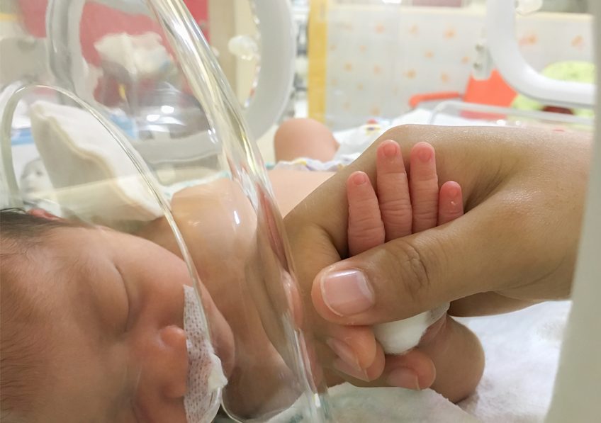
It’s good news. Over the last 20 years there has been a marked improvement in the survival rate of preterm infants, and rates of brain damage at the age of 2 years have halved. These are the latest results of the EPIPAGE-2 study carried out by researchers from the Inserm group EPOPé – Obstetrical, Perinatal, and Pediatric Epidemiology Team at the Center for Research in Epidemiology and Statistics, Sorbonne Paris Cité (CRESS, Unit 1153), AP-HP. These results from follow-up of 5,000 preterm infants were published in the British Medical Journal.

EPIPAGE-2 is a French study that initially included over 5,500 children, born preterm between 22 (5 months) and 34 weeks (7 and a half months) of pregnancy, between April and December 2011. The researchers’ main objective was to achieve a better understanding of the factors associated with children born preterm, in particular their neuromotor and sensory outcomes, and overall development at 2 years. The EPIPAGE-2 results were compared to those collected in 1997 by a similar study carried out in 9 French regions.

The data were collected from doctors who followed up with these children and families.

  • At 2 years, 52% of children born between 22 and 26 weeks of pregnancy, 93% of those born between 27 and 31 weeks, and 99% of those born between 32 and 34 weeks survived.
  • Rates of cerebral palsy (a motor disability often associated with preterm birth) were 7%, 4%, and 1% in these same groups by birth.
  • Less than 1% of children in the cohort had a severe sensory deficit (blindness or deafness).
  • Development, as assessed by parental questionnaires, was at the level expected for their age groups among 50% of children born between 24-26 weeks, 59% of children born between 27-31 weeks, and 64% of those born between 32-34 weeks.
  • Between 1997 and 2011, the rate of cerebral palsy was halved among very preterm babies. The rate of survival without severe motor or sensory impairment has thus increased in all groups by birth, particularly for the most preterm. Before 7 months of pregnancy, it was 74.5% in 1997, and 80.5% in 2011.

“Such studies are needed to better understand the impact of changes in medical practice on outcomes in children, and to adapt the organization of health care based on population-level data,” explains Pierre Yves Ancel. “Parental questionnaires, for example, have allowed children’s development to be evaluated by their own families, and enabled early identification of those requiring further investigation. For us, the challenge is therefore identifying at a very early stage the children most at risk of developmental delay in the future. These questionnaires represent a promising option for providing structured follow-up of babies while focusing resources on those who really need it,” adds Véronique Pierrat, who led the study.

However, the rates of overall and above all disability-free survival have only marginally improved among children born before 5 months of pregnancy. After adjusting for baseline characteristics, rates of survival and rates of survival without severe or moderate neuromotor and sensory disabilities at 2 years appeared to have increased between 1997 and 2011 for children born between 22 and 31 weeks of gestation. However, no change was observed in children born at 24 weeks of gestation or earlier. These statistics are noticeably poorer than in countries where management of the delivery room is more active than in France. Since publication of the initial EPIPAGE-2 results, a working group has been set up in France to consider the management of these extremely preterm children. As in seven other European countries, the recommendation in France is not to provide medical intervention for children born before 24 weeks, rather to provide them with “comfort” care.


Preterm birth

Children born between 22 and 26 weeks of gestation are considered to be extremely preterm.

Children born between 27 and 31 weeks of gestation are considered to be very preterm

Children born between 31 and 34 weeks of gestation are considered to be moderately preterm

Intestinal Viruses Predict the Risk of Graft-Versus-Host Disease

©Fotolia

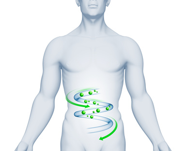
Teams from the virology, hematology-bone marrow transplant and biostatistics departments of the Saint-Louis Hospital, AP-HP, Université Paris Diderot and INSERM, in collaboration with scientists from the University of California-San Francisco (USA) discovered that a group of viruses in the intestine may trigger the onset of graft-versus-host disease. Researchers demonstrated the role of this intestinal “virome” (the population of viruses found in the intestine) in the onset of graft-versus-host disease by analyzing the intestinal virome of 44 patients receiving a bone marrow transplant.

These results appeared in a letter published in Nature Medicine.

Graft-versus-host disease (GVHD) affects up to 60% of patients who receive a stem cell graft (bone marrow transplant) with a high risk of death. Even though past studies have identified biomarkers associated with the severity of the disease, none of them have been used in practice to date in order to predict the onset of graft-versus-host disease.

The researchers carried out a longitudinal study analyzing the intestinal virome of 44 patients before a bone marrow transplant and six weeks after the transplant. By using high-throughput metagenomic techniques, they sequenced the DNA and RNA in the stool samples, in order to analyze and compare the flux over time in bacterial and viral populations.

The results show that GVHD is associated with significant changes in bacteria, bacteriophage (i.e. the viruses that infect bacteria) and virus populations in the stool.

In patients with intestinal GVHD, the persistent DNA viruses (Anelloviridae, Herpesviridae and Polyomaviridae) are absent from the intestinal tract in the first 2-3 weeks following the graft and then they increase in a significant manner. However, the presence of these viruses is stable as soon as the graft is performed in patients without GVHD.

In particular, the researchers detected the presence of a group of RNA viruses, picobirnavirus (PBV), in more than a third of patients after transplanting stem cells. As opposed to other viruses, the presence of PBV predicts the development of GVHD and is highly correlated to the severity biomarkers of intestinal graft-versus-host disease.

Metagenomic tools have made it possible to identify PBVs that have been little studied to date, since their extreme variability makes developing a test to detect them difficult. These viruses have been described in cases of intestinal gastroenteritis, notably in patients with AIDS, but their pathogenic role has not yet been determined. The potential utility of PBV as a predictive marker of GVHD has rekindled the interest in developing adapted to tools for clinical practice.

These results improve the state of knowledge of this complex disease, notably in terms of the role of viral infections in digestive inflammatory diseases and has opened the door to new therapeutic opportunities.

Testicular macrophages are guardians of fertility

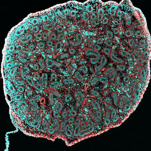
Cross section of newborn mouse’s testis (Ø = 20 µm), where we can see the seminiferous tubules (red) surrounded by macrophages (green).  Confocal micrograph.

© Noushine Mossadegh-Keller and Sébastien Mailfert / CIML

The origin, development, and characteristics of two types of testicular macrophage have been described by a CNRS team at the Centre d’Immunologie de Marseille-Luminy (CNRS / INSERM / Aix-Marseille University). To elucidate the nature of these immune cells, the researchers used a novel cell tracing method. Their findings were published on August 7, 2017, in the Journal of Experimental Medicine, and are of fundamental importance. They may help understand certain kinds of infertility in men and find new treatments for them.


From the start of life, an individual’s immune system learns to distinguish self—that is, native cells—from other, potentially pathogenic cells. But in males, as sperm only appear at puberty, they may be mistaken for foreign cells by certain elements of the immune system.  Testicular macrophages are special immune cells that rush to the defense of sperm. By releasing specific molecules, these guardians of fertility prevent other immune system agents from entering the testes.


Macrophages not only migrate to sites of infection and phagocytose pathogens, but also modulate immune system activity to ensure proper organ function and regeneration. They may arise from either embryonic progenitors or bone marrow cells in adults. Research with mice has enabled the team of Michael Sieweke from the Centre d’Immunologie de Marseille-Luminy (CNRS / INSERM / Aix-Marseille University) to describe both testicular macrophage populations in depth.


The testis is divided into two compartments. One kind of testicular macrophage is found in the interstitial spaces, where testosterone-producing Leydig cells are also located. These interstitial macrophages are of embryonic origin: they are present from the beginning of the individual’s life. The other kind is peritubular—that is, located on the surface of the seminiferous tubules that house sperm cell precursors. Each macrophage population has distinctive cellular markers.


The researchers used a new cell tracing method to follow the movement of peritubular macrophages from the bone marrow to the testes. They discovered that these macrophages only appear two weeks after the mice are born, which corresponds to the pubescent stage in human males. Surprisingly, once they have been established in the testes, macrophages of both populations remain there for the rest of their long lives. Sieweke’s team will next focus their research efforts on the relationships between macrophages, sperm, and testosterone production, which may yield innovative treatments for certain kinds of male infertility.

fermer