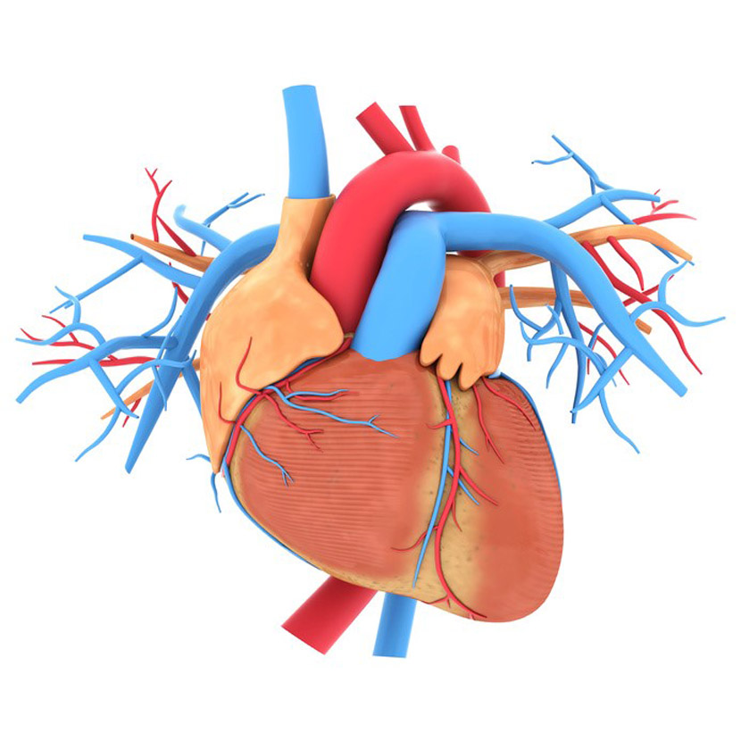
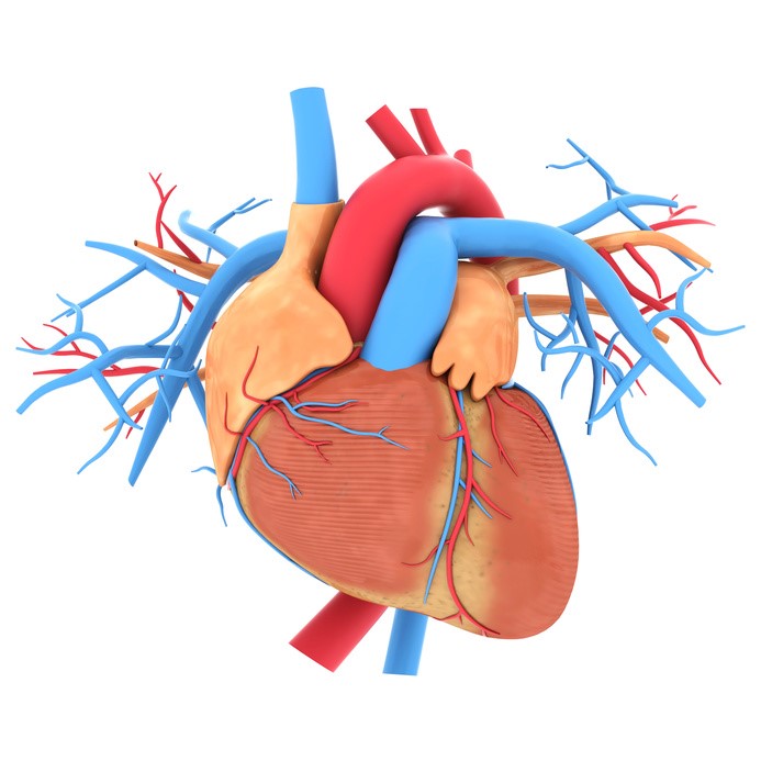
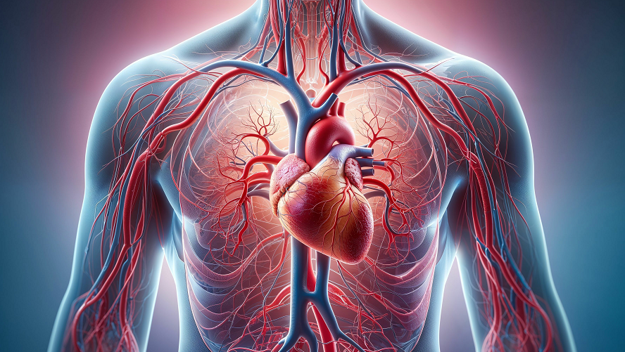
Menu

A promising vaccine against Nipah virus infection

NIPAH virusA scanning electron micrograph shows the Nipah virus (yellow) budding from the surface of a cell.© National Institute of Allergy and Infectious Diseases, NIH

The WHO recently classified the Nipah virus (NiV) as one of the eight main emerging pathogens likely to cause major epidemics in the future. In a context where no treatment or vaccine is yet available, a team comprising researchers from Inserm (Unit 955-VRI) and from the Université Paris-Est Créteil (UPEC) is presenting the preclinical results of an innovative vaccine against this virus. Most candidate vaccines target the viral surface proteins required for entry into human cells. To develop its new vaccine, the team at the VRI (Vaccine Research Institute of the ANRS MIE/Inserm) focused on the central role played by antigen-presenting cells (APCs) in the development of protective responses. The candidate vaccine, called CD40.NiV, carries specific parts of the surface proteins of the NiV-B virus, the Bangladesh strain. Following infection with the Nipah virus in animals, CD40.NiV demonstrated immunogenicity, neutralisation and complete protection, representing an important step towards the clinical development of a vaccine against the infection. The results of this work have just been published in the March 2024 issue of Cell Reports Medicine.

The Nipah virus (NiV) is a zoonotic virus, meaning it is transmitted from animals to humans. However, it can also be transmitted via contaminated food or directly between individuals. The clinical presentation can range from asymptomatic infection to acute respiratory infection to fatal encephalitis. First identified in Malaysia in 1999, the virus has since spread regularly through outbreaks in Bangladesh and India. Mortality linked to these outbreaks is estimated to be between 75% and 90%.

The virus has recently been included on the WHO list of priority emerging pathogens. There is currently no approved treatment or vaccine. Numerous candidate vaccines are under study or development. Most target the G and F proteins on the surface of the virus, which are necessary for it to enter human cells and spread throughout the body.

The teams at Inserm and UPEC have developed an original approach involving antigen-presenting cells (APCs), in particular dendritic cells, which play an important role in the immune response. To construct the CD40.NiV vaccine, specific parts (or epitopes) of the G, F and N proteins of the Bangladesh strain of Nipah virus (NiV-B) were attached to an antibody recognising the CD40 receptors on the surface of dendritic cells. The epitopes are thus presented directly to the cells of the immune system.

Immunogenicity (the ability to induce an immune response) of the vaccine was assessed in mice and non-human primates after two administrations of CD40.NiV vaccine (the so-called “prime-boost” strategy). As early as 10 days after the first vaccination with CD40.NiV (prime), NiV-specific IgG and IgA antibodies, as well as neutralising antibodies (specific antibodies that prevent infection by blocking viral entry into target cells) were produced. The neutralising antibody response is maintained for at least 100 days after the peak of the immune response. In addition, the team showed that the antibodies induced against NiV also neutralise various strains of NiV (Malaysia, Cambodia) and the Hendra virus (this is known as cross-neutralising immunity), an infectious agent transmitted by bats and causing a highly fatal infection in horses and humans.

To ensure that the vaccine was effective, the animals were infected with the NiV virus 60 days after the second injection of CD40.NiV (boost). Protection was complete.

This preclinical study demonstrated that the CD40.NiV vaccine candidate confers protection against the development of Nipah virus, with 100% survival of immunised animals until the end of the study, 28 days after infection. The absence of significant clinical signs or virus replication suggests that the candidate vaccine provides ‘sterilising immunity’, meaning that it can prevent the disease and its transmission.

Overall, results obtained with CD40.NiV are highly promising for fighting NiV infection and represent an important milestone towards the clinical development of a vaccine against this virus.

Improving the treatment of anaemia thanks to a new discovery in iron metabolism

globules rougesAn essential component of the haemoglobin in red blood cells, iron is crucial to many biological processes – including the transport and storage of oxygen in the body. © Inserm/Claude Féo

Anaemia is a major public health problem worldwide, affecting around one third of the population. Its causes are multiple, but the most common are a lack of red blood cell production, a lack of iron in the blood, and genetic diseases such as thalassaemia. A better understanding of iron metabolism is essential to improve the care of the many patients affected. In a new study, Inserm researchers at the Digestive Health Research Institute (Inserm/INRAE/Université Toulouse III – Paul-Sabatier/Toulouse National Veterinary School) identified the major role of a protein called FGL1 in iron metabolism. Their discovery paves the way for new clinical possibilities in the treatment of anaemia. These findings have been published in the journal Blood.

Anaemia is a disease in which the number of red blood cells – or the haemoglobin levels of the red blood cells – is lower than normal. A major factor in the morbidity and mortality of one third of the world’s population, anaemia is a major public health problem.

Anaemia can be caused by a deficit of iron in the blood resulting from dietary deficiencies, infections, chronic diseases, heavy menstruation, problems during pregnancy or by genetic diseases that affect the production of red blood cells (thalassaemia).

An essential component of the haemoglobin in red blood cells, iron is crucial to many biological processes – including the transport and storage of oxygen in the body. In other words, insufficient iron in the body means insufficient haemoglobin and red blood cells for transporting oxygen to the organs and tissues, which ultimately leads to organ failure.

For more information: C’est quoi l’hémoglobine ? (only available in French)

However, too much iron is also toxic to the body, meaning that its intake needs to be carefully regulated to avoid excessively high or low levels which are responsible for severe clinical complications.

Understanding iron metabolism

For several years, knowledge about anaemia and iron metabolism has been steadily increasing. It is now well known that iron levels in the body are regulated by a hormone called hepcidin.

We also now know that if the body needs more iron, as is the case with anaemia, a hormone called erythroferrone (ERFE) suppresses the expression of hepcidin in the liver. This process supplies the bone marrow with iron to synthesise new red blood cells and increase haemoglobin levels.

The identification of ERFE in 2014 by Inserm researcher Léon Kautz and his colleagues represented an important step in this field of research. However, these data obtained ten years ago were already suggesting that ERFE was not the only hormone controlling this process. The scientists hypothesised that a second protein, previously unknown, performed a similar function.


A new factor identified

This is what they have now confirmed by conducting new experiments in mouse models of anaemia, in two specific cases: one during an increased synthesis of red blood cells aimed at correcting induced anaemia in mice and the other in mice with thalassaemia.

The scientists started by studying the molecular mechanisms activated in the animals’ liver to identify the genes whose expression was increased during the anaemia. They observed that the expression of the gene coding for protein FGL1 was increased in the liver when the oxygen concentration decreased.

The researchers then produced different forms of protein FGL1 to test its mode of action in vivo in mice and in vitro in human liver cells. They were able to show that its mode of action is similar to that of the hormone ERFE, because FGL1 also represses hepcidin expression.

‘In addition to the fundamental aspects of this research in understanding anaemia, we believe that identifying the role of FGL1 will lead to the development of new therapeutic strategies to treat anaemia of various causes and for which the current treatments are ineffective,’ emphasises Léon Kautz, Inserm staff scientist.

For the moment, the team will start by conducting additional research to verify that FGL1 levels are indeed increased in the blood of patients with different types of anaemia. But the scientists plan to go further, with Inserm Transfert having already filed two patent applications for this study.

On the one hand, the first patent aims to better treat anaemia resulting from chronic diseases such as cancer. The objective is to identify analogous molecules or molecules that activate FGL1 synthesis, which would reduce hepcidin expression in these patients and increase their haemoglobin levels.

On the other hand, thalassaemia is characterised by very low levels of hepcidin, leading to excess iron that is harmful to the organs, causing high mortality. The team hypothesised that FGL1 is also involved in this process. The second patent therefore aims to achieve proof of concept that FGL1 inhibition could improve iron overloads in patients suffering from thalassaemia.

Eating meals early could reduce cardiovascular risk

© Freepik

A study led by scientists from INRAE, the Barcelona Institute for Global Health, Inserm, and the Université Sorbonne Paris Nord, has revealed that the time at which we eat could influence our risk of developing cardiovascular disease. This study, carried out on a sample of over 100,000 people from the NutriNet-Santé cohort, followed between 2009 and 2022, suggests that eating a late first or last meal is associated with a higher risk of cardiovascular disease. It also appears that a longer night-time fasting duration is associated with a reduced risk of cerebrovascular disease such as stroke. The findings, published in Nature Communications, suggest the importance of daily meal timing and rhythm in reducing cardiovascular disease risk.

Cardiovascular diseases are the leading cause of death in the world according to the Global Burden of Disease study, with 18.6 million annual deaths in 2019, of which around 7.9 are attributable to diet. This means that diet plays a major role in the development and progression of these diseases. The modern lifestyle of Western societies has led to specific eating habits such as eating dinner late or skipping breakfast. In addition to light, the daily cycle of food intake (meals, snacks, etc.) alternating with periods of fasting synchronizes the peripheral clocks, or circadian rhythms, of the body’s various organs, thus influencing cardiometabolic functions such as blood pressure regulation. Chrononutrition is emerging as an important new field for understanding the relationship between the timing of food intake, circadian rhythms and health.

Scientists used data from 103,389 participants in the NutriNet-Santé cohort (79% of whom were women, with an average age of 42) to study the associations between food intake patterns and cardiovascular disease. To reduce the risk of possible bias, the researchers accounted for a large number of confounding factors, especially sociodemographic factors (age, sex, family situation, etc.), diet nutritional quality, lifestyle and sleep cycle.

The results show that having a first meal later in the day (such as when skipping breakfast), is associated with a higher risk of cardiovascular disease, with a 6% increase in risk per hour delay. For example, a person who eats for the first time at 9 a.m. is 6% more likely to develop cardiovascular disease than someone who eats at 8 a.m. When it comes to the last meal of the day, eating late (after 9 p.m.) is associated with a 28% increase in the risk of cerebrovascular disease such as stroke compared with eating before 8 p.m., particularly in women. Finally, a longer duration of night-time fasting – the time between the last meal of the day and the first meal of the following day – is associated with a reduced risk of cerebrovascular disease, supporting the idea of eating one’s first and last meals earlier in the day.

These findings, which need to be replicated in other cohorts and through additional scientific studies with different designs, highlight a potential role for meal timing in preventing cardiovascular disease. They suggest that adopting the habit of eating earlier first and last meals with a longer period of night-time fasting could help to prevent the risk of cardiovascular disease.

The NutriNet-Santé study is a public health study coordinated by the Nutritional Epidemiology Research Team (EREN-CRESS, Inserm/INRAE/Cnam/Université Sorbonne Paris Nord/Université Paris Cité), which, thanks to the commitment and support of over 175,000 study participants, is advancing research into the links between nutrition (diet, physical activity, nutritional status) and health. The study was launched in 2009 and has already resulted in over 270 international scientific publications. There is still a call for new study participants living in France to continue advancing research into the relationship between nutrition and health.

By spending a few minutes a month responding via the etude-nutrinet-sante.fr secure online platform, participants help to advance knowledge of the relationship between diet and health.

Cardiovascular Diseases: Diet, Microbiota, Immunity, It Is All Linked!

prolifération des cellules immunitaires (lymphocytes)Visualization of immune cell (lymphocyte) proliferation in the mesenteric lymph nodes, under the influence of a microbiota modulated by a high-fat diet. © Soraya Taleb/PARCC

Although a high-fat, low-fiber diet is recognized as promoting cardiovascular diseases such as atherosclerosis, the mechanisms involved have not yet been fully identified. Researchers from Inserm and Université Paris Cité have studied the role of the gut microbiota in the development of atherosclerosis. Their work in mice reveals that the low fiber content of the high-fat diet leads to an imbalance in the gut microbiota, which itself causes systemic inflammation, worsening the development of atherosclerotic plaques in the arteries. These findings, published in Cell Reports, provide further evidence of the importance of fiber in the diet, both for good bowel function and for preventing the onset of cardiovascular diseases.

Cardiovascular diseases constitute one of the leading causes of death worldwide. Among them, atherosclerosis is characterized by the formation of an atherosclerotic plaque (atheroma), composed mainly of lipids, on the walls of the arteries. Over time, these plaques can cause damage to the arterial wall, obstruct the vessel, or rupture – often with serious consequences. Among the major risk factors for atherosclerosis is obesity, particularly when caused by a diet that is too high in fat and low in fiber. As such, it is not just diet but also its impact on the gut microbiota that are now avenues of interest for research into cardiovascular diseases.

A team led by Soraya Taleb, Inserm research director at the Paris-Center for Cardiovascular Research (Inserm/Université Paris Cité), looked at the influence of a high-fat, low-fiber diet on the gut microbiota of mice and how this could contribute to the development of atherosclerosis.

The researchers used a mouse model of diet-induced atherosclerosis to compare the effects of several diets on the metabolism, microbiota and development of atherosclerosis.

Unsurprisingly, in the mice fed a high-fat, low-fiber diet, their findings show an increase in metabolic risk factors linked to cardiovascular diseases (significant weight gain, hyperglycemia, insulin resistance, increased weight of the liver and its triglyceride content, etc.).

However, these are not the only effects observed of this diet, which also appears to be associated with an overall imbalance in the microbiota – in its composition and immune response –, reflected in the altered production of metabolic derivatives by its component bacteria. In particular, short-chain fatty acids, derived from the fermentation of fiber and recognized for their positive impact on health, are produced in smaller quantities.

However, this imbalance itself appears to be associated not only with metabolic risk factors but also with a worsening of the manifestations of atherosclerosis at vascular level, with an increase in atheromatous plaque size in the aorta as well as a systemic inflammatory phenomenon which results in an increase in the number of immune cells in these plaques. However, supplementation with fiber made it possible to counteract these effects.

“These findings show, in mice fed a high-fat diet, that a pathological gut microbiota accelerates the development of atherosclerosis,” comments Taleb. Our observations also show that, more than its high fat content, it is the small amount of fiber in this diet that causes the microbiotal imbalance and as such the worsening of the atherosclerosis. This further supports the idea of the essential role of fiber in structuring a healthy microbiota and in preventing systemic inflammatory diseases such as cardiovascular diseases”, she continues.

But how can we explain the surprising link that appears between the composition of the microbiota and the accumulation of immune cells in atheromatous plaques? In mice grafted with a gut microbiota initially modulated by a high-fat diet, the research team observed an increased proliferation of immune cells in the mesenteric lymph nodes[1], the site of their activation in the gastrointestinal tract.

Techniques used to track migration of the immune cells confirmed that it was indeed cells from the mesenteric lymph nodes that, after passing from the gut into the bloodstream, accumulated in atheromatous plaques, thereby contributing to the development of atherosclerosis.

The fact that we have seen that immune cells are capable of migrating from the gut to the periphery and thereby generate systemic inflammation that worsens the atheromatous plaques adds a new dimension to our understanding of the link between diet, gut, microbiota and atherosclerosis,” explains Taleb. Additional work is needed in order to identify which of the bacteria in the microbiota are involved in this mechanism, in order to envisage targeted therapeutic approaches and study these mechanisms in humans”, concludes the researcher.


[1] The mesenteric lymph nodes are located in the mesentery, a fold of the peritoneum (the membrane lining the abdominal cavity and covering the abdominal organs) that suspends the small intestine from the posterior abdominal wall.

In Primates, the Appendix Is Found to Have a Protective Effect Against Infectious Diarrhea

singe géladaThe gelada (Theropithecus gelada) is one of the primate species without an appendix referenced in this research. ©Vallée des singes

Although the cecal appendix is no longer considered a vestige of evolution with no particular role, its exact function remains to be discovered and several hypotheses are currently being explored. A research team from Inserm, CNRS, the French National Museum of Natural History (MNHN), Université de Rennes, Sorbonne Université and the Eugène Marquis Center looked at how the presence of an appendix affects the onset and severity of infectious diarrhea in primates, an animal order that is particularly affected by these diseases. Its research shows that the primate species with an appendix are less affected by infectious diarrhea and that it is less severe than in those without an appendix. They are also better protected against these infections during the first part of their lives, a period that is more vulnerable to severe diarrhea and crucial for reproduction. These findings, published in Scientific Reports provide new evidence supporting the advantageous role of the appendix in evolution.

The cecal appendix (more commonly referred to as the “appendix”) is a small blind-ended tube located in the lower part of the cecum, the first part of the large intestine. It is found in some mammals and particularly in some primate species, including humans. While it has long been considered an unnecessary vestige of evolution, research over the past decade has challenged this paradigm with scientists now tending to view it as a potential evolutionary advantage, although its function remains poorly understood.

One hypothesis concerning the role of the appendix is based on its composition of microorganisms. Different from that of the rest of the gut microbiota, it could constitute a reservoir of healthy flora safeguarded from the fecal flow, likely to recolonize the gut after a gut infection and enable faster remission. And it just so happens that primates are an animal order particularly affected by infectious diarrhea. In humans, mortality related to these infections was identified in 2015 as being the second leading cause of mortality in children between 1 month and 5 years of age. More specifically, in patients who have had their appendix removed (appendectomy), there has been an increased risk of the occurrence and/or severity of certain forms of infectious diarrhea, although no direct link has been demonstrated at this time.

A research team led by Éric Ogier-Denis, Inserm research director at the Oncogenesis Stress Signaling unit Inserm/Université de Rennes/Eugène Marquis Center), and Michel Laurin, CNRS research director at the Center for Research on Paleontology – Paris (CNRS/MNHN/Sorbonne Université), had shown in previous research that the mammalian species with an appendix had longer longevity than those without one[1]. As a continuation of this research, the team looked at how the presence of a cecal appendix could affect the frequency and severity of diarrhea in primates and thus be a determining factor in the life span of each species.

To do this, the researchers examined the veterinary records of 1 251 primates of 45 different species – 13 with an appendix, 32 without – living in semi-liberty in “La Vallée des Singes” zoological park in Romagne, France. They listed the frequency and severity of the diarrhea episodes that occurred between 1998 and 2018 in these animals.

gorille (Gorilla gorilla gorilla)The gorilla (Gorilla gorilla gorilla) is one of the species of primates with an appendix referenced in this research. ©Vallée des singes

Half of the primates had experienced at least one episode of diarrhea during the 20-year follow-up period, with 13% of the episodes qualifying as “severe”.

In the primates with an appendix, the frequency of diarrhea episodes was very much lower (by around 85%) than in those without one. The cases of severe diarrhea were also much less frequent, particularly during the first quarter of life when the risk is highest (but then gradually decreases throughout life).

In addition, in the species with an appendix, the median age of onset of diarrhea, whether severe or not, was significantly higher.

These findings support the hypothesis of a protective role of the cecal appendix against infectious diarrhea in primates, comments Jérémie Bardin, co-first author of the study. The observation of a particularly high protective effect in the first part of life, the period most vulnerable to severe diarrhea, but also the most optimal in terms of reproductive capacity, argues in favor of a selective advantage role in evolution”, adds Ogier-Denis.

The research should therefore be continued in order to gain deeper insights into the cecal appendix and a better understanding of the role of its specific flora. One of the next steps could be to compare the composition of the appendix microbiota between primate species in order to highlight possible similarities.

Finally, the last interesting observation in this study was that none of the primates with an appendix had been diagnosed with acute appendicitis over the 20-year period.

Although this is more common in humans than in other primate species, if the protection associated with the presence of the appendix in humans is of the same level as that observed in primates, it would very much counterbalance the risk related to fatal appendicitis“, concludes Maxime Collard, co-first author of the study.

[1]See our August 3, 2021 press release: https://presse.inserm.fr/en/the-appendix-is-not-an-unnecessary-organ-but-is-in-fact-correlated-with-a-longer-lifespan/60347/

Neurodevelopmental Disorders in Children: A New Gene Called Into Question

ADN© Double helix DNA – National Human Genome Research Institute, National Institutes of Health.

In the face of childhood neurodevelopmental disorders, how can we get out of the therapeutic “dead end”? The answer could well be found in the genes of the proteasome – an intracellular mechanism that is responsible for removing defective proteins from the cell. A research team from Inserm, CNRS, Nantes Université and Nantes University Hospital, at the Thorax Institute and in collaboration with international teams, studied the genome of 23 children with neurodevelopmental disorders. What they found were fifteen mutations in the PSMC3 gene of the proteasome, which may be involved in their disease. This research, published in Science Translational Medicine, opens up new research perspectives in order to better understand these diseases and identify treatments.

The origin of neurodevelopmental disorders in children remains difficult to identify, with patients and their families often having to wait several years for a diagnosis.

A research team from the Thorax Institute (Inserm/CNRS/Nantes Université/Nantes University Hospital), led by Stéphane Bézieau, Head of the Medical Genetics Department at Nantes University Hospital, has been working on the genetics of neurodevelopmental disorders in children for several years. In particular, its research has led to the identification of the role of a gene called PSMD12 in a childhood neurodevelopmental disease. This gene is expressed in a large complex of proteins located in the cells, which is called the proteasome.

The proteasome acts as a kind of “garbage collector” within the cell. By eliminating the defective proteins it contains, the proteasome plays a decisive role in a large number of cell processes. Alterations that may appear on some of its constituent genes are likely to affect its ability to break down defective proteins. Their accumulation results in the development of a wide variety of pathologies.

In new research[1] in collaboration with international teams, the team continued to explore the links between proteasome gene mutations and neurodevelopmental diseases. This time it was more specifically interested in the proteasome PSMC3 gene and its involvement in the neurodevelopmental disorders of 23 young European, U.S. and Australian patients with neurological symptoms (delayed speech, intellectual disability, or behavioral problems) frequently associated with abnormalities of the face and malformations of the skeleton, heart and other organs.

Thanks to the full sequencing of the genome of these patients, the researchers have revealed fifteen mutations in the PSMC3 gene likely to explain the origin of the symptoms.

“It quickly became apparent that the cells of patients with a defective PSMC3 gene were literally overloaded with unnecessary and toxic proteins,” explains Frédéric Ebstein, Inserm researcher and first author of the study.

He compares this phenomenon to that observed in some age-related neurodegenerative diseases, such as Alzheimer’s or Parkinson’s.

“The discovery of the involvement of a second gene in childhood neurodevelopmental disorders provides unprecedented insight into this group of rare diseases that had been unknown until recently, clarifies researcher Sébastien Küry, an engineer at Nantes University Hospital, who co-signed this research. This research, combined with the team’s recent discovery of other genes involved [but not published as yet, ed.], opens up major perspectives in the understanding of this group of neurodevelopmental diseases as well as prospects for their treatment,” he concludes.


[1]This research is supported by the French National Research Agency (ANR), the European Union (European Joint Programme on Rare Diseases), and the insurance company AXA.

A Major Advance in the Genetics and Risk Factors of a Form of Infarction That Mainly Affects Women

SCAD is a form of infarction that mainly affects women. © Fotalia


Spontaneous coronary artery dissection, more commonly known under the acronym SCAD, is a cause of infarction of which 9 out of 10 of its victims are women in their forties in apparent good health. Still poorly understood, it is often underdiagnosed, which complicates treatment despite the fact that it could represent up to one third of infarction cases in women under 60 years of age. In order to understand its genetic causes and biological mechanisms, a new international study led by Inserm Research Director Nabila Bouatia-Naji at the Paris-Cardiovascular Research Center – PARCC (Inserm/Université Paris Cité) was set up. Its findings show the genetic causes that define the risk of SCAD to be very numerous and distributed across the entire patient genome. The study has identified 16 genomic regions associated with a higher risk of SCAD, paving the way for a better understanding of the biological mechanisms that underlie this disease. The study was published on May 29, 2023 in Nature Genetics.

Unlike the majority of cardiovascular diseases, such as myocardial infarction, which mainly affect older and/or overweight men, spontaneous coronary artery dissection (SCAD) is a form of infarction that affects women in 9 out of 10 cases. Although these women are often in their forties, the disease can occur earlier – in the year after giving birth, or later – during the transition to menopause. Despite being increasingly recognized as a major form of infarction within this population, SCAD remains quite poorly documented due to a lack of data and a lack of knowledge of its specific risk factors – particularly genetic.

Over the past 20 years, considerable progress has been made in detailing the mechanisms of development of coronary diseases such as atherosclerosis and the very rare and syndromic forms of cardiovascular diseases. Such knowledge is essential in order to better understand these diseases and devise improved and personalized strategies for their prevention and treatment.

Nevertheless, research has lagged far behind in the understanding of diseases such as SCAD that affect women at key stages of their lives. It is therefore essential to now focus on this understudied cardiovascular disease and its own specific genetic risk.

The team of Inserm geneticist Nabila Bouatia-Najia conducted a large-scale study on the subject, coordinating a meta-analysis of 8 genome-wide association studies (GWAS)[1].  By comparing the genetic data of over 1900 patients with around 9300 healthy individuals, the scientists identified 16 genomic regions (or loci) of genetic predisposition to SCAD.


Towards a Better Understanding of the Biological Mechanisms

This study began by showing that the genetic variations most commonly found in patients having survived SCAD play a role in the composition of the “cement” that surrounds the coronary artery cells.

However, one of the genes identified is F3 and it encodes the tissue coagulation factor. Normally, the tissue factor initiates coagulation at cell level in order to resorb any hematomas. The results of the study suggest that a lack of F3 expression is often found in patients, constituting a potential cause of poor artery repair, which can lead to tearing. Poor hematoma resorption would therefore be a hitherto unknown genetic cause.

One of the other objectives of this study was to position SCAD in relation to other cardiovascular diseases in order to better understand its epidemiological particularities. Using the data that determine genetic cardiovascular risk factors and clever statistical methods, the scientists revealed a robust link between high blood pressure and risk of SCAD, while confirming that high cholesterol, overweight, and type 2 diabetes had no impact on this risk.

“This finding could therefore be clinically interesting in the longer term, to encourage doctors to closely monitor blood pressure changes in patients at increased genetic risk of SCAD,” explains Bouatia-Naji, Inserm Research Director and last author of the study.

Finally, this study reveals a genetic link between SCAD-related infarction and atherosclerosis-related infarction. Indeed, the researchers have shown that a large number of genomic regions predisposing to SCAD are shared with those of atherosclerosis-related infarction. However, even if these were the same genetic variants, the alleles[2] that are more common in SCAD patients are routinely described as being less common in subjects with atherosclerosis-related infarction.

“This finding is very surprising because they show that, depending on whether you are faced with a young woman with no risk factors, or an older man with risk factors, the genetic causes and biological mechanisms associated with their infarction can be opposed.  Our findings highlight the need to better understand the particularities of cardiovascular diseases in young women in order to improve their follow-up, which is currently identical to that of atherosclerosis-related infarction,” concludes Bouatia-Naji.

Building on these findings, the team is now working to develop new cell and animal models that better account for the genetic factors involved in the disease, particularly in order to better study their impact on the condition of the arteries. Always with a longer-term objective in mind: to shed the spotlight on a cardiovascular disease that is essentially female and all too often neglected, and to improve how it is understood and treated.


[1] Genome-wide association study, widely performed for several years now, which consists of analyzing the entire genome of thousands of healthy and sick individuals in order to identify the genomic regions which contain the genes influencing the vulnerability of people to the condition in question

[2] An allele is a version of a genetic variant resulting from a change in the DNA sequence. Any DNA sequence can have several alleles, which often determine the appearance of different hereditary characteristics.

A New Target to Regress Liver Fibrosis

foie

Cirrhosis is the final stage of fibrosis associated with chronic liver diseases. It affects 200,000 to 500,000 individuals in France and is responsible for 170,000 deaths per year in Europe. © Adobe Stock

Chronic liver diseases are characterized by persistent inflammation that contributes to their progression to more severe stages. They may progress to fibrosis and cirrhosis, and then require liver transplantation. Therefore, limiting the progression of fibrosis and bringing about its regression is a major therapeutic challenge. Several studies have recently suggested that one interesting approach could be to target the inflammatory response. In new research, scientists from Inserm and Université Paris-Cité at the Inflammation Research Center (CRI), in collaboration with teams from the Paris Public Hospitals Group (AP-HP)[1], have shown that blocking the activation of a particular population of T cells, the Mucosal-Associated Invariant T (MAIT) cells, could halt the progression of fibrosis and even bring about its regression. Targeting the MAIT cells involved in the inflammation seen in fibrosis and cirrhosis would therefore open up new avenues for better therapeutic care. This study has been published in Nature Communications.

Mainly alcoholic, viral, or metabolic in origin, cirrhosis is the last stage of fibrosis associated with chronic liver diseases. Cirrhosis affects between 200 000 and 500 000 people in France and is responsible for 170 000 deaths per year in Europe. Ultimately, it leads to liver failure for which the only cure is transplantation.

One characteristic of chronic liver diseases is persistent inflammation that contributes to their progression to more severe stages, particularly fibrosis and its final stage, cirrhosis. A better understanding of how to regulate this inflammatory response therefore constitutes a major challenge in developing new strategies to treat these diseases.

In 2018, the team of Inserm researcher Sophie Lotersztajn had shown that a population of T cells called MAIT promoted the progression of liver fibrosis. These immune cells are particularly abundant in the human liver and are involved in the inflammatory processes associated with fibrosis.

In their new study, the scientist and her colleagues worked on the basis of liver samples from cirrhotic patients as well as from mouse models of the disease.

They showed that the administration of a pharmacological agent to inhibit the activation of MAIT cells can limit liver inflammation and not only halt the progression of fibrosis, but also regress it.

It is now well known that other immune cells, such as the macrophages, play a central role in the progression and regression of fibrosis. Here, analysis of the mechanisms involved showed that blocking the activation of MAIT cells interrupts their dialog with profibrogenic macrophages, i.e. accelerators of fibrosis, and promotes the emergence of fibrosis-resolutive macrophages.

cellules MAIT

In the first image, the MAIT cells (in red, shown using arrows) are located near fibrogenic cells (in green) in the liver of cirrhotic patients. In the second image, the MAIT cells (in red) are activated (activation marker in green) in the liver of cirrhotic patients. This activation is blocked in the presence of a MAIT cell inhibitor. Credits: Sophie Lotersztajn


“Cirrhosis is a major public health problem. Even though the only treatment is liver transplantation, our research opens up other therapeutic avenues for targeting inflammation and halting or even regressing fibrosis. The research now needs to be continued, particularly in order to develop drug candidates that target and inhibit the MAIT cells,” concludes Lotersztajn.


[1] This research is the result of a collaboration between the team of Drs. Sophie Lotersztajn and Hélène Gilgenkrantz (Inflammation Research Center (CRI) Inserm-Université Paris Cité), the team of Dr. Valérie Paradis at the CRI (also Department of Pathology, Beaujon Hospital), Department of Anesthesiology and Critical Care (Prof. Emmanuel Weiss), the teams of Institut Curie (Dr. Olivier Lantz), Institut St Louis (Dr. Michèle Goodhardt) and Génosplice (Dr. Pierre de la Grange)

Research shows fatty liver disease endangers brain health

Liver cells invaded by lipid droplets (in white) from an animal on a diet rich in sugars and fats. © University Institute of Pathology of the University of Lausanne.


People with liver disease caused by eating too much sugar and fat could be at increased risk of developing serious neurological conditions like depression or dementia. In a study examining the link between non-alcoholic fatty liver disease (NAFLD) and brain dysfunction, scientists at the Roger Williams Institute of Hepatology, affiliated to King’s College London and the University of Lausanne, found an accumulation of fat in the liver causes a decrease in oxygen to the brain and inflammation to brain tissue – both of which have been proven to lead to the onset of severe brain diseases.

NAFLD affects approximately 25% of the population and more than 80% of morbidly obese people. Several studies have reported the negative effects of an unhealthy diet and obesity can have on brain function however this is believed to be the first study that clearly links NAFLD with brain deterioration and identifies a potential therapeutic target.

The research, conducted in collaboration with Inserm (the French National Institute of Health and Medical Research) and the University of Poitiers in France , involved feeding two different diets to mice. Half of the mice consumed a diet with no more than 10% fat in their calorie intake, while the other half’s calorie intake contained 55% fat; intended to resemble a diet of processed foods and sugary drinks.

After 16 weeks researchers conducted a series of tests to compare the effects of these diets on the body and more specifically, on the liver and the brain.

They found that all mice consuming the higher levels of fat were considered obese, and developed NAFLD, insulin resistance and brain dysfunction.

The study which was funded by the University of Lausanne and Foundation for Liver Research, published today in The Journal of Hepatology, also showed that the brain of mice with NAFLD suffered from lower oxygen levels. This is because the disease affects the number and thickness of the brain blood vessels, which deliver less oxygen to the tissue, but also due to specific cells consuming more oxygen while the brain is becoming inflamed. These mice were also more anxious and showed signs of depression.

By comparison, the mice consuming the healthy diet did not develop NAFLD or insulin resistance, they behaved normally, and their brain was completely healthy.

It is very concerning to see the effect that fat accumulation in the liver can have on the brain, especially because it often starts off mild and can exist silently for many years without people knowing they have it,” said lead author Dr Anna Hadjihambi, sub-team lead in the Liver-Brain Axis group at the Roger Williams Institute of Hepatology and honorary lecturer at King’s College London.

To try and combat the dangerous effect that NAFLD has on the brain, the scientists bred mice with lower levels of a whole-body protein known as Monocarboxylate Transporter 1 (MCT1) – a protein specialised in the transport of energy substrates used by various cells for their normal function.

When these mice were fed the same unhealthy fat- and sugar-rich diet as those in the initial experiment, they had no fat accumulation in the liver and exhibited no sign of brain dysfunction – they were protected from both ailments.

Identifying MCT1 as a key element in the development of both NAFLD and its associated brain dysfunction opens interesting perspectives,” said Professor Luc Pellerin, director of the Inserm U1313 research unit at the University of Poitiers in France and senior researcher in the study. “It highlights potential mechanisms at play within the liver-brain axis and points to a possible therapeutic target.

Dr Hadjihambi added: “This research emphasises that cutting down the amount of sugar and fat in our diets is not only important for tackling obesity, but also for protecting the liver to maintain brain health and minimise the risk of developing conditions like depression and dementia during ageing, when our brain becomes even more fragile.

The full paper is available to view online in The Journal of Hepatology.

Decoding a direct dialog between the gut microbiota and the brain

Diagram showing the direct dialog between the gut microbiota and the brain

© Institut Pasteur / Pascal Marseaud


Gut microbiota by-products circulate in the bloodstream, regulating host physiological processes including immunity, metabolism and brain functions. Scientists from the Institut Pasteur (a partner research organization of Université Paris Cité), Inserm and the CNRS have discovered that hypothalamic neurons in an animal model directly detect variations in bacterial activity and adapt appetite and body temperature accordingly. These findings demonstrate that a direct dialog occurs between the gut microbiota and the brain, a discovery that could lead to new therapeutic approaches for tackling metabolic disorders such as diabetes and obesity. The findings are due to be published in Science on 2022 04 15.

The gut is the body’s largest reservoir of bacteria. A growing body of evidence reveals the degree of interdependence between hosts and their gut microbiota, and emphasizes the importance of the gut-brain axis.

At the Institut Pasteur, neurobiologists from the Perception and Memory Unit (Institut Pasteur/CNRS)[1], immunobiologists from the Microenvironment and Immunity Unit (Institut Pasteur/Inserm), and microbiologists from the Biology and Genetics of the Bacterial Cell Wall Unit (Institut Pasteur/CNRS/Inserm)[2] have shared their expertise to investigate how bacteria in the gut directly control the activity of particular neurons in the brain.

The scientists focused on the NOD2 (nucleotide oligomerization domain) receptor which is found inside of mostly immune cells. This receptor detects the presence of muropeptides, which are the building blocks of the bacterial cell wall. Moreover, it has previously been established that variants of the gene coding for the NOD2 receptor are associated with digestive disorders, including Crohn’s disease, as well as neurological diseases and mood disorders. However, these data were insufficient to demonstrate a direct relationship between neuronal activity in the brain and bacterial activity in the gut. This was revealed by the consortium of scientists in the new study.

Using brain imaging techniques, the scientists initially observed that the NOD2 receptor in mice is expressed by neurons in different regions of the brain, and in particular, in a region known as the hypothalamus. They subsequently discovered that these neurons’ electrical activity is suppressed when they come into contact with bacterial muropeptides from the gut. “Muropeptides in the gut, blood and brain are considered to be markers of bacterial proliferation,” explains Ivo G. Boneca, Head of the Biology and Genetics of the Bacterial Cell Wall Unit at the Institut Pasteur (CNRS/Inserm). Conversely, if the NOD2 receptor is absent, these neurons are no longer suppressed by muropeptides. Consequently, the brain loses control of food intake and body temperature. The mice gain weight and are more susceptible to developing type 2 diabetes, particularly in older females.

In this study, the scientists have demonstrated the astonishing fact that neurons perceive bacterial muropeptides directly, while this task was thought to be primarily assigned to immune cells. “It is extraordinary to discover that bacterial fragments act directly on a brain center as strategic as the hypothalamus, which is known to manage vital functions such as body temperature, reproduction, hunger and thirst,” comments Pierre-Marie Lledo, CNRS scientist and Head of the Institut Pasteur’s Perception and Memory Unit.

The neurons thus appear to detect bacterial activity (proliferation and death) as a direct gauge of the impact of food intake on the intestinal ecosystem. “Excessive intake of a specific food may stimulate the disproportionate growth of certain bacteria or pathogens, thus jeopardizing intestinal balance,” says Gérard Eberl, Head of the Microenvironment and Immunity Unit at the Institut Pasteur (Inserm).

The impact of muropeptides on hypothalamic neurons and metabolism raises questions on their potential role in other brain functions, and may help us understand the link between certain brain diseases and genetic variants of NOD2. This discovery paves the way for new interdisciplinary projects at the frontier between neurosciences, immunology and microbiology, and ultimately, for new therapeutic approaches to brain diseases and metabolic disorders such as diabetes and obesity.


[1] This research unit is also known as the “Genes, Synapses and Cognition Laboratory” (Institut Pasteur/CNRS).
Paris Brain Institute (CNRS/Inserm/Sorbonne Université/AP-HP) also contributed to these findings.

[2] The CNRS unit’s name is the “Integrative and Molecular Microbiology Unit” and the Inserm unit’s name is the “Host-Microbe Interactions and Pathophysiology Unit” (Institut Pasteur/CNRS/Inserm).

fermer