Menu

TIM and TAM: 2 paths used by the Dengue virus to penetrate cells

A study carried out by Ali Amara’s team at the combined Inserm/CNRS- Université Paris Diderot “Molecular pathology and virology” unit in the Saint-Louis hospital in Paris, working jointly with the team from the Pasteur Institute in Paris and the team from the Salk Institute in San Diego, has identified two families of receptors that play an important part in the penetration of the Dengue virus into cells. By demonstrating that it is possible to inhibit the viral infection in vitro by blocking the bonding between the virus and these receptors, the researchers have opened the way to a new antiviral strategy. These works were published on line in the review “Cell Host & Microbe” of October 18, 2012.

The Dengue virus circulates in four different forms (four serotypes). It is transmitted to humans by mosquitoes. It is a major public health problem. Two billion people throughout the world are exposed to the risk of infection and 50 million cases of Dengue fever are recorded by the WHO every year. The infection is often asymptomatic, or resembles influenza symptoms, but its most serious forms can lead to fatal haemorrhagic fevers. At present, there is no preventive vaccine or efficient antiviral treatment for these four Dengue serotypes. So it is of vital importance that we develop new therapeutic strategies.

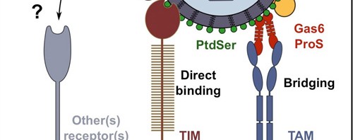
Ali Amara’s team performed genetic screening in order to identify cell receptors used by the virus to penetrate target cells[1]. The researchers have determined the important function played by the TIM receptors (TIM-1, 3, 4) and TAM receptors (AXL and TYRO-3) in the penetration process of the four Dengue serotypes. Mr. Amara’s team has succeeded in demonstrating that the expression of these 2 receptor families makes cells easier to infect. In addition, the researchers observed that interfering RNA or antibodies that target the TIM and TAM molecules considerably reduced the infection of the cells targeted by the Dengue virus. The TIM and TAM molecules belong to two distinct families of transmembrane receptors that interact either directly (TIM) or indirectly (TAM) with phosphatidylserine, an “eat-me” signal that allows the phagocytosis and the elimination of these apoptopic cells. Unexpectedly, the work of the Inserm researchers discovered that phosphatidylserine is abundantly expressed at the surface of virions and that it was essential that the TIM and TAM receptors recognize the phosphatidylserine to allow infection of target cells.

These results have helped to understand the first key stage in the Dengue virus infectious cycle, by discovering a new method of virus entry that works by mimicking the biological functions involved in the elimination of the apoptotic cells. The discovery of these new receptors has also opened the way for new antiviral strategies aimed at blocking bonding of the Dengue virus with the TIM and TAM molecules.

This research has been patent-protected by Inserrm Transfert.


[1] Up till present, only DC-SIGN receptors and L-SIGN receptors were known to play an active role in the penetration of the Dengue virus into target cells

Development of two tests for rapid diagnosis of resistance to antibiotics

Two new tests capable of rapidly diagnosing resistance to wide-spectrum antibiotics have just been developed by Inserm Unit 914 “Emerging resistances to antibiotics” (Bicêtre Hospital, Le Kremlin-Bicêtre) under the direction of Professor Patrice Nordmann. Thanks to these tests, it now takes only 2 hours to identify certain bacteria that are resistant to the most used and the most important antibiotics in hospitals. The main targeted bacteria are enterobacteriacae (such as E. Coli), that are responsible for infections. With their excellent sensitivity and specificity, the use of these extremely efficient tests on a world-wide scale would allow us to adapt antibiotic treatments to the individual’s needs and to be more successful in controlling antibiotic resistance, particularly in hospitals.

These works were published in September in two international reviews: Emerging Infectious diseases and The Journal of Clinical Microbiology.

These diagnostic tests will allow rapid identification of certain bacteria that are resistant to antibiotics and hence:

  • Allow us to better adapt the treatment to the infected patients
  • Avoid the inappropriate use of certain antibiotics, thus avoiding the over-use of certain wide-spectrum antibiotics
  • Isolate patients infected with these resistant bacteria and thus avoid the development of epidemics in hospitals

There is an ever-increasing number of emerging bacteria that cause cross-border epidemics. Researchers all agree on the fact that it is not the number of bacteria that is the problem, but their increasing resistance to antibiotics. The situation is particularly dramatic for certain species of bacteria, Gram-negative bacilli such as enterobacteriacae[1].


A worrying situation both for banal infections and for major treatments.

Whereas certain antibiotics such as wide-spectrum cephalosporins used to be reserved for the most serious cases, now there are cases where they are totally inactive against certain bacterial germs and consequently there is no effective antibiotic treatment for these. And so we are now faced with situations where the treatment of banal infection such as urinary or intra-abdominal infections has no effect. And this puts the life of the patients at risk. Every year, an estimated 25,000 deaths in Europe are due to multi-resistance to antibiotics.

Furthermore, the development of resistance to antibiotics affects an entire aspect of modern medicine that needs efficient antibiotics (grafts, transplants, major surgery, reanimation, etc.).

Undetected importation of multiresistant strains from foreign countries can also considerably accelerate the diffusion of this multiresistance phenomenon.

Two ultra-rapid tests: from Red to Yellow

In an attempt to slow down these increasing resistances, the Inserm researchers have developed a system that can rapidly detect the two enzymes responsible for causing resistance to the bacteria of two classes of common antibiotics: wide-spectrum cephalosprins and carpabenems. In these tests, the presence of an enzyme indicates the presence of a resistant bacteria.

These tests (Corba NP test and ESBL NDP test) are based on the acidification properties generated by the activity of the enzymes (ß-lactamases and carbapenemases) when they are in the presence of an antibiotic. If any one of these enzymes is present, the medium becomes acid and the acidity indicator (pH) turns from red to yellow (Figure, Corba NP test).

(Figure, le Carba NP test)

At present, these tests can be performed using bacteria isolated from urine samples taken during a detected infection, or from bacteria present in stools. The result is obtained in less than 2 hours (compared to 24 to 72 hours using current techniques). These tests are highly sensitive and highly reliable (100%). They are totally inoffensive since they are carried out on bacteria isolated from patients or on biological products such as urine, etc.

Patrice Nordmann, Inserm Research Director and main author of this work, points out that

“These tests are currently being assessed in order to ascertain their sensitivity directly from infected sites such as blood or urine”.

The invention of these tests is an important breakthrough in the fight against the resistance to antibiotics. These tests will provide a simple, inexpensive  means of very rapidly detecting the most serious cases of resistance to antibiotics in human medicine and will contribute to limiting international diffusion.

As Patrice Normann states “We can hope, in particular in many Western countries where the situation has not yet reached endemic proportions multi-resistances (France, in particular), to be able to preserve to a certain extent the efficiency of wide-spectrum cephalosporins and carbapenems, antibiotics used as a “last resource”.

Used straight at the patient’s bedside, these tests will help us to optimise the use of antibiotic treatment, in particular in the developing countries where the levels of resistance are extremely high.

Two international patent applications have been filed with Insert Transfert.


[1] Enterobaceriacae, including E. coli, usualy infest the human intestine. Given their proximity to the urinary and digestive tracts, urinary and digestive infections due to these germs are the most frequent.

Tuberculosis: newly discovered molecule in the immunity system plays a vital role in combating mycobacteria

Only 10% of individuals infected by Mycobacterium tuberculosis, which causes tuberculosis, go on to develop the disease. Why this should be is one of the questions Jean-Laurent Casanova and his team at Inserm Unit 980, “Human genetics of infectious diseases”/Université Paris Descartes, and their fellow researchers at New York’s Rockefeller University, asked themselves. To try and find an answer to this question, they set about studying the genetic components of human susceptibility to mycobacteria. The results of the study, published in this week’s issue of Science, reveal the key role played by a specific protein called ISG15 in immunity against mycobacteria

Tuberculosis is caused by a mycobacterium, chiefly Mycobacterium tuberculosis, also known as Koch’s bacillus. An estimated 25% of the world’s population is infected by tuberculosis. Of this number, 233 million men, women and children (10%) will develop clinical signs of the disease. Tuberculosis is currently responsible for 1.4 million deaths a year. Existing antibiotic treatments are becoming less effective and many vaccination campaigns end in failure. At least 50% of people who have been vaccinated do not develop any immunity. New strategies are therefore needed to combat tuberculosis effectively.

The question Jean-Laurent Casanova and his team have been trying to answer for more than 15 years is why all infected individuals do not go on to develop the disease. It was demonstrated a hundred years ago that identical twins, who share the same genetic material and an identical environment, are far more likely to both develop the disease than fraternal twins who are living in the same environment. That’s why the research team set out to prove that the likelihood of an infected individual developing the disease was determined by genetic factors.

The latest complete human genome sequencing techniques, combined with all the material resources at Rockefeller University, were put to work to identify these genetic components in children suffering from mycobacterial infections.

In 2010, the team identified the genetic etiology behind the illness in three children from two separate families. Two mutations in the ISG15 gene, resulting in a total loss of function, were observed.

Until then, the role played by ISG15 had primarily been described in vitro and in vivo in mice in antiviral immunity studies. ISG15-deficient laboratory mice were more likely to be infected by M. tuberculosis than wild mice.

In the article published in Science on 2 August, Jean-Laurent Casanova’s team explain how the ISG515 protein works. They show that it is a molecule secreted in response to the mycobacterial infection which induces the production of IFN-γ[1]. This research puts the spotlight on ISG15, a new player in the fight against mycobacterial diseases.

The new discovery opens up many new possibilities. From the medical angle, screening for new patients is underway and IFN-γ injections could provide an alternative therapeutic approach. From the scientific research point of view, gaining detailed insight into ISG15’s action mechanism and regulations will definitely teach us more about immunity against mycobacteria, which is a vital step forward in the fight against tuberculosis.


[1]   A “messenger” produced by the immunity system in response to a viral or bacterial attack. The crucial role of this messenger in combating mycobacteria has already been demonstrated.

(French) VIH : des globules blancs très actifs dans l’hyperactivation du système immunitaire

Sorry, this press release is only available in French.

(French) BCG et cancer de la vessie : vers un nouveau protocole pour les patients ?

Sorry, this press release is only available in French.

A new hope in the fight against tuberculosis

Researchers from the Institut Pasteur and Inserm, in collaboration with researchers from the University of Pisa, have uncovered the key role played by specific proteins in the virulence of the mycobacterium responsible for tuberculosis, Mycobacterium tuberculosis. They were able to create an attenuated strain of the mycobacterium, which confers a better protection against tuberculosis than the BCG vaccine to mice. This finding represents a major step forward in the scientific quest to develop a new vaccine, more efficient at fighting the disease. This study is being published today in the scientific journal Cell Host & Microbe.

© Institut Pasteur – Photograph of pulmonary tissues infected with Mycobacterium tuberculosis (in pink)

Tuberculosis is one of the most widespread diseases in the world. It is caused by an infection with the mycobacterium Mycobacterium tuberculosis, affecting one third of the world population. According to the World Health Organization (WHO), in 2010, 8.8 million people suffered from tuberculosis, of which 1.4 million died. Thus, M. tuberculosis remains to this day one of the most virulent and dangerous pathogens for man. Despite being effective for children, BCG does not protect adult men and women sufficiently against pulmonary tuberculosis. This type of tuberculosis is extremely contagious; hence, it is essential to create a new vaccine that is more successful at fighting this disease.

A study carried out by researchers from the Institut Pasteur and Inserm, coordinated by Dr Laleh Majlessi (1) and Pr Claude Leclerc (2) (Institut Pasteur/Inserm), in collaboration with Dr Roland Brosch (Institut Pasteur) and Dr Daria Bottai (University of Pisa), shows that a region of M. tuberculosis’ genome can be altered in order to obtain an avirulent strain of the bacterium in mice. This attenuated mycobacterium confers a strong protection against the onset of tuberculosis in a host organism.

The scientists were able to block, in the attenuated strain, the production and transport of certain proteins, called PE/PPE (3), linked to a specific region of the mycobacterium’s genome, the ESX-5 secretion system which can be found in all virulent strains of mycobacteria. They observed that the mice infected with the attenuated strain do not develop tuberculosis. Thus, they showed that the PE/PPE proteins produced by the ESX-5 system play a key role in the virulence of M. tuberculosis.

Furthermore, the scientists observed that the mice immunised with the attenuated strain are efficiently protected against infection by M. tuberculosis. This protection is correlated with the immune response specific to other PE/PPE proteins still present in this strain. Hence, the scientists demonstrated that the mutated strain of M. tuberculosis is a strong vaccine candidate against tuberculosis, triggering a more effective immune response than BCG in mice.

This major finding opens the door to new prospects for the development of a more efficient vaccine against the various pathologies caused by M. tuberculosis, especially against adult pulmonary tuberculosis. Numerous studies will need to be carried out before any of these research findings can be applied to man. The next step for the researchers of the Institut Pasteur, Inserm and the University of Pisa will be the creation of a strain in which other genetic mutations will be introduced in order to render it completely innocuous for man, with the aim to one day conduct clinical trials.

(1) Dr Laleh Majlessi, Immune Regulation and Vaccinology Unit (Institut Pasteur / Inserm U1041)

(2) Pr Claude Leclerc, director of the Immune Regulation and Vaccinology Unit (Institut Pasteur / Inserm U1041)

(3) The genes which encode the PE/PPE proteins belong to two large families that are unique to mycobacteria. They make up 10% of M. tuberculosis’ genome.

(French) Dengue : un anticorps prometteur caractérisé

Sorry, this press release is only available in French.

A new way to stimulate the immune system and fight infection

A study carried out by Eric Vivier and Sophie Ugolini at the Marseille-Luminy Centre for Immunology (Inserm/CNRS/Université Aix Marseille) has just reveal a gene in mice which, when mutated, can stimulate the immune system to help fight against tumours and viral infections. Whilst this gene was known to activate one of the body’s first lines of defence (Natural Killer, or ‘NK’ cells), paradoxically, when deactivated it makes these NK cells hypersensitive to the warning signals sent out by diseased cells. These new data are an essential step towards understanding the operation of these key cells in the immune system, and they could provide a new therapeutic approach to fighting infection. They also suggest that the operation of NK cells must be precisely regulated to guarantee an optimum immune reaction. Details of this work are published in the 20 January 2012 issue of the journal Science.

Our bodies are subject to attack by many different infectious particles (bacteria, viruses, etc.), which surround us in our everyday environment. Various immune cells are activated to fight off these attacks: the first response is from the innate immune cells (1), which gradually give way to the memory B and T lymphocytes of the adaptive immune system. The Natural Killer (NK) cells are a part of this first line of defence of the organism. They can selectively kill tumour cells or cells infected by microbes whilst secreting chemical messengers known as cytokines, which stimulate and direct the response of the B and T lymphocytes.

Following the launch of a major genetics programme a few years ago, scientists succeeded in revealing a gene whose deactivation causes heightened functioning of the NK cells (see figure below).

The normal mice all died within eight days following infection by a virus (cytomegalovirus), but all mutant mice were resistant to the same infection.

© With kind permission from the journal Science

The normal mice all died within eight days following infection by a virus (cytomegalovirus), but all mutant mice were resistant to the same infection.

This gene, called Ncr1, contributes to the manufacture of the receptor NKp46, which is present on the surface of NK cells. Surprisingly, its role in activating the NK cells has been known for several years.

‘NK cells go through various stages of development before combating microorganisms or tumour cells,’ explains Sophie Ugolini, joint author of the paper. ‘Without this receptor, the NK cells are more reactive and therefore more effective when they encounter the attackers of the organism.”

To test the therapeutic potential of their discovery, the scientists blocked the NKp46 receptor using a drug (in this case, a monoclonal antibody). As in the genetics experiments, this treatment that blocks NKp46 makes the NK cells much more effective.

‘Our aim now is to further explore the underlying biological mechanisms and to work in collaboration with the biopharmaceutical industry and the hospital to evaluate the medical potential of this new type of treatment, particularly for patients whose immune system has already been weakened, such as patients with an immunodeficiency and those who have had a bone marrow transplant or chemotherapy,’ concludes Eric Vivier.

Footnote

(1) Innate immunity is a front-line defence system against tumours and microbes. It immediately acts against microbial agents that come into contact with an organism. Innate immunity is present in all living organisms, and plays an essential role in activating the adaptive response in vertebrates. This compartment of immunity hit the headlines recently when Jules Hoffman of France, Canadian Ralph Steinman, and Bruce Beutler from the USA (another co-author of this paper) received the Nobel Prize for their work on innate immunity and its close links with the adaptive immune system.

fermer