Menu

Partially hydrolysed (hypoallergenic) baby formulas do not reduce allergy risks in infants

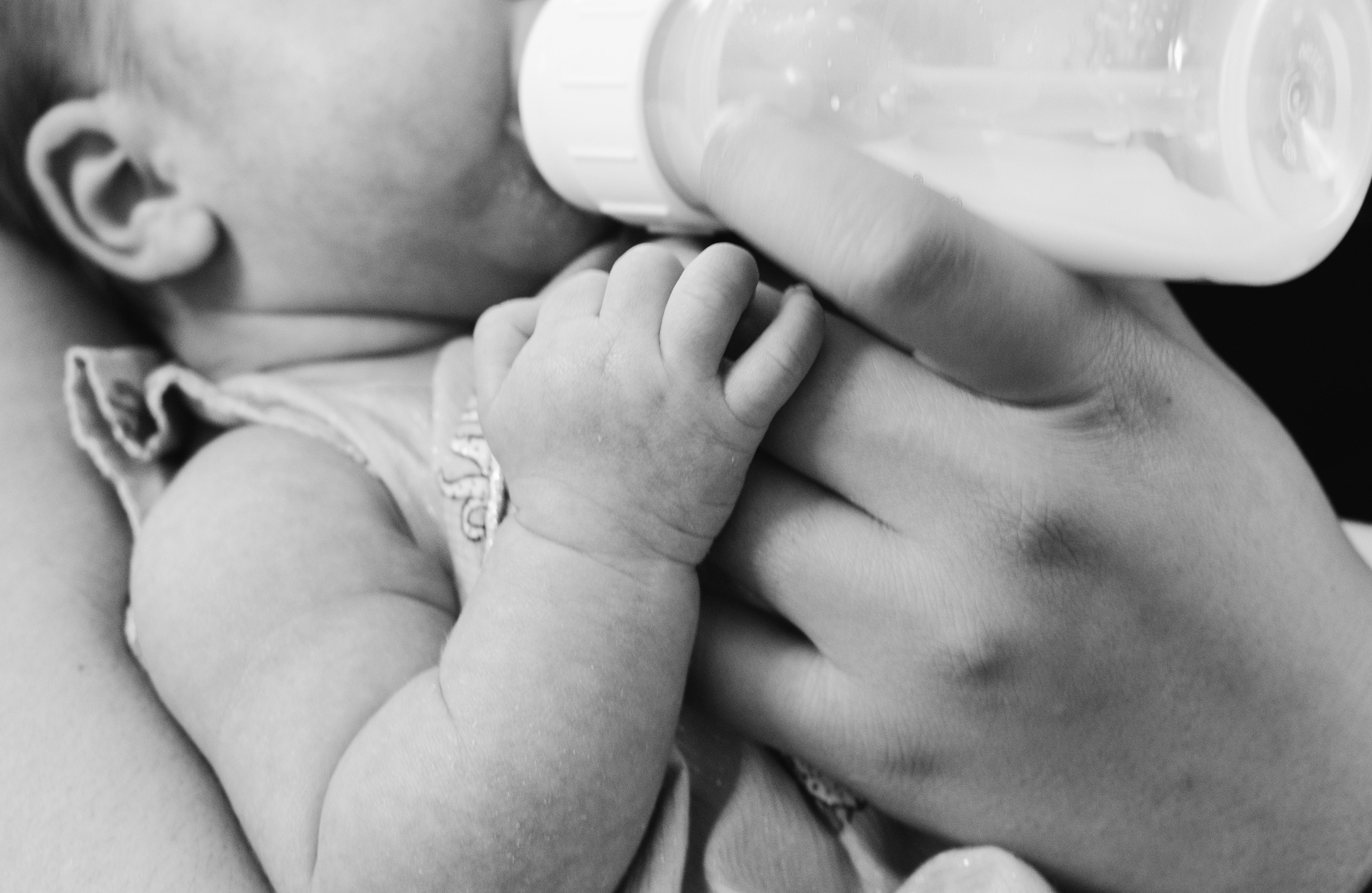
©Photo Kelly Sikkema on Unsplash

Often, hypoallergenic formulas are recommended for infants who are not exclusively breastfed and who are at risk of developing allergies. While it is assumed that such formulas reduce the development of allergies later in life, it is unclear whether this claim is true. A team of INRA and INSERM researchers have shown that the consumption of partially hydrolysed (hypoallergenic) formulas was not associated with a reduced risk of developing allergies, findings that were recently published in the journal Pediatric Allergy and Immunology. This research comes out of ELFE, the first large-scale longitudinal study in France that will track subjects from birth to adulthood. While this work cannot assess causality, it underscores the necessity of performing clinical studies before marketing such formulas as hypoallergenic.

Partially hydrolysed formulas are a class of hypoallergenic formulas that contain pre-processed proteins, which are smaller in size. These formulas are supposed to prevent infants from developing allergies. As a result, certain professional associations and health organisations recommend their use when an infant has at least one parent or sibling with allergies. However, the actual effectiveness of these formulas is the subject of debate. Little is known about their role in limiting the risk of allergies under real conditions of use. Certain professional associations of paediatricians, like the American Academy of Pediatrics and the Swiss Society of Paediatrics, have recently stated that they no longer recommend their usage.

INRA and INSERM researchers wanted to examine the relationship between the consumption of partially hydrolysed formulas and the occurrence of allergy-related conditions, such as eczema, wheezing, asthma, and food allergies. For two years, they followed 15,000 children participating in ELFE (the French Longitudinal Study of Children), which is being carried out by INED and INSERM. The first of its kind in France, this ambitious study seeks to broaden our understanding of how environmental factors affect the development, health, social lives, and academic trajectories of children. ELFE is particularly interested in examining the effects of diet in infants.

The researchers found that, among the two-month-old infants being fed formula, 5% were given partially hydrolysed formulas. However, in half of these children, their use was not justified because there was no familial history of allergies.

When these children were two years old, the researchers observed no evidence that partially hydrolysed formulas had reduced the development of allergies, as compared to what was observed in children who had received regular baby formula. In contrast, when two-month-old infants who did not display any allergies were given partially hydrolysed formulas, they were more likely to develop respiratory problems and food allergies in the years that followed.

New research should build on these epidemiological results. Indeed, they provide support for new EU legislation, which will come into effect in 2021 and require that clinical studies be carried out on such products before they can be marketed as reducing allergy risks.

New prebiotics: benefits without the downsides?

©Photo by Gesina Kunkel on Unsplash

Prebiotics are currently a preferred treatment for certain metabolic disorders, as they can restore the balance of dysfunctional gut microbiota, and improve the body’s metabolism. However, these substances have to be used at high doses, which can result in patients experiencing bloating and flatulence. A research group led by Matteo Serino, Inserm researcher at the Digestive Health Research Institute (Inserm/Inra/ENVT/UT3 Paul Sabatier), has recently shown three plant extracts to have prebiotic effects in obese/diabetic mice, with a shorter duration of treatment and at lower doses to the prebiotics currently in use. For Matteo Serino, this raises the question: do we need to reconsider the protocol for prebiotic administration? These results have been published in Molecular Nutrition & Food Research.

Dysbiosis of the gut microbiota corresponds to an imbalance in the populations of gut microorganisms and/or a change in their activity. This imbalance is often associated with metabolic disorders such as obesity and type 2 diabetes.

It has been shown that such dysbiosis can be targeted with prebiotics, long molecules that are not digested by the body but which increase the growth and activity of certain bacteria and thus improve patients’ carbohydrate and lipid metabolism. However, the duration of treatment is often greater than 4 weeks, and the effective dose at which an improvement in health starts to be observed can result in adverse effects such as bloating and flatulence.

A team of researchers from the Digestive Health Research Institute sought to identify other substances that might have a prebiotic effect. This led them to study extracts from three plants that had previously shown some beneficial effects: a red alga called Porphyra umbilicalis, the leaves of the lemon balm plant, Melissa officinalis L., and fruit extracts from an Indian plant, Emblica officinalis Gaertn.

The researchers administered these extracts to obese and diabetic mice for just two weeks, and at doses proportionally lower than the effective doses of traditional prebiotics.

The team considered a range of criteria in evaluating the individual prebiotic effects of the plant extracts.

The researchers first measured the impact of these substances on the diversity (the number of bacteria from different populations) and activity of the gut microbiota before and after treatment. “Our results were broadly positive for microbial diversity, with all the extracts increasing the abundance of at least one group of bacteria,” explains Matteo Serino, the Inserm researcher who led the study.

Beyond the impact on the microbiota, improved metabolism was one of the key criteria for evaluating the prebiotic efficacy of an extract. The research team observed that each of the three substances produced an increase in the gut of the quantity of the various short-chain fatty acids that are produced by bacterial metabolism of prebiotics and contribute to improving host metabolism.

The researchers also measured the presence of various markers in the blood that would enable them to demonstrate any metabolic improvements. The best result was obtained with the extract from the red alga P.umbilicalis. The researchers measured a 30% decrease in the level of blood triglycerides, large molecules with long-chain fatty acids that are present in excess levels in the blood of patients with metabolic disorders. This finding thus suggests an improvement in lipid metabolism among the treated animals.

“What’s interesting to note is that all of these effects were observed after just two weeks of treatment, and at low doses. This suggests the need for further studies to find out if this type of treatment could be effective in humans, and to test whether the extracts currently in use remain effective when given at low doses, thus limiting their side effects. That would mean reconsidering the existing protocol for prebiotic administration,” concludes Matteo Serino.

High-risk pregnancy: the interferon effect

Artist’s representation of cells and placenta © Fabrice Hyber – Organoïde-Institut Pasteur/Fluorescence microscopy of fused cells (in green) and nucleus (in blue). © Institut Pasteur

Discovery of a cellular mechanism involved in abnormal placental development during some high-risk pregnancies.

High-risk pregnancies occur frequently and may be caused by various factors. It is estimated that 10 to 20% of pregnant women miscarry during their first trimester of pregnancy. Slow fetal growth may also arise as a result of maternal infection with certain microbes, parasites or viruses (such as toxoplasmosis or infection with rubella virus, cytomegalovirus, herpes or Zika) or because of genetic or autoimmune diseases. Teams from the Institut Pasteur, the CNRS, Inserm, Necker-Enfants Malades Hospital (AP-HP) and Université de Paris have identified a new cellular mechanism that alters placental development, potentially causing serious complications during pregnancy. The mechanism is linked with the production of interferon, a molecule produced in response to infection, especially viral infection. The findings are published in Science on July 11, 2019.

The placenta is both a surface for exchange and a barrier between mother and fetus – it delivers nutrients needed for fetal growth, produces hormones and protects the fetus from microbes and the maternal immune system. The external layer of the placenta, known as the syncytiotrophoblast, is composed of cells which fuse together, forming giant cells that are optimized for the placenta’s barrier and exchange functions. Cell fusion is mediated by a protein known as syncytin. If the syncytiotrophoblast fails to form correctly, it can cause placental insufficiency and hinder fetal development. An abnormal syncytiotrophoblast can be observed in conditions such as slow intrauterine growth, the lupus and in women whose fetus has Down syndrome.

Interferon is a substance produced by immune cells during infection to combat viruses and other intracellular microbes. High levels of interferon are observed in autoimmune or inflammatory diseases such as lupus, and also in some infections. In this study, the scientists demonstrated that interferon is responsible for placental abnormality and that it acts by preventing syncytiotrophoblast formation. Specifically, interferon induces the production of a family of cellular proteins known as IFITMs (interferon-induced transmembrane proteins), which block the fusion activity of syncytin.

IFITM proteins are beneficial since they prevent viral fusion with cellular membrane, thereby stopping viruses from entering and multiplying within cells. The scientists used experimental models and human cells to demonstrate that this beneficial effect can nevertheless be harmful if IFITM proteins are produced in an important level in the placenta.

“Identifying the role of IFITMs gives us a better understanding of the mechanisms involved in placental development and how it may be disrupted during infections and other diseases,” comments Olivier Schwartz, Head of the Virus and Immunity Unit at the Institut Pasteur and joint last author of the paper. The scientists want to investigate whether placental pathologies of unknown etiology, such as some early spontaneous abortions and occurrences of preeclampsia, also involve IFITM proteins. In the longer term, blocking the effects of IFITMs could represent a new therapeutic strategy to prevent interferon-related placental abnormality.

In addition to the institutions mentioned above, this research was funded by the ANRS, Sidaction, the French Vaccine Research Institute (VRI), LabEx IBEID and the European Research Council (ERC).

Anaphylactic shock: IgG antibodies and neutrophils play an unexpected role

Antibodies © Adobe Stock

Anaphylactic shock, an exacerbated allergic reaction that can prove fatal, is sometimes caused by the use of drugs during surgery. In most of these extreme reactions, evidence can be provided that patients have anti-drug antibodies of the IgE class. In 10 to 20% of anaphylactic cases evidence for the involvement of anti-drug IgE is lacking. Anti-drug IgE enable activation of mast cells and basophils that release histamine, a potent mediator involved in anaphylaxis. Teams from the Institut Pasteur, Inserm, the Paris Public Hospital Network (AP-HP), the CNRS, Paris-Sud University and Sorbonne University have successfully identified a new pathological mechanism responsible for these previously unexplained cases, involving neutrophils activated by antibodies of the IgG class. These findings, published on July 10 in the journal Science Translational Medicine, will help improve diagnosis and treatment for patients with this type of shock.

Anaphylaxis is a hyperacute allergic reaction caused by an inappropriate immune response following the introduction of a usually harmless antigen into the body. When this antigen binds to antibodies already in the body, it triggers the secretion of large quantities of potent vasodilating mediators, sending the patient into a state of shock that may result in multiple organ failure and even death.

Anaphylaxis may be brought about by various substances, including drugs (antibiotics or neuromuscular blocking agents), food and insect venom. In this study, the scientists focused on allergic reactions to neuromuscular blocking agents (NMBAs), drugs used during general anesthesia to induce muscle relaxation.

The incidence of anaphylactic shock caused by NMBAs is one case in every 10,000 to 20,000 surgeries, representing approximately 3 to 5 cases each week in the Greater Paris area.

Although it was already known that IgE antibodies could cause anaphylaxis, teams from the Institut Pasteur, Inserm, the Paris Public Hospital Network (AP-HP), the CNRS, Paris-Sud University and Sorbonne University have demonstrated in a clinical study that IgG antibodies can also be involved in drug-induced anaphylactic shocks. This unexpected role of IgG antibodies had already been identified in mice in 2011 by some of the same authors.[1]

This multicenter study known as “NASA”, led by Bichat Hospital (part of the Paris Public Hospital Network), was launched in 2012 by a consortium of scientists, clinical biologists and anesthetists. The consortium monitored 86 patients with perioperative anaphylactic shock and 86 control patients in 11 hospitals in the Greater Paris area, coordinated at Bichat Hospital by immunologist Professor Sylvie Chollet-Martin (Paris-Sud University) and anesthetist Professor Dan Longrois. Blood samples were taken as soon as an anaphylactic shock occurred in the operating room, enabling the scientists to identify the alternative IgG-dependent mechanism. They demonstrated that IgG antibodies activate neutrophils (50-70% of our white blood cells), releasing high doses of harmful vasodilating mediators. Neutrophil activation was more pronounced in cases of severe shock than in cases of moderate shock. Interestingly, the IgG-neutrophil pathway was also identified in most cases of shock where the traditional IgE-dependent mechanism was observed, suggesting that IgGs and neutrophils may contribute to the severity of most cases of shock via an additive effect.

“These findings elucidate 10 to 20% of cases of anaphylactic shock that previously had no biological explanation. They will be extremely valuable in refining diagnosis in these patients and avoiding any future exposure with the drug that triggered the allergic reaction,” explains Professor Sylvie Chollet-Martin (Paris-Sud University), joint last author of the study and Head of the Immunology laboratory on Autoimmunity and Hypersensitivity at Bichat Hospital.

“Although IgG antibodies are known to protect the body from infection and to act as aggressors in some autoimmune diseases, this study reveals that they may be involved in humans in another reaction that is harmful for the body, anaphylaxis. We are currently carrying out experimental research to explore how we might block this new activation pathway for IgG antibodies so that we can propose a therapeutic solution,” comments Pierre Bruhns, joint last author of the study, Inserm Research Director and Head of the Institut Pasteur’s Antibodies in Therapy and Pathology Unit.

This research was funded by the organizations mentioned in the first paragraph and by the European Research Council (ERC – FP7; MyeloSHOCK project).

[1] Mouse and Human Neutrophils induce Anaphylaxis, The Journal of Clinical Investigation, March 23, 2011. doi: 10.1172/JCI45232

New Antibiotics Developed by Inserm and Université de Rennes 1

Résistances émergentes aux antibiotiques

Bacteria culture in a Petri dish© Inserm/Latron, Patrice

Not only are they effective against Gram-positive and negative multi-resistant bacteria, they also appear not to trigger resistance when used to treat infection in mice. Such are the promises of the two new antibiotics created by Prof. Brice Felden and his team at the Inserm and Université de Rennes 1 ‘Bacterial Regulatory RNAs and Medicine’ joint laboratory (U1230), in conjunction with a team from the  Rennes Institute of Chemical Sciences (CNRS/Université Rennes 1/ENSCR/INSA Rennes). This French advance could bring both fresh impetus and new possibilities for fighting antibiotic resistance worldwide. Details on this research will be published July 9 in Plos Biology.

Antibiotics have saved so many lives over the previous century of their use in humans that they are considered to be one of the major breakthroughs of contemporary medicine. Unfortunately, growing resistance is gradually rendering them ineffective, with the threat of catastrophic public health consequences should this trend continue much longer. The few new antibiotics being brought to market essentially consist of so-called me-too drugs – meaning that they are derived from existing classes of antibiotics.

Researchers from Inserm and Université de Rennes recently identified a new bacterial toxin which they transformed into potent antibiotics active against various bacteria responsible for human infections, whether Gram-positive or negative.

“It all started with a fundamental discovery made in 2011”, explains Brice Felden, Director of the Bacterial Regulatory RNAs and Medicine laboratory in Rennes. “We realized that a toxin produced by Staphylococcus aureus whose role is to facilitate infection is also capable of killing other bacteria present in our body. What we had identified was a molecule with dual toxic and antibiotic properties. We thought that if we could separate these activities, we would be able to create a new antibiotic non-toxic to the body. A challenge that we accepted!”.

In conjunction with the team of ISCR chemist Michèle Baudy Floc’h, a new family of so-called peptidomimetics was synthesized. As their name suggests, these peptides are inspired by the existing natural bacterial peptides but have been shortened and modified. Out of the twenty molecules created, two proved effective against resistant Staphylococcus aureus and Pseudomonas aeruginosa in mouse models of severe sepsis or skin infection. In addition, no toxicity to the other cells and organs, whether in animals or human cells was observed. These new compounds are well tolerated at their active doses – and even beyond – and are devoid of the renal toxicity issues often encountered with this type of compound. “We tested them at doses 10 to 50 times higher than the effective dose without seeing toxicity” specifies Felden, adding that “the participation and imagination of the team and our chemist colleagues was needed to devise the most active molecules possible”.

Little resistance observed under experimental conditions

Important to note was that the bacteria that the researchers had left in contact for several days in the animals with these antibiotics showed no signs of resistance. In order to go further, the researchers created conditions favorable to the development of resistance in vitro and in vivo – with nothing happening. However, caution is still required here given the short experimental time periods (up to 15 days).

The antibacterial activity of these peptidomimetics is partially due to the capacity of its non-natural amino acids to reinforce the association of these compounds with the membranes of the infectious bacteria. This strong binding leads to membrane permeability and the death of the bacteria. “We think these new molecules represent promising candidates for the development of new antibiotics that can provide alternative treatments to antimicrobial resistance.”

The next step involves launching phase I clinical trials in humans. The patent has been licensed and a start-up created.

Watch the interview of Prof. Brice Felden on YouTube (french with english subtitles).

In France, 2 out of every 100 women are subjected to violence during their pregnancy

kieferpix/iStock

Violence against women, including during pregnancy, constitutes a major public health problem. It can result in death, disability, and a range of mental and physical health issues, for both the mother and the unborn child. A joint team of researchers from Inserm and the University of Paris, working at CRESS (Center for Epidemiological and Statistical Research), has investigated the frequency of physical abuse during pregnancy, its consequences for the mother and child, and the characteristics associated with this violence using data from the 2016 French National Perinatal Survey, which included over 12,000 women. Their results, published in the Maternal and Child Health Journal, estimate that 1.8% of these women were subjected to physical violence during their pregnancy.

Although the figure is difficult to estimate and varies across different regions, approximately 1/3 of women around the world are victims of physical or sexual abuse, by their partner or another individual.

When these women are pregnant, this can have a highly damaging impact on both their health and that of their unborn child. But previously there was no data available on physical violence during pregnancy specifically, at a national level.

This gap has now been filled thanks to the work of a mixed research unit (EPOPé) bringing together Inserm and University of Paris researchers at CRESS. The researchers studied this phenomenon using data collected during the National Perinatal Survey, which was conducted in 2016 among 12,330 women in public and private maternity units in France. (See Rapport: Enquête nationale périnatale 2016. Les naissances et les établissements, situation et évolution depuis 2010 [National Perinatal Survey 2016 Report: Births and Institutions, Status and Change Since 2010]). The objective of this study, led by Marie-Josèphe Saurel-Cubizolles, was to understand the frequency of physical violence during pregnancy, the risk factors, the impact on the mother and child, and the key stages at which measures can be implemented to reduce the incidence of such violence.

The researchers estimated that 1.8% of all the women surveyed had been subjected to physical violence during their pregnancy, and found that this violence was associated with various characteristics.

In particular, women who were not in a cohabiting couple, whether they were in a non-cohabiting couple or had no partner, were more likely to be victims. The frequency of violence was also strongly linked to household income, and was higher among women living in households with fewer financial resources. Finally, certain behaviors, such as tobacco or cannabis consumption by the mother-to-be, were more common in cases of physical violence.

The researchers also showed that this violence had a highly damaging impact on the mother and child, with 62% of the women who had suffered physical abuse stating that they were psychologically distressed during the pregnancy, compared to 24% of other women. For the newborn child, this violence was associated with a higher risk of being born prematurely or transferred to an intensive care unit.

Improved understanding of the factors associated with situations of violence, and demonstration of the impact on the mother and child, should help healthcare professionals develop preventive and protective strategies, and prompt them to raise the issue during antenatal consultations.

fermer