Menu

A step closer to morphine without side effects?

Morphine has been used for centuries to relieve severe pain. However, its pain-relief properties are coupled with considerable side effects. Morphine imitates the action of molecules produced naturally by the brain (endorphins). So why does it have such harmful side effects? This question has recently been elucidated by Sébastien Granier, researcher at the Institute of Functional Genomics (Inserm/CNRS/Universités de Montpellier 1 and 2) and his team, in conjunction with collaborators in the US. The 3D structure of brain receptors is likely to differ, depending on the type of molecule bound upon it (morphine or endorphin): the body’s response will be completely and accordingly modified. Thanks to this discovery, the researchers have now set their sights on retaining the benefits of morphine while eliminating any undesired side effects.

The results were published in the Nature review on 21 March 12.

Opium is a natural poppy (papaver somniferum) extract and is one of the oldest drugs known to man, used for its psychotropic, sedative and analgesic properties. These effects are caused by its major component: morphine. Today morphine has widespread clinical pain-relief applications.

Morphine’s action is relayed by μ-opioid receptors expressed at the surface of cells in the central nervous system. These receptors are part of a superfamily of proteins, G protein-coupled receptors (GPCR), which are the target of around 30% of drugs currently on the market.

At a molecular level, morphine binds with μ-opioid receptors to imitate the action of molecules produced naturally in the brain: endorphins. However, its clinical use is limited by two effects. Firstly, the development of a tolerance phenomenon means that, in the case of repeated injections, the morphine dose must be increased to obtain the same therapeutic effect. Secondly, morphine consumption can lead to drug dependency (heroin, the acetylated form of morphine, is the most obvious example). Furthermore, morphine consumption has serious side effects: respiratory depression, constipation, physical and psychic dependency. These harmful effects can be explained by the fact that morphine triggers a cellular response that differs from that triggered by endorphins. Since morphine and endorphins bind with the same receptor, the two molecules stabilize the μ-opioid receptors in separate spatial conformations that cause the differences in biological responses.

It is therefore essential to understand the structural bases of morphine action, and opioids in general, if we are to develop molecules that retain the beneficial effects of morphine whilst eliminating the side effects,” explains Sébastien Granier, Inserm researcher and project leader.

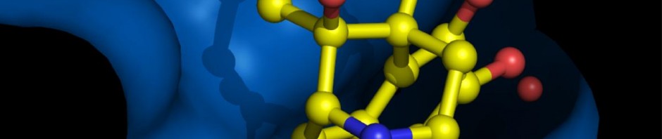
The research produced by Sébastien Granier (and Brian Kobilka’s team in Stanford) resolved the 3D structure of μ-opioid receptors when associated with a molecule that has a similar chemical structure to morphine.


© Kobilka Lab
3D representation of μ-opioid receptors when associated with morphine antagonistic.

© Kobilka Lab

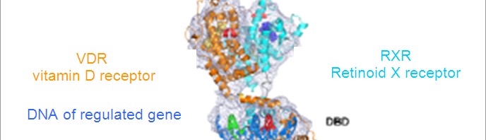
3D representation of a μ-opioid receptor dimer.

The images of the 3D structure above show that the specific area in the receptor where the opioid molecule binds is mostly open to the external medium, thus explaining the rapid action of these components.

The structure also reveals a very important characteristic of this receptor function: the formation of a receptor dimer (1). “This is the very first time that we have succeed in producing images of the 3D structure of a complex of this kind for this receptor family” he adds. This structure with a μ-opioid receptor dimer opens up avenues of research to study this phenomenon and improve understanding of these functional implications.

The 3D structure resolution of the μ-opioid receptor holds the key to treating pain and addictions, and could, in the long-run, lead to the production of new analgesic medicine with no side effects.

Footnote

(1) Molecule produced by ‘associating’ two identical molecules

Vitamin D receptor: first full 3D observation

For the first time, a team from the Institute of Genetics and Molecular and Cellular Biology (IGBMC, Université de Strasbourg/CNRS/Inserm) has succeeded in taking a full, 3D photograph in HD (1) of a small vital, molecule, enclosed at the heart of our cells: the vitamin D receptor (VDR). Published on 18 January 2012 in the EMBO Journal, this study provides key information regarding the 3D structure of the receptor and its action mechanism at a molecular level. This data is crucial for pharmaceutical research, since the VDR is involved in several diseases, such as cancer, rickets and type 1 diabetes.

The vitamin D receptor (VDR) is part of, and plays a crucial role in, what biologists refer to as the “large family of nuclear receptors”: proteins that are active in cell cores, including “steroid” receptors (sexual hormone receptors, etc.). It regulates the expression of genes involved in diverse and vital biological functions (cell growth, bone mineralisation, etc.).

© IGBMC (CNRS / Inserm / Université de Strasbourg)

Until now, researchers had only been able to study two parts of this receptor close-up: the region that interacts with DNA and the vitamin D-binding domain. These two parts were produced in a laboratory and their structu[]re was studied individually using the crystallography technique. This method did not make it possible to visualize VDR fully since it proved to be difficult to crystallize.

To overcome this challenge, by combining the skills of several teams from across the globe for more than 15 years, the teams led by Bruno Klaholz and Dino Moras, each CNRS research directors at the IGBMC, used an innovative technique: cryo-electron microscopy (cryo-EM), which requires the latest-generation electronic “high-definition” microscope. This marvel of technology can be used to view biological objects at the molecular, or even atomic, scale. In France, the first microscope of this kind was installed at the IGBMC (2) in 2008. Prior to this research, many people thought it was impossible to study VDR using cryo-EM. Until now, the smallest molecules that had been viewed using this technique weighed more than 300 kilo Dalton (3) (kDa), or even thousands of kDa, i.e. much more than the VDR, which weighs 100 kDa and measures just 10 nm (10 x 10-9 m).

In concrete terms, Bruno Klaholz and his colleagues have laboratory-produced large qualities of the human VDR receptor inEscherichia coli bacteria (one of the most commonly-used models in biology to produce proteins). They then isolated to receptor in a physiological solution containing water and a little salt. The sample containing VDR was then frozen and immersed in liquefied ethane, which produced extremely rapid cooling (in a fraction of a second, the sample passes from 25°C to approximately -184°C). Using the microscope, approximately 20,000 photos were required of VDR particles in different directions. It is these images, aligned and combined using a software program, which finally resulted in a 3D reconstruction of VDR.

This image has provided hitherto unknown information regarding how the receptor functions. It reveals that the VDR and its partner RXR (retinoid X receptor, a vitamin A derivative) form an open architecture, with the vitamin D-binding domain oriented almost perpendicularly to the DNA binding domain (see Figure below). This structure suggests cooperation between the two domains, which may work together to induce a more tight regulation of the expression of target genes.

This ground-breaking work paves the way for research into several other vital nuclear receptors, which are yet to be thoroughly investigated. In particular, biologists are now envisaging using cryo-EM to reveal the structure of steroid receptors.

View of 3D architecture of two receptors, the VDR (vitamin D receptor) and its partner RXR (retinoid X receptor, a derivative of vitamin A), after 3D reconstruction using images of individual particles. The purple mesh represents the experimental 3D map obtained through cyro-EM. The specific binding sites for DNA fragment are indicated in green and red, the ADN binding domains (BDB) and ligand binding domains (LBD) are indicated.

Footnotes

(1) 12 angstroms: 12 x10-10 metres (one angstrom corresponds to the average diameter of an atom).

(2) The second was inaugurated in February 2011 at the Institute of Structural Biology (CEA / CNRS / UJF) in Grenoble.

(3) One Dalton is, with relatively accurate precision, the mass of a hydrogen atom. A protein amino acid represents approximately 110 Da, an assembly of 100kDa contains approx. 900 amino acids.

fermer