Menu

Huntington’s disease: therapeutic potential of triheptanoin confirmed

A team of researchers from Inserm led by Fanny Mochel and located at the Brain and Spinal Cord Institute (Inserm/CNRS/UPMC/AP-HP) has just demonstrated the therapeutic potential of triheptanoin in ten patients with Huntington’s disease. Derivatives of this triglyceride, with its unique composition, might be able to slow the progression of the disease by improving the energy metabolism of the brain. This research is published in the journal Neurology.PhotoCP web

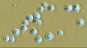
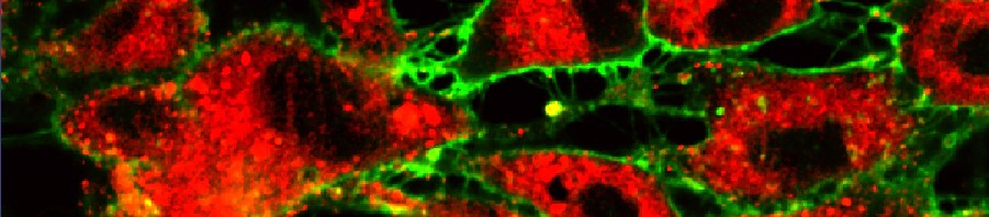
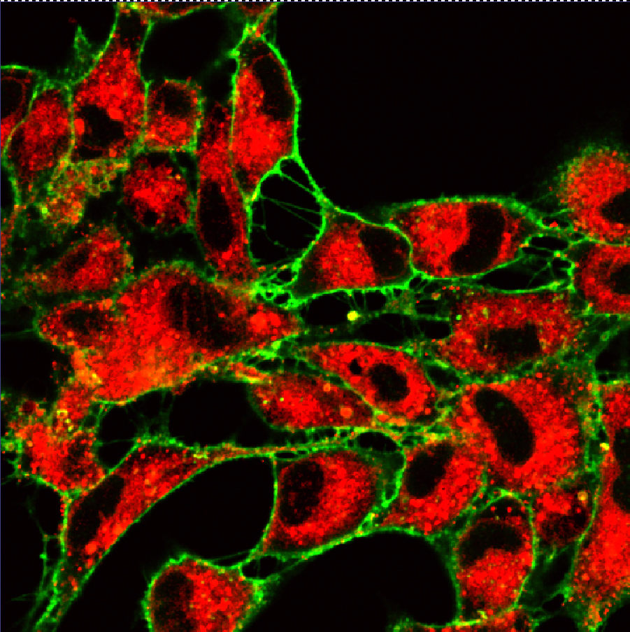
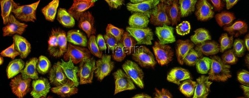
Study of the Huntington’s disease. © Inserm/Saudou, Frédéric

Huntington’s disease is a genetic disease that affects approximately 5,000 people in France, with another 10,000 individuals at risk of developing it. Mutation of the gene encoding the huntingtin protein results in a progressive degeneration of the neurons, especially in regions of the brain involved in the control of movement, thereby causing serious neurological, motor, cognitive and psychiatric problems. Weight loss is also observed in patients at an early stage in the disease, despite normal or even increased food intake. These two observations (degeneration of neurons and loss of weight) led the researchers to propose the hypothesis that an energy deficit in these patients might play an important role in the onset and progression of the disease symptoms.

In order to validate this hypothesis, the researchers had to succeed in measuring energy fluctuations in the brain during certain brain activation states. To do this, they needed to develop technologies enabling the measurement of compounds that reflect the energy metabolism of the brain in a dynamic manner.

“We observed that variations in the relative concentrations of two phosphorus-rich energy compounds (Pi/PCr) perfectly reflect this dysfunction in brain energetics,” explains Fanny Mochel, who coordinated this work.

In practical terms, this relationship was measured using NMR.[1] While energy production in their brains was being recorded, the nine patients and thirteen healthy volunteers were asked to perform a simple mental “effort” task causing their brains to consume energy. For this, they were subjected to a visual stimulus in the form of a flashing checkerboard for several minutes.

The Pi/PCr ratio was clearly reduced in patients compared with control subjects, indicating that their brain metabolism was impaired when faced with this effort. Once this biomarker was validated, the researchers were then able to test the efficacy of potential treatments in re-establishing normal brain metabolism.

A pilot clinical trial…

For one month, ten patients with Huntington’s disease underwent a treatment based on triheptanoin, a triglyceride (in the form of an oil to be taken with meals), in order to improve energy metabolism in their brains. At the end of this therapeutic trial, their energy profiles had become normal. This treatment was not chosen at random, since it had already proved effective in patients with rare diseases who were unable to use certain lipids present in their diet. The idea was to compensate for generalised energy dysfunction by supplementing the diet with specific metabolites that could restore the body’s energy metabolism to normal. The novelty of the approach proposed by Dr Mochel is the application of this energy treatment concept to diseases that affect the brain, such as Huntington’s disease.

…Prior to a larger-scale trial.

On the basis of these results, the researchers will launch a one-year double-blind trial in France and the Netherlands at the beginning of 2015, in approximately one hundred patients, using clinical and imaging parameters as assessment criteria.

“The complexity of the disease obliges us to design additional therapeutic approaches in order to target different aspects—genetic, metabolic, inflammatory, etc.—of the disease. Used together, these approaches will probably have the best chances of success, such as happens with other, more common diseases,” concludes Fanny Mochel.

These results, which are patent-protected, were the subject of a recent licensing agreement negotiated and signed by ICM (the Brain and Spinal Cord Institute) and Inserm Transfert with the American company Ultragenyx, which will launch a new clinical trial in the area of Huntington’s disease.


[1] Nuclear magnetic resonance (NMR) is a noninvasive method for studying the biochemistry and metabolism of the central nervous system. It enables the accurate quantification of several tens of molecules. (Source: French Atomic Energy Commission, CEA)

A mechanism for eliminating proteins accidentally localised to the cell nucleus

An international collaboration coordinated by the German Cancer Research Center (DKFZ) (University of Heidelberg), including French researchers from the Institute of Genetics and Development of Rennes (IGDR) (CNRS/University of Rennes 1) under the leadership of Gwenaël Rabut, Inserm Researcher, and teams from Sweden and Canada, has just demonstrated a new molecular mechanism that may allow cells to destroy proteins accidentally localised to the nucleus.

This research is published in the journal Nature.

Biological processes are far from perfect. Despite millions of years of refinement, the molecular mechanisms that help living beings to function make many errors, which can have serious consequences unless they are detected and corrected. For example, many cancers are caused by errors that occur while our genetic material is being copied. Similarly, incorrect folding of some neuronal proteins leads to the formation of toxic aggregates that disrupt nervous system function and cause neurodegenerative diseases, such as Alzheimer’s disease or Parkinson’s disease.

To prevent this happening, cells have established complex molecular mechanisms that control the quality of proteins and eliminate those that are defective. These mechanisms are localised and implemented mainly in the cytoplasm (the cellular compartment where the proteins are synthesised).

While working on several factors involved in protein quality control, researchers discovered that some of them are also localised in the cell nucleus (the compartment that contains the genetic material), and that they enable the degradation of proteins that are abnormally present in this compartment.

During this study, researchers from the Institute of Genetics and Development of Rennes (including Gwenaël Rabut, Inserm Researcher, project coordinator and manager in Rennes, and Ewa Blaszczak, doctoral student, joint first author of the article) were able to observe that these factors involved in protein quality control interact with each other in the nucleus and bring about the ubiquitination (the step preceding degradation) of a protein accidentally localised to the nucleus.

By using an observation method developed at the University of Heidelberg, based on fluorescence timing in the proteins of interest, the researchers were able to identify some twenty proteins the degradation of which depended on quality control factors localised in the nucleus. Since several of these proteins are normally localised to the cytoplasm, and accumulate in the nucleus when they are no longer degraded, the researchers propose that this quality control system serves to eliminate not only defective proteins, but also proteins accidentally localised to the nucleus.

These discoveries were made using a model organism, baker’s yeast, but it is likely that similar mechanisms also exist in humans.

Pirate viruses caught in their own trap ?

In order to infect a host cell and proliferate, some viruses, such as the hepatitis C virus, infiltrate the ribosomes, the molecular machines that assemble the proteins present in each of our cells. Viral proteins are thus produced to the detriment of cellular proteins. A group of scientists in Strasbourg has demonstrated that one of the 80 components of each ribosome is essential for infection by certain viruses without being necessary for normal cell functioning. This discovery, which may result in the development of new therapeutic strategies, was made by scientists in the Laboratoire Réponse Immunitaire et Développement chez les Insectes (CNRS) and the Institut de Recherche sur les Maladies Virales et Hépatiques (INSERM/Université de Strasbourg)1, with support from the ANRS, among others. It is the subject of an article published in Cell on 20 November 2014.

A viral infection can be treated by blocking certain viral components. However, these are far less numerous than the host cellular proteins with which they interact. Furthermore, these factors mutate much more easily and can therefore escape treatment. For these reasons, virologists are seeking to develop antiviral agents that can target these cellular proteins (or factors). But there is one downside, and it is considerable: the factors targeted by this strategy often play a crucial role in the cell, causing adverse effects.

In this context, a group of scientists in Strasbourg (France) has identified a promising cell component called RACK1 in the ribosome, the complex cellular machinery where proteins are  assembled. RACK1 could become the target for new types of antiviral therapies as it has been shown to be necessary for the infection of cells by certain viruses — but not essential for normal cell functioning.

The ribosome is an assembly line for proteins, made of amino acids whose alignment is determined by the genetic message the ribosome can read (contained in messenger RNA molecules). The strategy deployed by numerous viruses in order to replicate consists in entering the ribosome of an infected cell, thus forcing the manufacture of their own proteins, to the detriment of cellular proteins. This leads to the production of new viral particles that can infect other cells, and so on. The present study has shown that among the 80 or so sub-units that make up the ribosome, RACK1 is a portal of entry for several viruses, including hepatitis C. More remarkable still is the fact that most cellular mRNA can be translated into proteins in a RACK1-depleted ribosome, while this sub-unit is essential for the RNA translation — and hence the replication— of certain viruses.

The scientists made this discovery by working on the fruit fly (Drosophila melanogaster). RACK1 -depleted adult flies survived normally but could no longer be infected by certain insect viruses. The same observation was made relative to human cells in culture:the absence of RACK1 did not compromise their survival or replication, but prevented infection by the hepatitis C virus. And this may be valid for other viruses that adopt the same cell piracy strategy (such as polio and foot-and-mouth viruses or enteroviruses, etc.).

This discovery thus opens the way towards new therapeutic opportunities based on inhibiting this viral junction point on the cell ribosome. The fact that this mechanism can be used by viruses of markedly different types suggests that it may be possible to develop treatments with a broad spectrum of action that can be used to combat viral infections in insects, animals and humans.

However, if the RACK1 protein is conserved in species as different as the fruit fly and humans, it is probably because it has a function in these organisms. Indeed, although the adults are viable, RACK1- depleted fruit fly larvae and mouse embryos do not survive beyond a certain developmental stage. This means that cellular mRNA deployed in specific situations require RACK1 for their translation. Identifying the conditions under which RACK1 is useful to cells will therefore be crucial before it can be used as a therapeutic target.

At a fundamental level, these findings show that the translation of RNAs into proteins is more complex than previously thought. But they provide opportunities to elucidate the “ribosomal code” (superimposed on the genetic code and on other mechanisms that regulate gene expression); depending on the composition and structure of the ribosome, some RNAs may be selectively translated, and others not. The clues in favor of such a code are accumulating, but it now needs to be deciphered.

This work received support notably from the ANRS (France REcherche Nord&sud Sida – Hiv Hépatites), the FRM (Fondation pour la Recherche Médicale), the Fondation ARC for cancer research and the Institut Hospitalo-Universitaire de Strasbourg Mix-Surg.

1) in collaboration with the Laboratoire Architecture et Réactivité de l’ARN (CNRS) and the Laboratoire Spectrométrie de Masse Biologique et Protéomique (CNRS/ESPCI ParisTech).

2) These viruses have evolved in order to circumvent the antiviral strategies of cells. Their mRNAs contain an internal motif (called IRES:internal ribosome entry site),which recruits the ribosomes.
virus DCV drosophila CNRS - Jean luc imler


>Preparation of the DCV (Drosophila C virus) used during the study. © Jean-Luc Imler<


This image is available from the CNRS photo library (rf.srnc@euqehtotohp)

Key step in allergic reactions revealed

By studying the mode of action of the interleukin-33 protein, an alarmin for white blood cells, a team at the Institut de Pharmacologie et de Biologie Structurale (IPBS – CNRS/Université Toulouse III – Paul Sabatier) has been able to evidence truncated forms of the protein that act as potent activators of the cells responsible for triggering allergic reactions. This breakthrough in the understanding of the mechanisms underlying allergy could have important applications in the treatment of asthma and other allergic diseases such as eczema and allergic rhinitis. Co-directed by CNRS researcher Corinne Cayrol and INSERM senior researcher Jean-Philippe Girard, this work is published in PNAS on 13 October 2014.


Interleukin-33 (IL-33), discovered in 2003 by Jean-Philippe Girard’s team, is a protein in the family of interleukins, soluble messengers that enable communication between cells in the immune system and play a crucial role in tissue inflammation. This protein, which is stored in the blood vessels, lungs, skin or intestine, is only released in the event of cell damage. It acts as an alarmin that warns the body of trauma or infection by stimulating numerous cells in the immune system.

For several years, researchers have been trying to understand how the activity of interleukin-33 is regulated. They have now discovered that the protein is released by damaged cells and is then truncated by enzymes secreted by mastocytes, white blood cells that are key factors in allergy. By amplifying the danger signal to the immune system, these truncated forms have been shown to be 30 times more potent than the original form of interleukin-33.

The scientists have demonstrated that this highly potent signal is able to alert cells related to lymphocytes, group 2 innate lymphoid cells (ILC2). By triggering the chain reactions responsible for the allergic symptoms of asthma, eczema or allergic rhinitis, these cells have an essential role in allergy.

For the research team, preventing production of the truncated forms of interleukin-33 in order to reduce the allergic reactions triggered by ILC2 represents a promising strategy for the treatment of asthma and other allergic diseases.

This work notably received funding from the French National Research Agency (ANR), the Fondation ARC for cancer research and the French Medical Research Foundation (Fondation pour la Recherche Médicale – FRM)

Lipids serving the brain

Consuming oils rich in polyunsaturated fatty acids, especially ‘omega 3’, is good for our health. But the mechanisms explaining these effects are poorly understood. Researchers from the Institute of Molecular and Cellular Pharmacology (CNRS/Nice Sophia Antipolis University), the Compartmentation and Cellular Dynamics Unit (CNRS/Curie Institute/UPMC) of Inserm and Poitiers University1 were interested in the effect of lipids carrying polyunsaturated chains when they are taken into cell membranes. Their study shows that the presence of these lipids makes them more malleable and thus much more sensitive to the action of proteins that deform and split them. These results, published in the journal Science on 8 August 2014, offer a route to explaining the extraordinary efficiency of endocytosis2 in neuronal cells.

Membrane plasmique en vert, transferrine en rouge

©Inserm/Barelli Hélène


The consumption of polyunsaturated fatty acids (such as ‘omega 3’ fatty acids) is beneficial for health. These effects go from neuronal differentiation to protection against cerebral ischemia3. However, the molecular mechanisms responsible for their effects are quite poorly understood. The researchers therefore looked into the role of these fatty acids in cell membrane function.

To ensure that a cell functions properly, its membrane must be able to deform and split up to form small vesicles. This phenomenon is called ‘endocytosis’. These vesicles generally allow cells to encapsulate molecules and transport them. For neurones, these synaptic vesicles perform the role of drive belt to the synapse for nerve impulses. They are formed inside the cell then move to its edge and merge with its membrane, in order to transfer the neurotransmitters that they contain. They are then reformed in less than one-tenth of a second: this is synaptic recycling.

In this work published in Science, the researchers showed that cellular or artificial membranes rich in polyunsaturated lipids are much more sensitive to the action of two proteins, dynamine and endophiline, which deform and split membranes. Other measurements from the study and simulations suggest that these lipids also make membranes more malleable. By facilitating the deformation and fission steps needed for endocytosis, the presence of polyunsaturated lipids could explain the speed of recycling for these synaptic vesicles.

The abundance of these lipids in the brain could therefore represent a major advantage for cognitive functions.

This work partially lifts the shroud covering the mode of action of omega 3s. Even if we know that our body cannot make them and that only suitable foods (rich in fish oil, etc.) provide them to us, it seems important to continue this work to understand the link between the functions these lipids perform in the neuronal membrane and their beneficial effects on health.

endocytose

Membranes containing monounsaturated lipids (left) and polyunsaturated lipids (right) after adding dynamine and endophiline. In a few seconds, the membranes rich in polyunsaturated lipids undergo multiple splits.© Mathieu Pinot

Transferrin endocytosis (iron transport) in cells containing polyunsaturated lipids in their membranes (right) compared to cells deprived of them (left). In 5 minutes, the number of endocytosis vesicles formed (internalised transferrin in red) is increased nearly 10 times, reflecting facilitated endocytosis.© Hélène Barelli

(1) This study was carried out in collaboration with teams from the Joint Applied Microscopy Centre (Nice Sophia Antipolis University) and the Signalling and Membrane Ionic Transport laboratory (CNRS/Poitiers University/Tours François Rabelais University).

(2) Endocytosis is the process by which cells absorb various substances present in the surrounding environment by encapsulating them in a lipoprotein membrane. It plays a role in various physiological functions.

(3) For example, see previous work by the Institute of Molecular and Cellular Pharmacology on this type of cerebro-vascular accident: Polyunsaturated fatty acids are potent neuroprotectors. Lauritzen I, Blondeau N, Heurteaux C, Widmann C, Romey G, Lazdunski M; EMBO J. (2000) 19:1784-93.

Cancer du col de l’utérus : première résolution 3D d’une oncoprotéine du virus à papillome humain

The skin aging regulator


Despite progress in regenerative medicine, with age, the skin loses its properties in an irreversible manner. The ATIP-Avenir team “Epidermal homeostasis and tumorigenesis” directed by Chloé Féral, an Inserm researcher at the French Cancer and Aging Research Institute (Inserm/CNRS/Université Sophia Antipolis), has just defined the cellular and molecular mechanisms involved in maintaining skin cells and skin healing in advanced years. These mechanisms, described in vivo in mice, engage molecule CD98hc, which is involved in epidermis renewal and could be an indicator of the skin’s capacity for regeneration.

The results were published in the Journal of Experimental Medicine review.


©Fotolia

The epidermis, the surface layer of the skin, is mainly composed of keratinocytes cells, which, in humans, are renewed continuously over a 21-day cycle. These cells are located on a membrane made up of components from the extracellular matrix that provides the junction with the dermis, the deep layer of the skin (see diagram).  The epidermis is renewed by cell proliferation and differentiation that maintains the balance of adult tissues. This balance, known as “homeostasis”, is essential for tissues to function correctly and any alterations to it are responsible for the physical changes associated with aging: sagging skin due to reduced skin cell proliferation, wound healing defects, loss of hair, etc.

The ATIP-Avenir team “Epidermal homeostasis and tumorigenesis” directed by Inserm researcher Chloé Féral, studied the numerous cellular factors involved in maintaining this balance. Particular attention was paid to CD98hc, a molecule known for its interaction with receptors that cause skin aging. With age, the activity of the transporter CD98hc and integrins (the receptors connected to the components in the extra cellular matrix) is disturbed. However, until now the mechanisms involved had never been identified.

Through their work, the researchers showed in vivo in mice that removing the gene CD98hc (coding gene for transporter CD98hc) disturbs skin balance and the healing process. By modifying cell proliferation and migration, removing this gene also causes a fault in the hair follicle cycle. The researchers have deciphered all the complex mechanisms associated with CD98hc, particularly integrin deregulation caused by this missing molecule in vivo. They confirm what was described in vitro: the amino acid transporter CD98hc modules the integrin signal, which is essential for skin renewal. As such, CD98hc actively participates in skin renewal through the efficient and widespread recruitment of skin cells when needed (healing a wound, for example).

“CD98hc appears to be necessary for rapid and effective skin renewal. Its reduced expression, observed in vivo in elderly mice, confirms its role in maintaining tissues, the hair follicle cycle and healing, which are disturbed with age,” states Chloé Féral. “The status of carrier CD98hc in vivo could be an indicator of the skin’s capacity to renew itself” she concludes.

© I-STEM

Skin renewal is driven by stem keratinocytes. The latter have two properties: active division and differentiation. Each keratinocyte produces two identical daughter cells. One remains static and divides again, whereas the other migrates to the upper layer, the differentiation layer, where it will provide the different types of skin cellls.

Towards the development of bisphenol A substitutes

Bisphenol A (BPA), which is present to a great extent in our domestic environments, is suspected of inducing hormonal effects in humans. Researchers from Inserm and CNRS in Montpellier have studied the interaction of BPA and its derivatives with the oestrogen receptor, one of its main targets, at molecular level. In this study published in PNAS, the researchers describe for the first time how this compound acts at molecular level and present a bioinformatic tool that can predict in 3D its interaction with the receptor and can evaluate whether potential substitutes bind to this receptor. These results will ultimately guide the synthesis of new compounds that keep their industrial characteristics but do not have hormonal properties.

Bisphenol A (BPA) is a chemical compound used in the composition of plastics and resins. For example, it is used in the manufacture of food containers such as plastic bottles and babies’ feeding bottles. It can also be found in the protective film inside beverage cans and in till receipts where it is used as a developing agent. Recent studies have shown that this industrial compound has adverse effects on the reproduction, development and metabolism of laboratory animals. BPA is currently strongly suspected of having the same effects on humans. The manufacture and sale of babies’ bottles produced using bisphenol A has been banned in Europe since January 2011. The studies are continuing and some have already revealed significant levels of BPA in blood, urine and the human placenta.

Although considered to be an endocrine disruptor that can upset certain cellular receptors by mimicking the action of natural hormones, the molecular action that causes these adverse effects remained obscure. By combining complementary approaches from cellular and structural biology, two teams from Montpellier  (Inserm/UM1/CRLC Unit 896 ‘Montpellier Cancer Research Institute’ and Inserm/CNRS/UM1 Unit 1054 ‘Centre for Structural Biochemistry’) have shown how BPA and its derivatives interact with the oestrogen receptor and modulate its activity.

The researchers first showed using biological tests that the regions of the receptor activated by the binding of bisphenols A, AF and C differ from those activated by estradiol, the hormone that naturally binds with it.

These results suggest that bisphenols might not reproduce all the effects of estradiol in the various target tissues and underline the importance of choosing the right tests when assessing whether bisphenols are ‘endocrine disruptors’,”

explain Patrick Balaguer and William Bourguet, the two Inserm researchers who are the main authors of the study.

To find out how bisphenols bind to the oestrogen receptor, they then characterised the interaction at atomic level using X-ray crystallography. This technique, which uses state-of-the-art instruments like the ESRF Synchrotron in Grenoble, consists of obtaining a crystal made of proteins (in this case measuring 1/10 mm) from the compounds to be analysed and lighting it up with an X-ray beam to find out its atomic structure.

Unlike the theoretical models designed on the basis of the structure of the receptor when estradiol is present, the results of the X-ray crystallography analysis give a very precise picture in 3D of the actual structures present when bisphenol binds to the receptor. From these results, the researchers developed a bioinformatic tool that can predict the interaction between bisphenols and their various target receptors (oestrogen receptors, androgen receptors and the oestrogen-related receptor gamma).

Cette structure cristallographique montre comment le BPA (atomes de carbone gris et atomes d’oxygène rouge) 
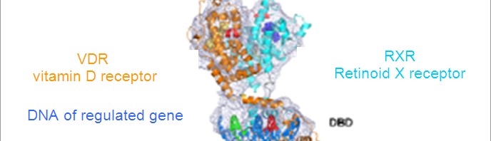
se lie au récepteur des estrogènes (surface moléculaire bleue clair). Certains acides aminés importants pour l’interaction sont représentés en orange.

Figure 1: This crystallography structure shows how BPA (grey carbon atoms and red oxygen atoms) binds to the oestrogen receptor (light blue molecule surface). Certain amino acids that are important for the interaction are shown in orange. 

©Inserm CNRS / V. Delfosse et W.Bourguet

“The results of our study and the cellular, biophysical and bioinformatic tools we have developed will make it possible to guide the synthesis of new compounds so they keep their industrial characteristics but do not have hormonal properties,” explain Patrick Balaguer and William Bourguet, research directors at Inserm.

“We are currently continuing our work to identify the crystallographic structures formed with other endocrine disruptors such as alkylphenols, pesticides, parabens and benzophenones, and to extend our IT programme to these environmental pollutants. The availability of these results should also make it easier to evaluate all the molecules referred to by the REACH Regulation (140,000 compounds) to find out whether they are ‘endocrine disruptors’,” conclude the researchers.


A step closer to morphine without side effects?

Morphine has been used for centuries to relieve severe pain. However, its pain-relief properties are coupled with considerable side effects. Morphine imitates the action of molecules produced naturally by the brain (endorphins). So why does it have such harmful side effects? This question has recently been elucidated by Sébastien Granier, researcher at the Institute of Functional Genomics (Inserm/CNRS/Universités de Montpellier 1 and 2) and his team, in conjunction with collaborators in the US. The 3D structure of brain receptors is likely to differ, depending on the type of molecule bound upon it (morphine or endorphin): the body’s response will be completely and accordingly modified. Thanks to this discovery, the researchers have now set their sights on retaining the benefits of morphine while eliminating any undesired side effects.

The results were published in the Nature review on 21 March 12.

Opium is a natural poppy (papaver somniferum) extract and is one of the oldest drugs known to man, used for its psychotropic, sedative and analgesic properties. These effects are caused by its major component: morphine. Today morphine has widespread clinical pain-relief applications.

Morphine’s action is relayed by μ-opioid receptors expressed at the surface of cells in the central nervous system. These receptors are part of a superfamily of proteins, G protein-coupled receptors (GPCR), which are the target of around 30% of drugs currently on the market.

At a molecular level, morphine binds with μ-opioid receptors to imitate the action of molecules produced naturally in the brain: endorphins. However, its clinical use is limited by two effects. Firstly, the development of a tolerance phenomenon means that, in the case of repeated injections, the morphine dose must be increased to obtain the same therapeutic effect. Secondly, morphine consumption can lead to drug dependency (heroin, the acetylated form of morphine, is the most obvious example). Furthermore, morphine consumption has serious side effects: respiratory depression, constipation, physical and psychic dependency. These harmful effects can be explained by the fact that morphine triggers a cellular response that differs from that triggered by endorphins. Since morphine and endorphins bind with the same receptor, the two molecules stabilize the μ-opioid receptors in separate spatial conformations that cause the differences in biological responses.

It is therefore essential to understand the structural bases of morphine action, and opioids in general, if we are to develop molecules that retain the beneficial effects of morphine whilst eliminating the side effects,” explains Sébastien Granier, Inserm researcher and project leader.

The research produced by Sébastien Granier (and Brian Kobilka’s team in Stanford) resolved the 3D structure of μ-opioid receptors when associated with a molecule that has a similar chemical structure to morphine.


© Kobilka Lab
3D representation of μ-opioid receptors when associated with morphine antagonistic.

© Kobilka Lab

3D representation of a μ-opioid receptor dimer.

The images of the 3D structure above show that the specific area in the receptor where the opioid molecule binds is mostly open to the external medium, thus explaining the rapid action of these components.

The structure also reveals a very important characteristic of this receptor function: the formation of a receptor dimer (1). “This is the very first time that we have succeed in producing images of the 3D structure of a complex of this kind for this receptor family” he adds. This structure with a μ-opioid receptor dimer opens up avenues of research to study this phenomenon and improve understanding of these functional implications.

The 3D structure resolution of the μ-opioid receptor holds the key to treating pain and addictions, and could, in the long-run, lead to the production of new analgesic medicine with no side effects.

Footnote

(1) Molecule produced by ‘associating’ two identical molecules

Vitamin D receptor: first full 3D observation

For the first time, a team from the Institute of Genetics and Molecular and Cellular Biology (IGBMC, Université de Strasbourg/CNRS/Inserm) has succeeded in taking a full, 3D photograph in HD (1) of a small vital, molecule, enclosed at the heart of our cells: the vitamin D receptor (VDR). Published on 18 January 2012 in the EMBO Journal, this study provides key information regarding the 3D structure of the receptor and its action mechanism at a molecular level. This data is crucial for pharmaceutical research, since the VDR is involved in several diseases, such as cancer, rickets and type 1 diabetes.

The vitamin D receptor (VDR) is part of, and plays a crucial role in, what biologists refer to as the “large family of nuclear receptors”: proteins that are active in cell cores, including “steroid” receptors (sexual hormone receptors, etc.). It regulates the expression of genes involved in diverse and vital biological functions (cell growth, bone mineralisation, etc.).

© IGBMC (CNRS / Inserm / Université de Strasbourg)

Until now, researchers had only been able to study two parts of this receptor close-up: the region that interacts with DNA and the vitamin D-binding domain. These two parts were produced in a laboratory and their structu[]re was studied individually using the crystallography technique. This method did not make it possible to visualize VDR fully since it proved to be difficult to crystallize.

To overcome this challenge, by combining the skills of several teams from across the globe for more than 15 years, the teams led by Bruno Klaholz and Dino Moras, each CNRS research directors at the IGBMC, used an innovative technique: cryo-electron microscopy (cryo-EM), which requires the latest-generation electronic “high-definition” microscope. This marvel of technology can be used to view biological objects at the molecular, or even atomic, scale. In France, the first microscope of this kind was installed at the IGBMC (2) in 2008. Prior to this research, many people thought it was impossible to study VDR using cryo-EM. Until now, the smallest molecules that had been viewed using this technique weighed more than 300 kilo Dalton (3) (kDa), or even thousands of kDa, i.e. much more than the VDR, which weighs 100 kDa and measures just 10 nm (10 x 10-9 m).

In concrete terms, Bruno Klaholz and his colleagues have laboratory-produced large qualities of the human VDR receptor inEscherichia coli bacteria (one of the most commonly-used models in biology to produce proteins). They then isolated to receptor in a physiological solution containing water and a little salt. The sample containing VDR was then frozen and immersed in liquefied ethane, which produced extremely rapid cooling (in a fraction of a second, the sample passes from 25°C to approximately -184°C). Using the microscope, approximately 20,000 photos were required of VDR particles in different directions. It is these images, aligned and combined using a software program, which finally resulted in a 3D reconstruction of VDR.

This image has provided hitherto unknown information regarding how the receptor functions. It reveals that the VDR and its partner RXR (retinoid X receptor, a vitamin A derivative) form an open architecture, with the vitamin D-binding domain oriented almost perpendicularly to the DNA binding domain (see Figure below). This structure suggests cooperation between the two domains, which may work together to induce a more tight regulation of the expression of target genes.

This ground-breaking work paves the way for research into several other vital nuclear receptors, which are yet to be thoroughly investigated. In particular, biologists are now envisaging using cryo-EM to reveal the structure of steroid receptors.

View of 3D architecture of two receptors, the VDR (vitamin D receptor) and its partner RXR (retinoid X receptor, a derivative of vitamin A), after 3D reconstruction using images of individual particles. The purple mesh represents the experimental 3D map obtained through cyro-EM. The specific binding sites for DNA fragment are indicated in green and red, the ADN binding domains (BDB) and ligand binding domains (LBD) are indicated.

Footnotes

(1) 12 angstroms: 12 x10-10 metres (one angstrom corresponds to the average diameter of an atom).

(2) The second was inaugurated in February 2011 at the Institute of Structural Biology (CEA / CNRS / UJF) in Grenoble.

(3) One Dalton is, with relatively accurate precision, the mass of a hydrogen atom. A protein amino acid represents approximately 110 Da, an assembly of 100kDa contains approx. 900 amino acids.

fermer