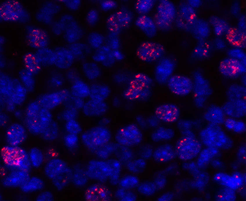
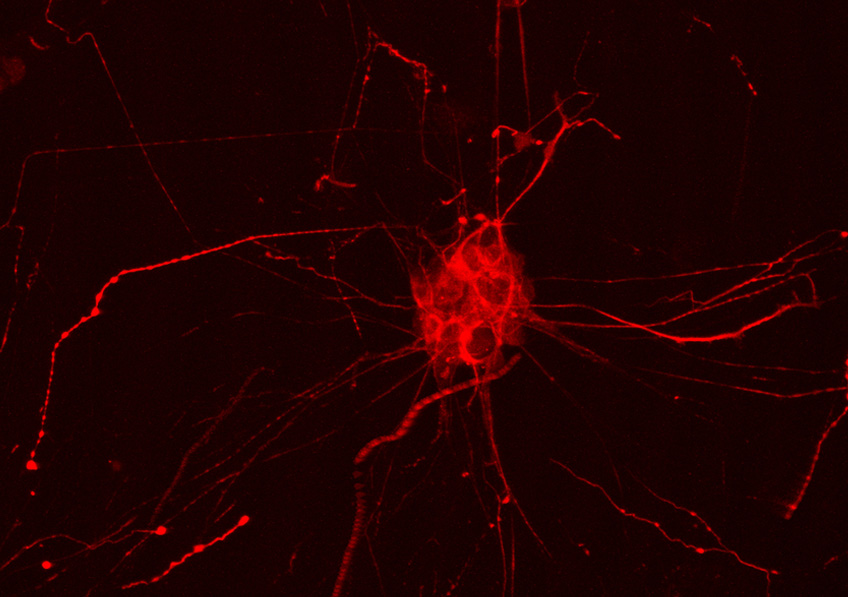
Menu

Questioning the Universal Application of Neurocognitive Tests

Crowd

Human interactions are enabled by a set of neurocognitive mechanisms defined by the concept of “social cognition”. © Ryoji Iwata /Unsplash

Human interactions are enabled by a set of neurocognitive mechanisms defined by the concept of “social cognition”. In order to identify social cognition disorders, specialists use internationally validated evaluation tests. However, these are most often developed in western, industrialized countries, which could question the relevance of applying them to all humanity. A research team from Inserm, University Hospital Lille, and Université de Lille within the Lille Neuroscience & Cognition laboratory looked at the impact of cultural differences on the performances on two of the neurocognitive tests most used worldwide, comparing the results of around 600 healthy participants across 12 countries. Their study, to be published in Neuropsychology, highlights notable differences in performance from one country to another and calls for more consideration to be given to the social sciences when developing such tests.

The concept of “social cognition” designates the various cognitive processes (perception, memorization, reasoning, emotion…) involved in social interactions. Disorders of social cognition are encountered in many diseases, such as schizophrenia and Parkinson’s, and in neurodevelopmental disorders, such as autism. They are responsible for highly incapacitating interpersonal difficulties that strongly impact the lives of patients and those around them. Consequently, the detection, quality of evaluation, and treatment of such disorders represent a major challenge for mental health specialists.

To evaluate social cognition capacities and diagnose a potential disorder, there are tests used internationally that measure “cognitive functions” – namely the capacities that enable us to interact effectively with others.

However, these reference cognitive tests are most often developed in western, industrialized, and democratic countries and their “norms” for the most part based on profiles of well-educated, rich, white people. Insofar as such individuals constitute only 12 % of humanity, their overrepresentation in the development of neuropsychological tests calls into question the relevance of their application to other populations.

A team of scientists led by Inserm researcher Maxime Bertoux within the Lille Neuroscience & Cognition laboratory (Inserm/University Hospital Lille/Université de Lille) sought to determine whether cultural differences have a notable impact on the results of the most commonly used social cognition tests. This involved conducting a vast international study on 587 healthy participants, from 18 to 89 years of age, across 12 countries (Germany, England, Argentina, Brazil, Canada, Chili, China, Colombia, Spain, France, Italy, and Russia). Neuropsychologists subjected the participants to two types of test evaluating the capacities considered essential in social cognition.

The first, devised in the UK, evaluated the capacity to decode social rules and infer others’ mental state by asking the participants to identify, in various short scenes, whether one of the characters was committing a social faux pas (for example, mistaking a customer for a waiter in a restaurant). The second test, devised in the US, evaluated the capacity to recognize the expression of emotions by asking the participants to identify various facial expressions on photographs.

The results of the study show that a large proportion of the divergences in performance on these two tests (around one quarter for the faux pas test and over 20 % for the emotions recognition test) is attributable to the differences in nationality of the participants.

The best performances in the faux pas test were obtained by the English participants, without the literal translation from English to the other participants’ native languages having an impact on the results.

For example, 100 % of the English participants considered it a faux pas to mistake a customer for a waiter in a restaurant versus only 65 % of the Canadian participants. Furthermore, while 100 % of the English participants considered it normal to give up their seat on a bus for an elderly person, 21 % of the Chinese participants considered it a faux pas.

In the results of the emotional facial expression recognition test, the comparison between the countries reveals that some emotions were not identified in the same way by all participants. While positive expressions such as joy were interpreted unambiguously from one country to another, the interpretation of negative emotions was much more variable. For example, fear was confused with surprise by the majority of the Canadian and Brazilian participants, whereas the English and Argentinians had virtually no difficulty differentiating them.

This study shows that individual and cultural factors have a strong impact on social cognition measures, declares Bertoux. Beyond the effect of age, gender, and education, there is an influence of local concepts, norms and habits on the categorization of emotions, inferring others’ intentions and understanding their behavior. As such, the use of tests devised by rich, white US or UK scientists, would favor the performance of the participants from the same country, culture, and social level.

Of course, this does not mean that the inhabitants of one country are superior or inferior to those of another, adds the researcher. Our study shows that a test devised in a specific context favors those who are familiar with that context. For example, identifying a faux pas requires detecting that an implicit social rule has been broken – but social rules fluctuate from one country to another.” 

These findings therefore question the international applicability of a neuropsychological test devised and validated in one country, particularly when it comes to evaluating and diagnosing cognitive disorders. 

For its future studies, the research team would like to enrich its data by including more participants and countries – particularly regions of the world not represented in this research, such as Africa and the Middle East –, but also by exploring the neurocognitive and cultural variations within vast countries such as China and Canada. “The neurosciences need to interact more with the social sciences in exploring and considering cultural diversity in order to build a more rigorous, relevant, and inclusive neuropsychology,” concludes Bertoux.

Inserm and CNRS to Lead Large-Scale Program on Psychiatry Research

dépression

Psychiatric illnesses constitute a major public health problem. © Danie Franco/Unsplash

On July 18, 2022, France’s minister for Higher Education and Research Sylvie Retailleau announced an unprecedented psychiatry research effort in the form of 80 million euros allocated over five years to the precision-psychiatry project-program PROPSY, led by Inserm and CNRS as part of the Priority Research and Resources Programs (PEPRs). With this ambitious research program centered around four of the most incapacitating psychiatric disorders, the two institutes aim to develop precision psychiatry in order to revolutionize both their diagnosis and patient care.

Led jointly by Inserm and CNRS, the project will involve partners whose competences are both renowned and complementary, such as Fondation FondaMental, CEA, Sorbonne Université, Université de Bordeaux, Université de Lille, Université de Paris, and Université Paris Est Créteil.

Psychiatric illnesses constitute a major public health problem, affecting 20 % of French people on a daily basis – and very often their loved ones. Developing early in life, these illnesses often strike young adults and are sadly linked to a 10-to-20-year reduction in life expectancy. In response to the diagnoses – that are sometimes long in coming – the therapeutic options are often thin on the ground, with those that are available having incapacitating side effects. Not to mention the considerable socioeconomic impact, with recent estimations putting the direct and indirect costs at 160 billion euros in 2019 – representing over 5 % of GDP.

Faced with these two observations, it would appear imperative to improve the coordination of France’s psychiatry research forces and increase the research-care continuum. These are the proposals of PROPSY, a project led by Inserm and CNRS which has recently been selected to receive 80-million-euro funding over five years.

Centered around four of the most incapacitating disorders, bipolar disorder, major depressive disorders, schizophrenia, and autism spectrum disorders, this exploratory PEPR will broaden the field of precision medicine in psychiatry.

This will make it possible to provide each patient with the best care. In order to achieve this ambition, the multiple challenges will be to:

  • Better understand the causes and mechanisms behind mental illnesses
  • Discover prognostic markers for these disorders and identify homogenous patient subgroups
  • Develop targeted therapeutic strategies that include eHealth, immunomodulators, brain stimulation, and biotherapies
  • Reduce stigma and false representations
  • Support the development of a French biomedical sector in mental health, including pharma, MedTech and digital, through public-private partnerships
  • Create a new generation of scientists and medical staff in psychiatry by renewing the approach to these illnesses and thanks to training initiatives

Selected as part of the call for Priority Research and Resources Programs, PROPSY will include the funding of projects already identified – such as the French Minds longitudinal cohort of 3 000 adult patients who will undergo exhaustive clinical, behavioral, and environmental evaluation with the help of digital tools, biological markers, and brain imaging – as well as open calls for proposals.

We are proud that the government has selected our research program,” confide Gilles Bloch, Inserm CEO and Marion Leboyer, 2021 Inserm Grand Prize recipient, Executive Director of the FondaMental foundation and project scientific lead. “With the negative impact of the health crisis on mental health (an over 30 % increase in cases of depression), this extremely important decision represents hope for millions of patients, their families, as well as researchers and medical staff. “


With the benefit of better support for patients, the PROPSY PEPR will enable research and patient care to join forces to meet the public health challenges related to psychiatric disorders,” adds Antoine Petit, Chairman and CEO of CNRS.

Researchers described how the cerebellum modulates our ability to socialize

This image of the cerebellum of a mouse expressing a fluorescent protein in Purkinje cells expressing dopamine D2 receptors © Emmanuel Valjent, Institut de Génomique Fonctionnelle (Montpellier)

The cerebellum is essential for sensorimotor control but also contributes to higher cognitive functions including social behaviors. In a recent study, an international research consortium including scientists from Inserm – University of Montpellier (France), the Institut de Neurociències Universitat Autònoma de Barcelona (INc-UAB) (Spain), and the University of Lausanne (Switzerland) uncovered how dopamine in the cerebellum modulates social behaviors via its action on D2 receptors (D2R). By using different mouse models and genetic tools, the researchers’ work shows that changes in D2R levels in a specific cerebellar cell type, the Purkinje cells, alter sociability and preference for social novelty without affecting motor functions. These new findings pave the way to determine whether socially related psychiatric disorders, such as autism spectrum disorders (ASD), bipolar mood disorder, or schizophrenia, are also associated with altered dopamine receptors expression in specific cerebellar cell types.

Dopamine (DA) neurons are a major component of the brain reward system. By encoding motivational value and salience, they tighly regulate motivation, emotional states and social interactions. Although the regulation of these processes has been largely ascribed to neural circuits embedded in limbic regions, recent evidence indicate that the cerebellum, a region primarily involved in motor control, may also contribute to higher cognitive functions including social behaviors. However, whether cerebellar dopamine signaling could participate to the modulation of these functions remained unexplored. Researchers from Inserm – Montpellier University (France), the Institut de Neurociències UAB (Spain), and the University of Lausanne (Switzerland) uncovered a new role for dopamine as modulator of social behaviors in the mouse cerebellum.

By combining cell type-specific transcriptomics, immunofluorescence analyses and 3D imaging, researchers first demonstrated the presence of dopamine D2 receptors (D2R) in Purkinje cells (PCs), the output neurons of the cerebellar cortex. Using patch-clamp recordings, they were able show D2R modulated synaptic excitation onto PCs. “This first set of results was already determinant for us, as they unveiled that D2R were present in the cerebellum and that, despite their low expression level, they were functional”, highlights Dr Emmanuel Valjent, research director at Inserm (France), and coordinator of the study.

The researchers then went on to study their functions. By using genetic approaches to invalidate or overexpress D2R selectively in PCs, they analyzed the impact of these alterations on motor and non-motor cerebellar functions. “We have uncovered an unexpected causal link between PCs D2R expression levels right in the center of the cerebellum, the Crus I/II lobules, and the modulation of social behaviors. Reducing the expression of this specific dopamine receptor impaired the sociability of mice as well as their preference for social novelty, while  their coordination and motor functions remained unaffected” explains Dr. Laura Cutando, Marie-Curie researcher at the Mitochondrial Neuropathology research group at INc-UAB, and first author of the article.

This study constitutes a first step towards a better understanding of the role of dopamine in the cerebellum and the mechanisms underlying psychiatric disorders such as schizophrenia, ADHD and anxiety disorders, which have all in common aberrant DA signaing and altered social behaviors.

Do Some Cognitive Biases Contaminate Even Our Simplest Mental Mechanisms?

Enfants apprenant à écrire à l'école

©woodleywonderworks via Flickr


When we implement complex cognitive processes, for example when making decisions, we are subject to cognitive bias. But what about simpler processes, such as those involved in the most basic learning? In a new study analyzing data from all previous research in the field, researchers from Inserm and ENS-PSL show that not only are positivity and confirmation biases present even in the simplest human and animal cognitive processes, but also that incorporating them into learning algorithms would enhance their performance. This research, published in Trends in Cognitive Sciences, suggests that these biases could initially have been a very old evolutionary advantage.

Cognitive biases, such as positivity and confirmation biases, are known to influence our beliefs and decisions. Until recently, it was assumed that they were specific to so-called “high-level” cognitive processes, namely those that come into play when we reason about complex and uncertain proposals.  For example, it is well known that people overestimate the likelihood of desirable events (France winning the World Cup) and underestimate that of undesirable events (a marriage ending in divorce). 

In a study published in Trends in Cognitive Sciences, Stefano Palminteri, Inserm researcher at the ENS-PSL/Inserm Laboratory for Cognitive and Computational Neuroscience and Maël Lebreton, researcher at the Paris School of Economics, challenge this conception of the involvement of positivity and confirmation biases.

The researchers drew on the various existing data in the scientific literature on “reinforcement learning”. This is a basic cognitive process of learning through rewards and punishments that humans share with many animals. This literature review reveals that very simple reinforcement learning tests can reveal behavioral signatures of positivity and confirmation biases in people who are subjected to them. These biases appear to be much more common than previously thought and are even present in the simplest cognitive processes such as learning to make good decisions through trial and error (reward and punishment).

What is more, these biases do not seem to be exclusive to humans: the behavioral signatures also appear in similar tests conducted in animals. This suggests that these biases may have emerged during evolution in a common ancestor, long before the appearance of Homo sapiens, raising the question of why evolution has selected and maintained what may at first glance be perceived as processes that can generate seemingly irrational behavior.

Palminteri and Lebreton believe that they have identified part of the answer to this question through the findings of studies based on computerized simulations. These studies compared the performance of reinforcement learning algorithms – with some algorithms incorporating positivity and confirmation biases and others not. These simulations show that the presence of a confirmation bias in the algorithm actually enables it to learn more effectively in a wide range of situations. These biases could therefore actually promote survival, which would explain why they have not been corrected during the course of evolution.

The article opens up new avenues of research that would allow us to deepen our understanding of the cognitive biases and processes related to reinforcement learning. In particular, the researchers suggest exploring the role of these biases in the development and maintenance of pathological states, such as addiction and depression. These findings also suggest that including these biases in artificial intelligence algorithms could paradoxically improve their performance.

Il existerait un lien entre la taille du réseau social et la structure du cerveau

Mère macaque toilettant son jeune singe.

A macaque grooming her offspring. An image that illustrates how macaques form relationships. © Noah Snyder-Mackler

The more social relationships we have, the more certain structures in our brain are developed. This has been the hypothesis of various neuroscience research projects for several years. With previous findings having highlighted the role of our social environment as one of the key factors behind the expansion of the cerebral cortex, researchers from Inserm and Université Lyon Claude Bernard Lyon 1, in collaboration with the University of Pennsylvania, went one step further in elucidating this link. They were more specifically interested in a species of macaques whose brain architecture is comparable to that of humans. By observing these non-human primates in their natural state and by analyzing images of their brains, they discovered that the number of companions they have is predictive of the size of certain brain regions associated with social cognition and empathy. The findings of this study have been published in Science Advances.

The links between social network and brain size have already been the subject of neuroscientific studies. For example, scientists have already looked at variations in the size of the human brain amygdala in relation to the number of Facebook friends that a person has1.

In order to build on this research and attempt to find out more about the organization and functions of the neural networks in humans, research teams have worked with an animal species whose brain characteristics are similar to those of humans: the Rhesus macaque.

In a new study, researchers from Inserm and Université Claude Bernard Lyon 1 at the Stem Cell and Brain Research Institute, in collaboration with the University of Pennsylvania, studied a group of these non-human primates in their natural state for several months before taking images of their brains. The fact that the animals were free ranging enabled the scientists to understand the social group in its full complexity. They were therefore able to measure the intensity2 of the interactions with other individuals and identify an animal’s position within the group social hierarchy.

For example, some of the observations focused on grooming partners, which represent direct, important relationships for macaques.

Alongside these behavioral observations, the scientists analyzed brain scans of individuals from the group, which consisted of 103 rhesus macaques, including 68 adults and 21 young macaques under 6 years of age.

In the adults, they found that the greater the number of social partners, the greater the size of some regions of the brain’s temporal lobe – the ventral dysgranular insula and the mid-superior temporal sulcus3 – regions considered essential for understanding emotions and the perception of how others behave.

In order to better understand how this phenomenon occurs, the scientists also collected brain scans from 21 newborn macaques. The research showed that they are not born with these differences in brain structure size but that they are established during their development.

According to the researchers’ observations, there is therefore no correlation between social network size and brain volume at birth. These findings suggest that exposure to the social environment over the course of life contributes to the maturation of brain networks.

“Which is interesting because if we had seen the same correlation in young macaques, this could have meant that having a very popular mother (with many interactions with the group) could have predisposed the newborn to also become popular. But in fact our data suggest that the differences we see in adults are strongly determined by our social environments, perhaps more than by our innate predisposition,” explains Jérôme Sallet, Inserm Research Director.

Following this study, the researchers now wish to look at anatomical changes at cell level, in order to reveal the mechanisms behind the increased size of the brain regions that were identified using brain imaging.


1 Kanai R., Bahrami B., Roylance R. and Rees G. 2012. Online social network size is reflected in human brain structure, Proc. R. Soc. B.

2 The researchers measured the number of interactions between the animals, their duration and whether these interactions were cooperative or aggressive.

3 The mid-superior temporal sulcus is involved in social cognition and perception.

Decoding a direct dialog between the gut microbiota and the brain

Diagram showing the direct dialog between the gut microbiota and the brain

© Institut Pasteur / Pascal Marseaud


Gut microbiota by-products circulate in the bloodstream, regulating host physiological processes including immunity, metabolism and brain functions. Scientists from the Institut Pasteur (a partner research organization of Université Paris Cité), Inserm and the CNRS have discovered that hypothalamic neurons in an animal model directly detect variations in bacterial activity and adapt appetite and body temperature accordingly. These findings demonstrate that a direct dialog occurs between the gut microbiota and the brain, a discovery that could lead to new therapeutic approaches for tackling metabolic disorders such as diabetes and obesity. The findings are due to be published in Science on 2022 04 15.

The gut is the body’s largest reservoir of bacteria. A growing body of evidence reveals the degree of interdependence between hosts and their gut microbiota, and emphasizes the importance of the gut-brain axis.

At the Institut Pasteur, neurobiologists from the Perception and Memory Unit (Institut Pasteur/CNRS)[1], immunobiologists from the Microenvironment and Immunity Unit (Institut Pasteur/Inserm), and microbiologists from the Biology and Genetics of the Bacterial Cell Wall Unit (Institut Pasteur/CNRS/Inserm)[2] have shared their expertise to investigate how bacteria in the gut directly control the activity of particular neurons in the brain.

The scientists focused on the NOD2 (nucleotide oligomerization domain) receptor which is found inside of mostly immune cells. This receptor detects the presence of muropeptides, which are the building blocks of the bacterial cell wall. Moreover, it has previously been established that variants of the gene coding for the NOD2 receptor are associated with digestive disorders, including Crohn’s disease, as well as neurological diseases and mood disorders. However, these data were insufficient to demonstrate a direct relationship between neuronal activity in the brain and bacterial activity in the gut. This was revealed by the consortium of scientists in the new study.

Using brain imaging techniques, the scientists initially observed that the NOD2 receptor in mice is expressed by neurons in different regions of the brain, and in particular, in a region known as the hypothalamus. They subsequently discovered that these neurons’ electrical activity is suppressed when they come into contact with bacterial muropeptides from the gut. “Muropeptides in the gut, blood and brain are considered to be markers of bacterial proliferation,” explains Ivo G. Boneca, Head of the Biology and Genetics of the Bacterial Cell Wall Unit at the Institut Pasteur (CNRS/Inserm). Conversely, if the NOD2 receptor is absent, these neurons are no longer suppressed by muropeptides. Consequently, the brain loses control of food intake and body temperature. The mice gain weight and are more susceptible to developing type 2 diabetes, particularly in older females.

In this study, the scientists have demonstrated the astonishing fact that neurons perceive bacterial muropeptides directly, while this task was thought to be primarily assigned to immune cells. “It is extraordinary to discover that bacterial fragments act directly on a brain center as strategic as the hypothalamus, which is known to manage vital functions such as body temperature, reproduction, hunger and thirst,” comments Pierre-Marie Lledo, CNRS scientist and Head of the Institut Pasteur’s Perception and Memory Unit.

The neurons thus appear to detect bacterial activity (proliferation and death) as a direct gauge of the impact of food intake on the intestinal ecosystem. “Excessive intake of a specific food may stimulate the disproportionate growth of certain bacteria or pathogens, thus jeopardizing intestinal balance,” says Gérard Eberl, Head of the Microenvironment and Immunity Unit at the Institut Pasteur (Inserm).

The impact of muropeptides on hypothalamic neurons and metabolism raises questions on their potential role in other brain functions, and may help us understand the link between certain brain diseases and genetic variants of NOD2. This discovery paves the way for new interdisciplinary projects at the frontier between neurosciences, immunology and microbiology, and ultimately, for new therapeutic approaches to brain diseases and metabolic disorders such as diabetes and obesity.


[1] This research unit is also known as the “Genes, Synapses and Cognition Laboratory” (Institut Pasteur/CNRS).
Paris Brain Institute (CNRS/Inserm/Sorbonne Université/AP-HP) also contributed to these findings.

[2] The CNRS unit’s name is the “Integrative and Molecular Microbiology Unit” and the Inserm unit’s name is the “Host-Microbe Interactions and Pathophysiology Unit” (Institut Pasteur/CNRS/Inserm).

Alzheimer’s Disease: The Identification of 75 Genetic Risk Factors Brings New Insights

Alzheimer

In Alzheimer’s disease, two brain pathological phenomena have already been well documented: the accumulation of beta-amyloid peptides and the modification of Tau, a protein, which is found as aggregates in neurons.© NIH/domaine public

Identifying genetic risk factors for Alzheimer’s disease is essential if we are to improve our understanding and treatment of it. Progress in human genome analysis along with genome-wide association studies[1] are now leading to major advances in the field. Researchers in Europe, the US and Australia have identified 75 regions of the genome that are associated with Alzheimer’s disease. Forty-two of these regions are novel, meaning that they have never before been implicated in the disease. The findings, published in Nature Genetics, bring new knowledge of the biological mechanisms at play and open up new avenues for treatment and diagnosis.

Alzheimer’s disease is the most common form of dementia, affecting around 1,200,000 people in France. This complex, multifactorial disease, which usually develops after the age of 65, has a strong genetic component. The majority of cases are thought to be caused by the interaction of different genetic predisposition factors with environmental factors.

Although our understanding of the disease continues to improve, there is no cure at this time. The medications available are mainly aimed at slowing cognitive decline and reducing certain behavioral disorders.

In order to better understand the origins of the disease, one of the major challenges of research is to better characterize its genetic risk factors by identifying the pathophysiological processes at play[2], and thereby propose novel therapeutic targets.

As part of an international collaboration, researchers from Inserm, Institut Pasteur de Lille, Lille University Hospital and Université de Lille conducted a genome-wide association study (GWAS) on the largest Alzheimer’s patient group set up until now[3], under the coordination of Inserm Research Director Jean-Charles Lambert.

Encouraged by advances in genome analysis, these studies consist of analyzing the entire genome of tens of thousands or hundreds of thousands of individuals, whether healthy or sick, with the aim of identifying genetic risk factors associated with specific aspects of the disease.

Using this method, the scientists were able to identify 75 regions (loci) of the genome associated with Alzheimer’s, 42 of which had never previously been implicated in the disease. “Following this major discovery, we characterized these regions in order to give them meaning in relation to our clinical and biological knowledge, and thereby gain a better understanding of the cellular mechanisms and pathological processes at play,” explains Lambert.

Highlighting pathological phenomena

In Alzheimer’s disease, two pathological brain phenomena are already well documented: namely, the accumulation of amyloid-beta peptides and the modification of the protein Tau, aggregates of which are found in the neurons.

Here, the scientists confirmed the importance of these pathological processes. Their analyses of the various genome regions confirm that some are implicated in amyloid peptide production and Tau protein function.

Furthermore, these analyses also reveal that a dysfunction of innate immunity and of the action of the microglia (immune cells present in the central nervous system that play a “trash collector” role by eliminating toxic substances) is at play in Alzheimer’s disease.

Finally, this study shows for the first time that the tumor necrosis factor alpha (TNF-alpha)-dependent signaling pathway is involved in disease[4].

These findings confirm and add to our knowledge of the pathological processes involved in the disease and open up new avenues for therapeutic research. For example, they confirm the utility of the following: the conduct of clinical trials of therapies targeting the amyloid precursor protein, the continuation of microglial cell research that was initiated a few years ago, and the targeting of the TNF-alpha signaling pathway.

Risk score

Based on their findings, the researchers also devised a genetic risk score in order to better evaluate which patients with cognitive impairment will, within three years of its clinical manifestation, go on to develop Alzheimer’s disease. “While this tool is not at all intended for use in clinical practice at present, it could be very useful when setting up therapeutic trials in order to categorize participants according to their risk and improve the evaluation of the medications being tested,” explains Lambert.

In order to validate and expand their findings, the team would now like to continue its research in an even broader group. Beyond this exhaustive characterization of the genetic factors of Alzheimer’s disease, the team is also developing numerous cellular and molecular biology approaches to determine their roles in its development.

Furthermore, with the genetic research having been conducted primarily on Caucasian populations, one of the considerations for the future will be to carry out the same type of studies in other groups in order to determine whether the risk factors are the same from one population to the next, which would reinforce their importance in the pathophysiological process.


[1] These studies consist of analyzing the entire genome of thousands or tens of thousands of people, whether healthy or sick, to identify genetic risk factors associated with specific aspects of the disease.

[2] All functional problems caused by a particular disease or condition.

[3] Here, the researchers were interested in the genetic data of 111,326 people who were diagnosed with Alzheimer’s disease or had close relatives with the condition, and 677,663 healthy “controls”. These data are derived from several large European cohorts grouped within the European Alzheimer & Dementia BioBank (EADB) consortium.

 [4] Tumor necrosis factor alpha is a cytokine: an immune system protein implicated in the inflammation cascade, particularly in tissue lesion mechanisms.

New Brain Abnormalities Associated with Child Abuse

Immunolabeling of parvalbumin neurons (in green) surrounded by perineuronal nets (in red) in the human prefrontal cortex. © Arnaud Tanti/Inserm


In collaboration with a Canadian team, scientists from Inserm and Université de Tours, at Unit 1253 Imaging & Brain1, have shown in post-mortem brain samples that victims of child abuse present specific brain characteristics. The teams have revealed for the first time in humans an increase in the number and maturation of perineuronal nets, dense protein structures surrounding the neurons. In animals, this phenomenon regulates brain plasticity by inhibiting the remodeling of neural networks. This research suggests that abuse could cause lasting changes to the developmental trajectories of certain brain regions with potential effects on psychological health. The study has been published in Molecular Psychiatry.

Child abuse has effects on psychological development, including an increased risk of depression and suicide during the course of life. Sexual violence, physical violence or chronic neglect during childhood or adolescence are suspected to cause lasting structural and functional changes in the brain. It is during these periods that personality traits, attachment patterns, cognitive functions and emotional responses are shaped by our experiences, including traumatic ones.

To better understand the neurobiological changes associated with child abuse, researchers from Inserm and Université de Tours, in collaboration with McGill University – Douglas Mental Health University Institute in Montreal, Canada, investigated the ventromedial prefrontal cortex, a brain region that regulates emotional responses. They took a closer look at “perineuronal nets” – particularly compact and dense structures that encircle certain neurons, especially parvalbumin neurons, whose inhibitory action plays a role in controlling the activity of large groups of neurons.

Perineuronal nets emerge in early childhood and continue to develop until the end of adolescence, increasing in size and number. In animals, their development represents an important stage of brain maturation, with their emergence marking the closure of “critical periods” of plasticity during which the development of the neural circuitry can be easily modified by experiments.

The researchers estimate that these perineuronal nets could play a role in childhood trauma by immobilizing the neural networks associated with these memories, predisposing the individual to future depressive or behavioral disorders.

Denser perineuronal nets

To study these structures, the researchers analyzed post-mortem brain sections from adults (from brain donations with informed consent from the next of kin) who had committed suicide an episode of major depression. Of the 28 subjects concerned, 12 had a marked history of child abuse. These sections were also compared with those of control subjects having died of natural causes and with no history of abuse or psychiatric illness. The different types of analyses performed by the researchers led them to make several observations.

Firstly, in the subjects who were abused during childhood, the perineuronal nets were denser and more numerous than those of the other individuals. They also presented more marked maturation characteristics, including increased structural development around the parvalbumin neurons. Finally, the researchers showed that the cells producing the main proteins that make up the perineuronal nets are oligodendrocyte progenitors, cells which are found throughout the brain.

The researchers will now use mice to explore the consequences of these observations in more detail, particularly on the persistence of traumatic memories linked to early adversity.

These observations reinforce the hypothesis of a correlation between early stress and increased perineuronal net development. It remains to be discovered whether there is a causal link, that is to say whether these changes contribute to the development of behaviors associated with abuse, and how. Perhaps in the longer term we could consider a procedure targeting the perineuronal nets to help restore some plasticity and reduce the impact of trauma and the subsequent psychiatric risk,” explains Arnaud Tanti, Inserm researcher and first author of this study.


1 JRU 1253, iBrain, Inserm, Université de Tours

Does Falling Asleep Boost Creativity?

Sommeil _ Dali© Wiki Commons – Fair Use

Salvador Dali liked to use short phases of sleep to stimulate his creativity. © Wiki Commons – Fair Use

What if a few minutes of sleep could trigger creativity? This is what suggests a study by researchers from Inserm and Sorbonne Université at the Brain Institute and the department of sleep medicine at Pitié-Salpêtrière Hospital AP-HP. Their findings have been published in Science Advances.

The inventor Thomas Edison is said to have taken short naps to spark his creativity. During them, he would hold a metal ball in each hand. Upon falling asleep, the balls would crash to the floor, waking him up just in time so that he could note his flashes of creativity. Other famous people also liked to use short phases of sleep to stimulate their creativity, such as Albert Einstein and Salvador Dali.

Inspired by this, the team of Inserm researcher Delphine Oudiette and her colleague Célia Lacaux at the Brain Institute and Pitié-Salpêtrière Hospital AP-HP wished to explore this very specific phase of sleep, to determine whether or not it affected creativity.

As part of their study, the team set the 103 participants math problems that could all be instantly solved using the same rule – of which the participants were unaware when starting the test. The subjects were allowed a first attempt at solving the problems. Those who had not found the hidden rule were invited to take a twenty minute nap inspired by Edison, holding an object in their right hand, before repeating the math tests.

Spending at least 15 seconds in this very first phase of sleep after falling asleep tripled the chances of finding the hidden rule, due to the effect of the famous ‘Eureka!’ moment. ” An effect that disappeared if the subjects plunged into a deeper sleep,” explains Lacaux, first author of the study.

In parallel, the researchers revealed several key neurophysiological markers of this sleep phase that generates creativity.

During the onset of sleep, there is indeed a phase that is conducive to creativity. Activating it requires finding the right balance between falling asleep quickly and not falling asleep too deeply. These “creative naps” could be an easy and accessible way of stimulating our creativity in everyday life.

“The sleep onset phase has so far been relatively neglected by the cognitive neurosciences. This discovery opens up an extraordinary new avenue for future studies, particularly of the brain mechanisms of creativity. Sleep is also often seen as a loss of time and productivity. By showing that it is in fact essential to our creative performance, we hope to reiterate its importance to the general public. ” concludes Oudiette, Inserm researcher and last author of the study.

The Impacts on Reproductive Function of Early Exposure to Endocrine Disruptors

Astrocyte né pendant la période infantile

Astrocyte born during the infancy period (red) (7 to 20 days after birth) adhering to a GnRH neural cell body (green). The astrocytes’ processes are shown in white. © Vincent Prévot, Inserm.


Researchers from Inserm, Lille University Hospital and Université de Lille, at the Lille Neuroscience and Cognition laboratory, have discovered one of the mechanisms by which endocrine disruptors can alter reproductive function development from birth. At the neural level, they saw in animals how exposure to low doses of bisphenol A – a known endocrine disruptor – a few days after birth disrupts the integration of GnRH neurons within their neural circuit and alters their reproductive function regulation activity. The findings of this study have been published in Nature Neuroscience.

In mammals, reproduction is regulated by the GnRH neurons, a population of neurons that, during embryonic development, appears in the nose and then migrates to the hypothalamus in the brain. Being well established in the brain at birth, these neurons go on to control the various processes associated with reproductive function: puberty, acquisition of secondary sexual characteristics, and fertility in adulthood.

To perform their functions, the GnRH neurons must be surrounded by another type of neural cell: the astrocytes. The adhesion of the astrocytes to the GnRH neurons plays a decisive role in their integration within the neural network. The encounter between these two cell types takes place during the so-called “mini-puberty” period that begins one week after birth in mammals, when the GnRH neurons are first activated, and which is when the first sex hormone secretions occur.

“Failure of GnRH neurons to integrate during mini-puberty may lead to a predisposition to developing puberty and/or fertility disorders, and also potentially affect brain development, thereby leading to learning disorders or metabolic disorders, such as being overweight,” explains Vincent Prévot, Inserm Research Director and last author of the study.

But how does this encounter between GnRH neurons and astrocytes take place? According to the findings of this research, the astrocytes do not get there by chance but respond to molecular signals emitted by the GnRH neurons, which recruit them as soon as they appear in the hypothalamus.

Early bisphenol A exposure prevents communication between GnRH neurons and astrocytes

Going further, the researchers wanted to understand the importance of this meeting between astrocytes and GnRH neurons in the development of mammalian reproductive functions during the mini-puberty period. With recent studies having shown that the GnRH neural network is particularly sensitive to endocrine disruptors and that there is a link between the latter and puberty disorders, the researchers investigated the impact of exposure to one of these endocrine disruptors, bisphenol A, in rats.

Bisphenol A is a compound used in the industrial manufacturing of plastics whose endocrine disruptive nature is now widely recognized. It is used for example in the production of food containers such as drinks bottles and baby feeding bottles. It is also found in the protective linings of drinks cans and tinned food, or on till receipts where it is used as a developer. In France, bisphenol A has been banned from use in baby feeding bottles and other  food containers since 2015. It has been replaced with substitutes such as bisphenol S (BPS) and bisphenol B (BPB) which also raise questions regarding their safety.

“Despite its ban, bisphenol A continues to remain present in our environment due to the slow degradation of plastic waste, and also because people are still using food containers they had purchased before 2015. With the recycling of waste, bisphenol A from plastics produced before 2015 has also found itself in new products,” explains Prévot.

During the 10 days following their birth, the female rats received low-dose injections of bisphenol A. Using an astrocyte labelling technique, the researchers saw that, under the effect of bisphenol A, the astrocytes were unable to permanently adhere to the GnRH neurons. The absence of such a phenomenon

occurring between these nerve cells then led to delayed puberty and the absence of estrous cycles in the adult female rats (equivalent to the menstrual cycle in women), suggesting that reproductive functions are affected.

Our findings suggest that early exposure to chemicals in contact with food, such as bisphenol A, can disrupt the onset of puberty and have a lasting impact on reproductive functions, by preventing GnRH neurons from building an appropriate and necessary environment in the hypothalamus for their role as fertility coordinator,” explains Ariane Sharif, lecturer at Université de Lille, who co-led the study.

Taking this research further, the scientists are now seeking to understand the exact mechanism by which bisphenol A prevents communication between GnRH neurons and astrocytes. One hypothesis is that bisphenol A acts directly on the astrocytes’ receptors, preventing them from adhering to the GnRH neurons. The research team is also interested in the action of bisphenol A on DNA and the traces it may leave.

fermer