Menu

Alzheimer’s Disease: The Identification of 75 Genetic Risk Factors Brings New Insights

Alzheimer

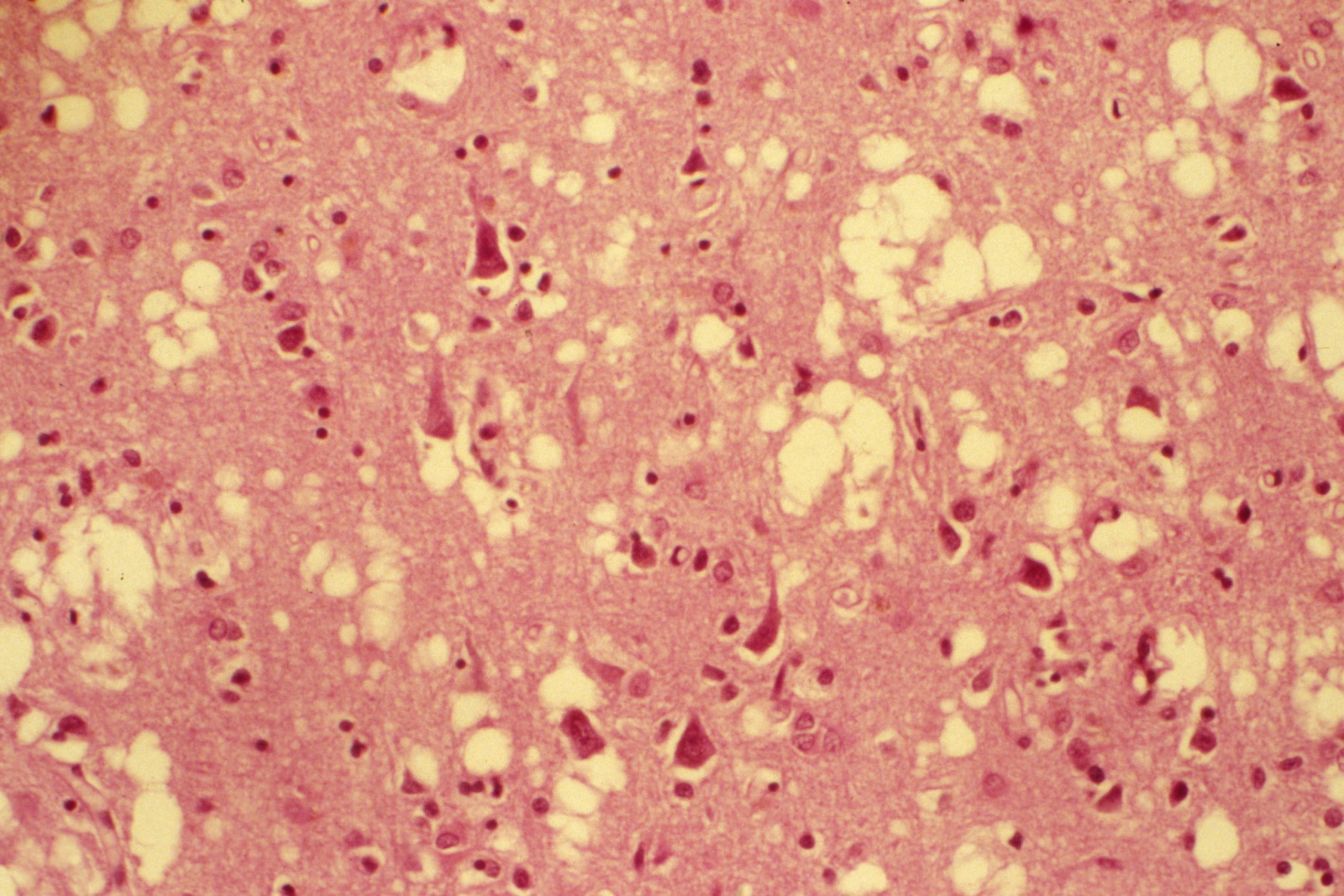
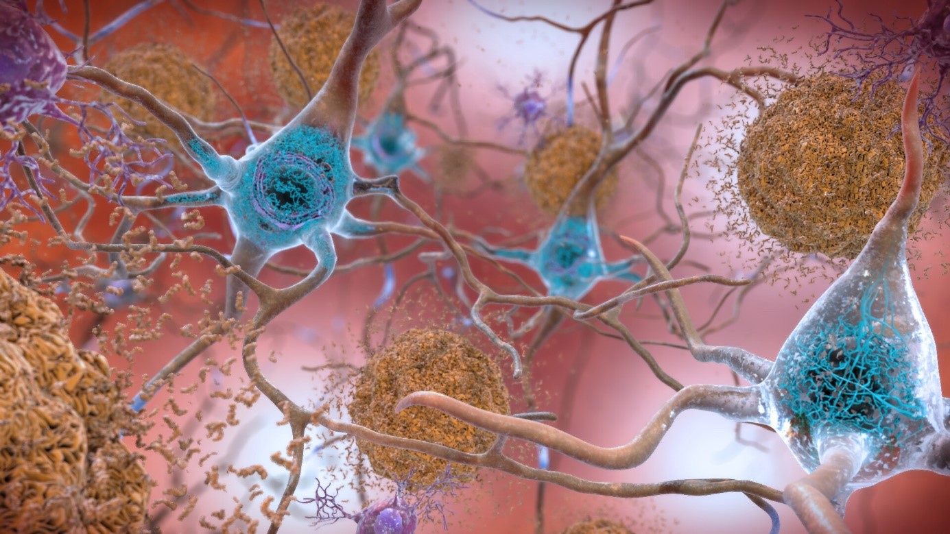
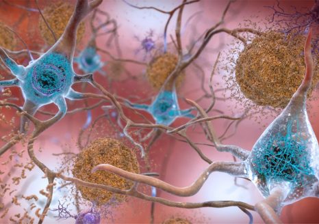
In Alzheimer’s disease, two brain pathological phenomena have already been well documented: the accumulation of beta-amyloid peptides and the modification of Tau, a protein, which is found as aggregates in neurons.© NIH/domaine public

Identifying genetic risk factors for Alzheimer’s disease is essential if we are to improve our understanding and treatment of it. Progress in human genome analysis along with genome-wide association studies[1] are now leading to major advances in the field. Researchers in Europe, the US and Australia have identified 75 regions of the genome that are associated with Alzheimer’s disease. Forty-two of these regions are novel, meaning that they have never before been implicated in the disease. The findings, published in Nature Genetics, bring new knowledge of the biological mechanisms at play and open up new avenues for treatment and diagnosis.

Alzheimer’s disease is the most common form of dementia, affecting around 1,200,000 people in France. This complex, multifactorial disease, which usually develops after the age of 65, has a strong genetic component. The majority of cases are thought to be caused by the interaction of different genetic predisposition factors with environmental factors.

Although our understanding of the disease continues to improve, there is no cure at this time. The medications available are mainly aimed at slowing cognitive decline and reducing certain behavioral disorders.

In order to better understand the origins of the disease, one of the major challenges of research is to better characterize its genetic risk factors by identifying the pathophysiological processes at play[2], and thereby propose novel therapeutic targets.

As part of an international collaboration, researchers from Inserm, Institut Pasteur de Lille, Lille University Hospital and Université de Lille conducted a genome-wide association study (GWAS) on the largest Alzheimer’s patient group set up until now[3], under the coordination of Inserm Research Director Jean-Charles Lambert.

Encouraged by advances in genome analysis, these studies consist of analyzing the entire genome of tens of thousands or hundreds of thousands of individuals, whether healthy or sick, with the aim of identifying genetic risk factors associated with specific aspects of the disease.

Using this method, the scientists were able to identify 75 regions (loci) of the genome associated with Alzheimer’s, 42 of which had never previously been implicated in the disease. “Following this major discovery, we characterized these regions in order to give them meaning in relation to our clinical and biological knowledge, and thereby gain a better understanding of the cellular mechanisms and pathological processes at play,” explains Lambert.

Highlighting pathological phenomena

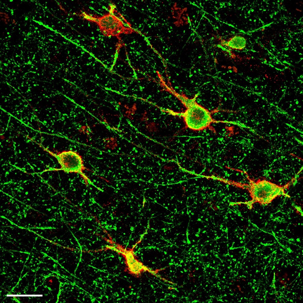
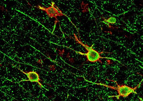
In Alzheimer’s disease, two pathological brain phenomena are already well documented: namely, the accumulation of amyloid-beta peptides and the modification of the protein Tau, aggregates of which are found in the neurons.

Here, the scientists confirmed the importance of these pathological processes. Their analyses of the various genome regions confirm that some are implicated in amyloid peptide production and Tau protein function.

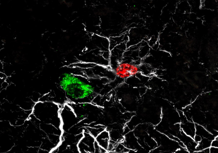
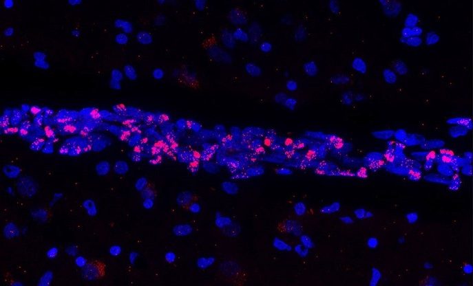
Furthermore, these analyses also reveal that a dysfunction of innate immunity and of the action of the microglia (immune cells present in the central nervous system that play a “trash collector” role by eliminating toxic substances) is at play in Alzheimer’s disease.

Finally, this study shows for the first time that the tumor necrosis factor alpha (TNF-alpha)-dependent signaling pathway is involved in disease[4].

These findings confirm and add to our knowledge of the pathological processes involved in the disease and open up new avenues for therapeutic research. For example, they confirm the utility of the following: the conduct of clinical trials of therapies targeting the amyloid precursor protein, the continuation of microglial cell research that was initiated a few years ago, and the targeting of the TNF-alpha signaling pathway.

Risk score

Based on their findings, the researchers also devised a genetic risk score in order to better evaluate which patients with cognitive impairment will, within three years of its clinical manifestation, go on to develop Alzheimer’s disease. “While this tool is not at all intended for use in clinical practice at present, it could be very useful when setting up therapeutic trials in order to categorize participants according to their risk and improve the evaluation of the medications being tested,” explains Lambert.

In order to validate and expand their findings, the team would now like to continue its research in an even broader group. Beyond this exhaustive characterization of the genetic factors of Alzheimer’s disease, the team is also developing numerous cellular and molecular biology approaches to determine their roles in its development.

Furthermore, with the genetic research having been conducted primarily on Caucasian populations, one of the considerations for the future will be to carry out the same type of studies in other groups in order to determine whether the risk factors are the same from one population to the next, which would reinforce their importance in the pathophysiological process.


[1] These studies consist of analyzing the entire genome of thousands or tens of thousands of people, whether healthy or sick, to identify genetic risk factors associated with specific aspects of the disease.

[2] All functional problems caused by a particular disease or condition.

[3] Here, the researchers were interested in the genetic data of 111,326 people who were diagnosed with Alzheimer’s disease or had close relatives with the condition, and 677,663 healthy “controls”. These data are derived from several large European cohorts grouped within the European Alzheimer & Dementia BioBank (EADB) consortium.

 [4] Tumor necrosis factor alpha is a cytokine: an immune system protein implicated in the inflammation cascade, particularly in tissue lesion mechanisms.

New Brain Abnormalities Associated with Child Abuse

Immunolabeling of parvalbumin neurons (in green) surrounded by perineuronal nets (in red) in the human prefrontal cortex. © Arnaud Tanti/Inserm


In collaboration with a Canadian team, scientists from Inserm and Université de Tours, at Unit 1253 Imaging & Brain1, have shown in post-mortem brain samples that victims of child abuse present specific brain characteristics. The teams have revealed for the first time in humans an increase in the number and maturation of perineuronal nets, dense protein structures surrounding the neurons. In animals, this phenomenon regulates brain plasticity by inhibiting the remodeling of neural networks. This research suggests that abuse could cause lasting changes to the developmental trajectories of certain brain regions with potential effects on psychological health. The study has been published in Molecular Psychiatry.

Child abuse has effects on psychological development, including an increased risk of depression and suicide during the course of life. Sexual violence, physical violence or chronic neglect during childhood or adolescence are suspected to cause lasting structural and functional changes in the brain. It is during these periods that personality traits, attachment patterns, cognitive functions and emotional responses are shaped by our experiences, including traumatic ones.

To better understand the neurobiological changes associated with child abuse, researchers from Inserm and Université de Tours, in collaboration with McGill University – Douglas Mental Health University Institute in Montreal, Canada, investigated the ventromedial prefrontal cortex, a brain region that regulates emotional responses. They took a closer look at “perineuronal nets” – particularly compact and dense structures that encircle certain neurons, especially parvalbumin neurons, whose inhibitory action plays a role in controlling the activity of large groups of neurons.

Perineuronal nets emerge in early childhood and continue to develop until the end of adolescence, increasing in size and number. In animals, their development represents an important stage of brain maturation, with their emergence marking the closure of “critical periods” of plasticity during which the development of the neural circuitry can be easily modified by experiments.

The researchers estimate that these perineuronal nets could play a role in childhood trauma by immobilizing the neural networks associated with these memories, predisposing the individual to future depressive or behavioral disorders.

Denser perineuronal nets

To study these structures, the researchers analyzed post-mortem brain sections from adults (from brain donations with informed consent from the next of kin) who had committed suicide an episode of major depression. Of the 28 subjects concerned, 12 had a marked history of child abuse. These sections were also compared with those of control subjects having died of natural causes and with no history of abuse or psychiatric illness. The different types of analyses performed by the researchers led them to make several observations.

Firstly, in the subjects who were abused during childhood, the perineuronal nets were denser and more numerous than those of the other individuals. They also presented more marked maturation characteristics, including increased structural development around the parvalbumin neurons. Finally, the researchers showed that the cells producing the main proteins that make up the perineuronal nets are oligodendrocyte progenitors, cells which are found throughout the brain.

The researchers will now use mice to explore the consequences of these observations in more detail, particularly on the persistence of traumatic memories linked to early adversity.

These observations reinforce the hypothesis of a correlation between early stress and increased perineuronal net development. It remains to be discovered whether there is a causal link, that is to say whether these changes contribute to the development of behaviors associated with abuse, and how. Perhaps in the longer term we could consider a procedure targeting the perineuronal nets to help restore some plasticity and reduce the impact of trauma and the subsequent psychiatric risk,” explains Arnaud Tanti, Inserm researcher and first author of this study.


1 JRU 1253, iBrain, Inserm, Université de Tours

Does Falling Asleep Boost Creativity?

Sommeil _ Dali© Wiki Commons – Fair Use

Salvador Dali liked to use short phases of sleep to stimulate his creativity. © Wiki Commons – Fair Use

What if a few minutes of sleep could trigger creativity? This is what suggests a study by researchers from Inserm and Sorbonne Université at the Brain Institute and the department of sleep medicine at Pitié-Salpêtrière Hospital AP-HP. Their findings have been published in Science Advances.

The inventor Thomas Edison is said to have taken short naps to spark his creativity. During them, he would hold a metal ball in each hand. Upon falling asleep, the balls would crash to the floor, waking him up just in time so that he could note his flashes of creativity. Other famous people also liked to use short phases of sleep to stimulate their creativity, such as Albert Einstein and Salvador Dali.

Inspired by this, the team of Inserm researcher Delphine Oudiette and her colleague Célia Lacaux at the Brain Institute and Pitié-Salpêtrière Hospital AP-HP wished to explore this very specific phase of sleep, to determine whether or not it affected creativity.

As part of their study, the team set the 103 participants math problems that could all be instantly solved using the same rule – of which the participants were unaware when starting the test. The subjects were allowed a first attempt at solving the problems. Those who had not found the hidden rule were invited to take a twenty minute nap inspired by Edison, holding an object in their right hand, before repeating the math tests.

Spending at least 15 seconds in this very first phase of sleep after falling asleep tripled the chances of finding the hidden rule, due to the effect of the famous ‘Eureka!’ moment. ” An effect that disappeared if the subjects plunged into a deeper sleep,” explains Lacaux, first author of the study.

In parallel, the researchers revealed several key neurophysiological markers of this sleep phase that generates creativity.

During the onset of sleep, there is indeed a phase that is conducive to creativity. Activating it requires finding the right balance between falling asleep quickly and not falling asleep too deeply. These “creative naps” could be an easy and accessible way of stimulating our creativity in everyday life.

“The sleep onset phase has so far been relatively neglected by the cognitive neurosciences. This discovery opens up an extraordinary new avenue for future studies, particularly of the brain mechanisms of creativity. Sleep is also often seen as a loss of time and productivity. By showing that it is in fact essential to our creative performance, we hope to reiterate its importance to the general public. ” concludes Oudiette, Inserm researcher and last author of the study.

The Impacts on Reproductive Function of Early Exposure to Endocrine Disruptors

Astrocyte né pendant la période infantile

Astrocyte born during the infancy period (red) (7 to 20 days after birth) adhering to a GnRH neural cell body (green). The astrocytes’ processes are shown in white. © Vincent Prévot, Inserm.


Researchers from Inserm, Lille University Hospital and Université de Lille, at the Lille Neuroscience and Cognition laboratory, have discovered one of the mechanisms by which endocrine disruptors can alter reproductive function development from birth. At the neural level, they saw in animals how exposure to low doses of bisphenol A – a known endocrine disruptor – a few days after birth disrupts the integration of GnRH neurons within their neural circuit and alters their reproductive function regulation activity. The findings of this study have been published in Nature Neuroscience.

In mammals, reproduction is regulated by the GnRH neurons, a population of neurons that, during embryonic development, appears in the nose and then migrates to the hypothalamus in the brain. Being well established in the brain at birth, these neurons go on to control the various processes associated with reproductive function: puberty, acquisition of secondary sexual characteristics, and fertility in adulthood.

To perform their functions, the GnRH neurons must be surrounded by another type of neural cell: the astrocytes. The adhesion of the astrocytes to the GnRH neurons plays a decisive role in their integration within the neural network. The encounter between these two cell types takes place during the so-called “mini-puberty” period that begins one week after birth in mammals, when the GnRH neurons are first activated, and which is when the first sex hormone secretions occur.

“Failure of GnRH neurons to integrate during mini-puberty may lead to a predisposition to developing puberty and/or fertility disorders, and also potentially affect brain development, thereby leading to learning disorders or metabolic disorders, such as being overweight,” explains Vincent Prévot, Inserm Research Director and last author of the study.

But how does this encounter between GnRH neurons and astrocytes take place? According to the findings of this research, the astrocytes do not get there by chance but respond to molecular signals emitted by the GnRH neurons, which recruit them as soon as they appear in the hypothalamus.

Early bisphenol A exposure prevents communication between GnRH neurons and astrocytes

Going further, the researchers wanted to understand the importance of this meeting between astrocytes and GnRH neurons in the development of mammalian reproductive functions during the mini-puberty period. With recent studies having shown that the GnRH neural network is particularly sensitive to endocrine disruptors and that there is a link between the latter and puberty disorders, the researchers investigated the impact of exposure to one of these endocrine disruptors, bisphenol A, in rats.

Bisphenol A is a compound used in the industrial manufacturing of plastics whose endocrine disruptive nature is now widely recognized. It is used for example in the production of food containers such as drinks bottles and baby feeding bottles. It is also found in the protective linings of drinks cans and tinned food, or on till receipts where it is used as a developer. In France, bisphenol A has been banned from use in baby feeding bottles and other  food containers since 2015. It has been replaced with substitutes such as bisphenol S (BPS) and bisphenol B (BPB) which also raise questions regarding their safety.

“Despite its ban, bisphenol A continues to remain present in our environment due to the slow degradation of plastic waste, and also because people are still using food containers they had purchased before 2015. With the recycling of waste, bisphenol A from plastics produced before 2015 has also found itself in new products,” explains Prévot.

During the 10 days following their birth, the female rats received low-dose injections of bisphenol A. Using an astrocyte labelling technique, the researchers saw that, under the effect of bisphenol A, the astrocytes were unable to permanently adhere to the GnRH neurons. The absence of such a phenomenon

occurring between these nerve cells then led to delayed puberty and the absence of estrous cycles in the adult female rats (equivalent to the menstrual cycle in women), suggesting that reproductive functions are affected.

Our findings suggest that early exposure to chemicals in contact with food, such as bisphenol A, can disrupt the onset of puberty and have a lasting impact on reproductive functions, by preventing GnRH neurons from building an appropriate and necessary environment in the hypothalamus for their role as fertility coordinator,” explains Ariane Sharif, lecturer at Université de Lille, who co-led the study.

Taking this research further, the scientists are now seeking to understand the exact mechanism by which bisphenol A prevents communication between GnRH neurons and astrocytes. One hypothesis is that bisphenol A acts directly on the astrocytes’ receptors, preventing them from adhering to the GnRH neurons. The research team is also interested in the action of bisphenol A on DNA and the traces it may leave.

Using Mechanical Tools Improves Our Language Skills


aires cérébrales liées au langage

The brain regions associated with language have increased during periods of technological boom, when the use of tools became more widespread. © Adobe stock


Our ability to understand the syntax of complex sentences is one of the most difficult language skills to acquire. In 2019, research had revealed a correlation between being particularly proficient in tool use and having good syntactic ability. A new study, by researchers from Inserm, CNRS, Université Claude Bernard Lyon 1 and Université Lumière Lyon 2 in collaboration with Karolinska Institutet in Sweden, has now shown that both skills rely on the same neurological resources, which are located in the same brain region. Furthermore, motor training using a tool improves our ability to understand the syntax of complex sentences and – vice-versa – syntactic training improves our proficiency in using tools. These findings could be applied clinically to support the rehabilitation of patients having lost some of their language skills. This study is published in November 2021 in the journal Science.

Language has long been considered a very complex skill, mobilizing specific brain networks. However, in recent years, scientists have revisited this idea.

Research suggests that brain areas, which control certain linguistic functions, such as the processing of word meanings, are also involved in controlling fine motor skills. However, brain imaging had not provided evidence of such links between language and the use of tools. Paleo-neurobiology[1] has also shown that the brain regions associated with language had increased in our ancestors during periods of technological boom, when the use of tools became more widespread.

When considering this data, research teams couldn’t help wondering: what if the use of certain tools, which involves complex movements, relies on the same brain resources as those mobilized in complex linguistic functions such as syntax?


Syntax exercises and use of tongs

In 2019, Inserm researcher Claudio Brozzoli in collaboration with CNRS researcher Alice C. Roy and their team had shown that individuals who are particularly proficient in the use of tools were also generally better at handling the finer points of Swedish syntax.

In order to explore the subject in greater depth, the same team, in collaboration with CNRS researcher Véronique Boulenger[1], developed a series of experiments that relied on brain imaging techniques (functional magnetic resonance imaging or MRI) and behavioral measurements. The participants were asked to complete several tests consisting of motor training using 30 cm-long pliers and syntax exercises in French. This enabled the scientists to identify the brain networks specific to each task, but also common to both tasks.

They discovered for the first time that the handling of the tool and the syntax exercises produced brain activations in common areas, with the same spatial distribution, in a region called the “basal ganglia”.


ganglions de la base

The handling of the tongs and the syntax exercises proposed to the participants produced activations in a region called “basal ganglia”. © Claudio Brozzoli


Cognitive training

Given that these two skill types use the same brain resources, is it possible to train one in order to improve the other? Does motor training with the mechanical tongs improve the understanding of complex phrases? In the second part of their study, the scientists looked at these issues and showed that this is indeed the case.

This time, the participants were asked to perform a syntactic comprehension task before and after 30 minutes of motor training with the pliers (see box for details of the experiment). With this, the researchers demonstrated that motor training with the tool leads to improved performance in syntactic comprehension exercises.

In addition, the findings show that the reverse is also true: training of language faculties, with exercises to understand sentences with complex structure, improved motor performance with the tool.

Motor training and syntax exercises

The motor training involved using the pliers to insert small pegs into holes that matched their shape but with differing orientations.

The syntax exercises which were completed before and after this training consisted of reading sentences with a simple syntax, such as “The scientist who admires the poet writes an article” or with a more complex syntax, such as “The scientist whom the poet admires writes an article.” Then the participants had to decide whether statements such as “The poet admires the scientist” were true or false. Sentences with the French object relative pronoun “que” are more difficult to process and therefore performance was generally poorer.

These experiments show that after motor training, the participants did better with the sentences that were considered to be more difficult. The control groups, which performed the same linguistic task but after motor training using their bare hands or no training at all, did not show such an improvement.

The scientists are now thinking about how to best apply these findings in the clinical setting. “We are currently devising protocols that could be put in place to support the rehabilitation and recovery of language skills of patients with relatively preserved motor faculties, such as young people with developmental language disorders. Beyond these innovative applications, these findings also give us an insight into how language has evolved throughout history. When our ancestors began to develop and use tools, this proficiency profoundly changed the brain and imposed cognitive demands that may have led to the emergence of certain functions such as syntax,” concludes Brozzoli.


[1] A field in which scientists study the evolution of our ancestors’ brain anatomy.

[2] Involved in these findings are the Lyon Neuroscience Research Center (Inserm/CNRS/Université Claude Bernard Lyon 1) and the Dynamics of Language laboratory (CNRS/Université Lumière Lyon 2).

COVID-19: How Does SARS-CoV-2 Infection Affect Vascular Irrigation of the Brain?

tissu cérébral humain post-mortem

Fluorescent image of post-mortem human brain tissue showing cell nuclei (blue) that reveal a blood vessel in which the vascular endothelial cells express the genetic material of SARS-CoV-2 (red). © Vincent Prévot/Inserm


A large number of researchers are currently mobilized to increase knowledge of SARS-CoV-2 in order to improve the treatment of infected patients and try to predict the future health impacts of infection with the virus. As part of an international collaboration, researchers from Inserm, Université de Lille, Lille University Hospital, and Pasteur Institute Lille within the Lille Neuroscience & Cognition laboratory, along with their colleagues from the CNRS1, have been the first to identify a direct effect of SARS-CoV-2 on the brain’s blood vessels. Certain cells, namely the cerebral vascular endothelial cells – essential components of the blood-brain barrier that protects the brain – are affected by a phenomenon of cell death. These findings, published in the journal Nature Neuroscience, particularly question the long-term impacts of the disease.

The blood vessels are comprised of endothelial cells. These include the vascular endothelial cells in the brain that make up the blood-brain barrier (BBB). The primary function of the BBB is to isolate the central nervous system from the bloodstream, preventing foreign substances or potentially toxic molecules from entering the brain and spinal cord while allowing the transfer of nutrients essential to their activity. As part of this effort, the vascular endothelial cells in the brain therefore play a key role in the proper irrigation of the organ, with their survival being essential for it to function correctly.

Within the framework of an international collaboration funded by the European Research Council3, the authors of the study looked at the vascular endothelial cells of the brain and the consequences of SARS-CoV-2 infection on their functioning.

Using preclinical research models and also by studying the cortex of patients who died as a result of SARS-CoV-2 infection, the researchers have shown that infection leads to the death of endothelial cells in the brain, resulting in the appearance of “ghost vessels” in the brain (empty tubes with no endothelial cells).

As a result, these essential cells can no longer perform their function in the BBB.

How does this endothelial cell death occur? What are the mechanisms involved? Thanks to state-of-the-art techniques2, the team has discovered that SARS-CoV-2 generates the manufacturing, from its own genetic material, of molecular scissors by the endothelial cells it infects. These scissors cleave a protein called NEMO which, being necessary for the endothelial cells to survive, therefore leads to their death.


The impacts of endothelial cell death on brain function

According to the scientists, the death of vascular endothelial cells in the brain can have two major consequences:

  • A temporary rupture of the BBB causing microbleeds in regions where the blood is not meant to have free access.
  • Hypoperfusion of some brain regions (due to the presence of non-functional ghost vessels), which is a decrease in blood flow that in the most serious cases can be fatal.

However, the study shows that the situation is reversible.

Furthermore, the scientists are interested in the long-term impacts of this phase of vulnerability during which brain irrigation is decreased. According to them, even if this hypothesis remains to be verified, this window of time could predispose certain people with the disease to develop cognitive or neurodegenerative disorders, or even dementia.

“This awareness of the severity of SARS-CoV-2 infection and its impacts on proper brain function is vital to enable the best possible management of infected patients in the years to come,” concludes Vincent Prévot, Inserm Research Director.


1 At the Center for Infection and Immunity of Lille (CNRS/Inserm/Institut Pasteur Lille/Université de Lille/Lille University Hospital)

2 Such as transgenesis, single-cell RNA sequencing, mass spectrometry and super-resolution microscopy.

3 Program funded by the European Research Council (ERC Synergy), with the participation of Drs. Prévot (Inserm, France), Nogueiras (University of Santiago de Compostela, Spain), and Schwaninger (University of Lübeck, Germany).

Regenerating Lost Neurons: A Successful Bet for Research

neurones

Confocal microscopy image showing induced neurons (red with a yellow nucleus) expressing the NeuN neuronal marker (green) within an epileptic mouse hippocampus. © Extract from: Lentini et al., Cell Stem Cell, 2021.


Many central nervous system diseases are associated with the death of neurons without the brain being able to regenerate them. A phenomenon that is observed particularly in Parkinson’s disease, Alzheimer’s disease, following stroke, and in some forms of epilepsy. How can lost neurons be regenerated? This question has been tackled by a team of researchers from Inserm, CNRS and Université Claude Bernard Lyon 1 at the Stem Cell and Brain Research Institute, in collaboration with King’s College London. Using an animal model of epilepsy, the researchers have succeeded in transforming non-neuronal cells in the brain into new inhibitory neurons that reduce chronic epileptic activity by half. This research will in time make it possible to envisage a therapeutic application of this strategy. The findings of this study have been published in Cell Stem Cell.

Our brain generally lacks the regenerative capacity to replace lost or damaged neurons. The goal of regenerative medicine is to replace lost cells in order to correct the functional disorders associated with that loss. Direct cell reprogramming (as opposed to induced pluripotent stem cell reprogramming) has emerged as an innovative strategy that consists of “reprogramming” the identity of certain non-neuronal cells present within the affected brain to transform them into neurons. If this strategy is to be effective, many challenges need to be addressed. The new neurons must be integrated into the networks of surviving neurons and take over from those they replace in order to correct the pathological disorders.

This was the strategy explored in a new study published in the journal Cell Stem Cell. A team of researchers from Inserm, CNRS and Université de Lyon have succeeded in transforming glial cells of the brain into new neurons in a mouse model with mesial temporal lobe epilepsy, the most common form of drug-resistant epilepsy in humans.


Proliferation of glial cells: a cell source from which to generate neurons

In neuronal death, as observed in mesial temporal lobe epilepsy, the most common form of adult focal epilepsy, the glial cells present in the direct environment of the damaged neurons react by multiplying themselves, albeit without resolving the problem.

In the study, the researchers had the idea of taking advantage of this proliferation and using these extra glial cells. First, they had to identify genes making it possible to transform these glial cells into inhibitory neurons, whose loss plays a key role in the onset of seizures, in order to restore the balance of the neuronal activities that had been affected. The researchers therefore selected genes known for being involved in the genesis of these inhibitory neurons during development.

By forcing the expression of these genes, they were able to reprogram the identity of the glial cells to make them so-called “induced neurons” whose properties are comparable to those lost in the disease. Through stereotactic surgery[1], the genes were inserted directly into the brains of the mice at the sites of origin of the epilepsy using deactivated viral vectors that induce reprogramming of the glial cells. Within a few weeks, the vast majority of these gene-treated glial cells had transformed into new neurons.


Functional neurons integrated into the epileptic network

The results of the study show that the induced neurons adopt an identity of inhibitory neurons that present a set of molecular characteristics comparable to those of neurons having degenerated in epilepsy.

Using electrophysiological recordings, the scientists were able to confirm that they were indeed functional neurons, capable of inhibiting the neighboring neurons responsible for seizures, thereby reducing their activity. Then, by tracing connections between the neurons, they were able to determine that the induced neurons were fully integrated into the epileptic network but also more broadly in the brain.

Finally, thanks to electroencephalographic (EEG) recordings in the sites of origin of the seizures, the researchers were able to show, in the reprogrammed mice, that the seizures had reduced by half.

“These findings reveal the therapeutic potential of this cell reprogramming strategy in fighting a pathology such as mesial temporal lobe epilepsy. This represents a blessing in the specific case of this disease at a time when 30% of patients are refractory to pharmacological treatments,” explains Christophe Heinrich, the study’s designer.

Even if much remains to be done before this research can truly be applied to the treatment of patients, this study highlights the reprogramming of glial cells into neurons as a new strategy capable of modifying not just a disease such as epilepsy, but which also could be expanded to include other devastating brain diseases.


[1] Neurosurgery technique that uses a system of 3D coordinates in space for the precision-access of brain regions. 

Demonstration of the major role of mutations in the PIK3CA gene in sporadic cavernomas

Brain scan, X-ray

Brain scan, X-ray© Adobe Stock


Teams from Inserm, CNRS, AP-HP and Sorbonne University, grouped together within the Brain Institute at Pitié-Salpêtrière AP-HP hospital and coordinated by Dr Matthieu Peyre and Prof. Michel Kalamarides, studied the presence of mutations in the PIK3CA genes in cavernomas. This work was published on September 09, 2021 in the New England Journal of Medicine .

Cavernomas are low-flow cerebrovascular malformations that consist of abnormally enlarged capillary cavities with no visible brain parenchyma between the dilated vascular cavities; this condition affects 1 in 200 to 250 people. Although it is characterized mainly by bleeding visible on MRI but not causing any clinical symptoms, cavernomas can lead to seizures and hemorrhagic strokes with significant neurological complications, especially when localized in the brainstem.

Cavernomas can occur in isolation or as part of a familial genetic disease. Mutations occurring in a family context concern the CCM genes in 80% of cases . The genetics of sporadic cavernomas, which represent up to 90% of cases, are however poorly understood.

In order to study meningeal tumorigenesis and meningiomas (the most common tumor of the central nervous system of which they are experts), Dr Peyre and Pr Kalamarides have generated two new genetically modified murine models of meningiomas by activating mutation of PIK3CA and AKT1 genes in the PI3K-AKT-mTOR pathway.

The unexpected observation of typical cavernomas identical to human lesions prompted them to investigate the possible involvement of PIK3CA and AKT1 mutations in sporadic human cavernomas. They identified 39% mutations in the PIK3CA gene in a series of 88 sporadic cavernomas. Moreover, their results shed new light on the cell of potential origin of the cerebral cavernous malformations which was until now considered to be of endothelial lineage. They have in fact shown that it is in fact the PGDS-positive pericytes which in their models are at the origin of cavernomas by disorganization of the neurovascular unit.

Their results may provide a better understanding of the biology of sporadic cavernous cerebral malformations by highlighting the major role of PIK3CA mutations in them, rather than that of CCM genes , initially considered to be predominant.

This result, which was corroborated by a preclinical model, opens up new perspectives, yet to be validated, for the development of targeted therapies for the treatment of sporadic human PIK3CA mutated cavernomas which are refractory to surgery and radiotherapy or radiosurgery. and lead to frequent complications. PIK3CA inhibitors have indeed shown promising results in patients with CLOVES syndrome (PIK3CA overgrowth syndrome) as well as in patients with a wide range of tumors.

Diabetes: Study Of Satiety Mechanism Yields New Knowledge

Diabète

In purple, the tanycytes that form the brain’s cellular gateway to the hormone leptin; in yellow, the appetite-inducing neurons and, in blue, the appetite-suppressing neurons. Leptin targets both neuron types, inhibiting the former and using its appetite-suppressant signal to activate the latter. © Vincent Prévot


Diabetes, a disease in which blood sugar levels remain too high for too long, can lead to health complications in the long term. Type 2 diabetes (T2D) accounts for 90% of cases. Patients are usually obese or overweight, with risk factors that include sedentary lifestyle and unbalanced diet. To increase their understanding of the disease, a team of researchers from Inserm, Université de Lille, and Lille University Hospital in the Lille Neuroscience and Cognition laboratory[1] has for several years studied the role of leptin, a hormone involved in appetite control that sends satiety signals to the brain. In a new study published in the journal Nature Metabolism, in addition to furthering scientific knowledge of the mechanism of satiety, the scientists developed a new mouse model of diabetes that will be useful for and relevant to future research in this area.

Leptin, the satiety or appetite-suppressant hormone, is secreted by the adipose tissue at levels proportional to the body’s fat reserves and regulates appetite by controlling the feeling of fullness.  It is transported to the brain by tanycytes – cells which it enters by attaching to the LepR receptors. Tanycytes are therefore leptin’s gateway to the brain, helping it to cross the blood-brain barrier and deliver satiety information to the neurons.

Previous research has revealed that such transport is impaired in subjects who are obese or overweight. This goes some way to explaining their dysfunctional appetite regulation given that it is more difficult for the information on satiety to reach the brain. In their new study, the researchers took a closer look at this transport mechanism, and more precisely the role played by the LepR receptors.


The key role of satiety hormone receptors in glucose management

In mouse models, the researchers removed the LepR receptor that is located on the surface of the tanycytes. After three months, the mice experienced a marked increase in their fat mass (which doubled over the period) as well as a loss of muscle mass (reduced by more than half). The total amount of weight gained was only fairly moderate. The scientists also regularly measured the animals’ blood sugar levels following the injection of glucose.

They found that in order to maintain blood sugar at normal levels (between 0.70 and 1.10 g/L), the mice secreted more insulin during the first four weeks of the experiment. Three months after removing the receptor, their ability to secrete insulin from the pancreas appeared to be exhausted.

Removing the LepR receptors and impairing leptin transport to the brain therefore led the mice to initially develop a pre-diabetic state. This occurs when the body releases more insulin than usual in order to control blood sugar. Then, in the longer term, the mice became unable to secrete insulin and as such unable to control their blood sugar levels. These data therefore suggest that impaired leptin transport to the brain via the LepR receptors plays a role in the development of type 2 diabetes.

In a healthy animal or person, blood sugar levels rise slightly after the ingestion of glucose and then decrease rapidly. In order to bring blood sugar back to within normal limits, the pancreas secretes insulin that helps the glucose to penetrate the body’s cells. 

In animals deprived of the LepR receptor where leptin enters the brain, blood sugar levels are abnormally high in the fasting state and even more so after ingesting glucose. The pancreas becomes unable to secrete the insulin needed for the body to absorb the glucose. The brain’s “deafness” to the information conveyed by leptin thereby renders the pancreas ineffective.

In the last part of their research, the scientists reintroduced leptin to the brain and observed the immediate resumption of its pancreatic function-promoting action – particularly the ability of the pancreas to secrete insulin to regulate blood sugar. The mice quickly regained a healthy metabolism.

This study therefore elucidates the brain’s role in type 2 diabetes and also helps to further research into a disease that until then had not been considered to involve the central nervous system.

“We show that the brain’s perception of leptin is essential for the management of energy homeostasis[2] and blood sugar. We also show that blocking the transport of leptin to the brain impairs the functioning of the neurons that control pancreatic insulin secretion,” concludes Vincent Prévot, research director at Inserm and last author of the study.

Another interesting finding of this study: by removing the LepR receptor where leptin enters the brain, the animal model obtained exhibits the characteristics of so-called East Asian Diabetes, still little studied by researchers. This diabetes phenotype mainly affects the populations of Korea and Japan.

While so-called Western Diabetes is mainly associated with people who are markedly overweight (BMI >25) or morbidly obese (BMI >30), this other type 2 diabetes phenotype is often associated with people who are slightly overweight, have increased levels of abdominal fat, and insulin insufficiency related to deficient insulin secretion by the pancreas.

According to the scientists, the development of this new animal model will make it possible to further research into this disease that affects millions of people.


The research team started by describing the mechanism by which leptin passes through the cell gate: tanycytes (Figure opposite: cells in yellow). These cells capture circulating leptin from the blood vessels which at that location have the particularity of letting it through (step 1). Whilst in the tanycyte, the leptin captured by LepR activates the EGF receptor (or EGFR) which itself activates an ERK signaling pathway (step 2), triggering its release into the cerebrospinal fluid (step 3). The leptin then activates the brain regions that convey its anorectic (appetite suppressant) action, as well as control of pancreatic function (step 4).


[1] This research was performed in collaboration with two laboratories at Institut Cochin and Université de Strasbourg as part of a project funded by the French National Research Agency (ANR) and two European laboratories, one at Lübeck University in Germany and the other at the University of Santiago de Compostela in Spain, within the framework of European Community funding. In addition, the Lille Neuroscience and Cognition laboratory is a member of LabEx EGID (European Genomic Institute for Diabetes) and DISTALZ (Development of Innovative Strategies for a Transdisciplinary approach to ALZheimer’s disease).

[2] Stabilization, regulation in living organisms, of certain physiological characteristics (food intake, energy expenditure, etc.).

Suspension provisoire des travaux sur les prions dans les laboratoires de recherche publics français

The Key Role of Astrocytes in Cognitive Development

Primary culture of astrocytes © Inserm/Ruiz, Anne-Laure

Astrocytes are cells in the brain which have long been considered only as mere support cells for neurons. In recent years, the study of astrocytes has grown, gradually revealing their importance in brain function. Researchers from Inserm, CNRS and Collège de France at the Center for Interdisciplinary Research in Biology have now uncovered their crucial role in closing the period of brain plasticity that follows birth, finding them to be key to the development of sensory and cognitive faculties. Over the longer term, these findings will make it possible to envisage new strategies for reintroducing brain plasticity in adults, thereby promoting rehabilitation following brain lesions or neurodevelopmental disorders. This research has been published in Science.


Brain plasticity is a transient key period after birth in which the brain remodels the “wiring” of the neurons according to the external stimulations it receives (environment, interactions, etc.). The end – or “closure” – of this period marks the stabilization of the neural circuits, associated with efficient information processing and normal cognitive development. Plasticity is still possible in the future, although at a much lower level than at the beginning of life.


Problems occurring during the brain plasticity period could have major long-term consequences. For example, in the event of an eye condition preventing an individual from seeing correctly, such as strabismus (crossed eyes), the corresponding brain wiring will be permanently altered if it is not treated in time.


To remedy this, the researchers aim to remodel this wiring by identifying a therapy that would reintroduce brain plasticity, even once closure has occurred. To achieve this, they also seek to better characterize the biological mechanisms that underlie this closure.

Pioneering studies from the 1980s showed that transplanting immature astrocytes into the brains of adult animals reintroduced a period of major plasticity. The team of Inserm researcher and study coordinator Nathalie Rouach at the Center for Interdisciplinary Research in Biology (Inserm/CNRS/Collège de France)[1] took inspiration from this procedure to reveal the hitherto unknown cellular process responsible for the closure of plasticity.

Transplanting immature astrocytes to reintroduce brain plasticity

Through experiments on the mouse visual cortex, the researchers show that the presence of immature astrocytes is the key to brain plasticity. The astrocytes are then later involved in developing interneuron maturation[1] during the plasticity period, ultimately leading to its closure. This maturation process occurs via a novel mechanism involving the protein Connexin 30, of which the researchers found high levels in mature astrocytes during closure.


Could transplanting astrocytes into adult mice reintroduce brain plasticity?


To find out, the researchers cultured immature astrocytes from the visual cortex of young mice (1 to 3 days’ old). These immature astrocytes were transplanted into the primary visual cortex of adult mice, following which the activity of the visual cortex was evaluated after four days of monocular occlusion – a standard technique used to assess brain plasticity. They found that the mice transplanted with the immature astrocytes presented a high level of plasticity, unlike the control mice which did not receive the transplant.

 “This study is a reminder that in the neurosciences we must not only focus on neurons. The glial cells, of which the astrocytes are a subtype, regulate most of the brain’s functions. We realized that these cells have active roles. Glial cells are less fragile than neurons and so represent a more accessible means of acting on the brain, ” emphasizes Rouach.

[1]  The interneurons establish connections between an afferent neural network (which sends information to the central nervous system) and an efferent neural network (which sends this information to the organs responding to the stimulation)

fermer