Menu

Regenerative medicine: clinical trials launched for the treatment of delayed union fractures

50% of fractures do not heal alone and require surgical bone grafting; this figure equates to around one million patients in Europe. The REBORNE project (Regenerating Bone Defects using New biomedical Engineering approaches), funded by the European Commission and coordinated by Inserm, has just received the green light from the French Medicinal Agency ANSM to begin orthopaedic surgery clinical trials. The clinical trials aim to repair bones using adult stem cells combined with biomaterials. It will be conducted in France in the Créteil University Hospital Centre and the Regional University Hospital Centre in Tours, in collaboration with the French Blood Establishment.

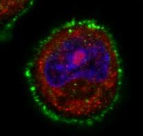
The REBORNE project began three years ago and its originality lies in using mesenchymal stem cells (MSC), found in bone marrow, to help heal fractures. Adult stem cells are amplified in culture, then are associated with biomaterial before being implanted on the fracture. Promising pre-clinical trials have now paved the way for the launch of these new clinical trials.

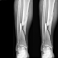
Following trauma (fall or road accident), there is a high incidence of delayed union or non-union in tibia, femur or humerus bones requiring a surgical apposition of an autologous bone graft (patient’s own bone). However, the bone graft available is limited and complications are often observed at the second surgical site.

The partners of the European project REBORNE propose an alternative treatment using autologous mesenchymal stem cells associated with a synthetic bone substitute.

© P. Layrolle, Inserm

Thanks to a bone marrow aspiration under local anaesthesia, the mesenchymal stem cells are isolated and amplified in culture for 21 days. During the surgical intervention stem cells are associated with biphasic calcium phosphate ceramic granules and implanted at the site of the fracture. In this way, the biomaterial is used as “scaffolding” and encourages stem cell proliferation. The stem cells are then differentiated into bone cells and regenerate the bone tissue around the fracture.

© P. Layrolle, Inserm

Bone formation (in green) and bone barrow formation (in red) further to a mixture of human stem cells and biomaterial granules (grey/white) being implanted at a subcutaneous site in nude mice. This histological section shows osteoinduction by the cell/biomaterial mix with the formation of around 41% of bone tissue four weeks after implantation.

The French Medicinal Agency ANSM gave its green light on 3 January 2013 so that these Inserm-sponsored trials can now begin in France, with seven patients receiving treatment in the Créteil University Hospital Centre and the Regional University Hospital Centre in Tours. Ultimately, thirty patients will be included in France, Spain, Germany and Italy to be part of this multi-centre European study.

“The objective of this trial is to show that biomaterials and stem cells are safe and are at least equivalent to standard treatments, without their disadvantages. This surgery is less invasive and preserves the patients’ bone stock. For these reasons, it is preferable to bone grafts in terms of triggering bone healing”.

 explains Pierre Layrolle, director of Research, Inserm and coordinator of REBORNE.

For further information:

REBORNE – Regenerating Bone Defects using New biomedical Engineering approaches

The objective of REBORNE is to develop new biomaterials that stimulate bone tissue formation with a view to correcting bone regeneration defects in orthopaedic and maxillofacial surgery. Biomaterials, combined with the use of stem cells, are interesting alternatives to biological grafts.

The REBORNE project kicked-off in January 2010 and will run for five years using an overall budget of roughly 12 million Euros provided by the European Union (PC7). The project involves 24 partners, based in eight European countries: http://www.reborne.org/presentation.html

La protéine à l’origine de la maladie de Huntington impliquée dans les tumeurs mammaires

Detrimental effect of obesity on lesions associated with Alzheimer’s disease

Researchers from Inserm and the Université Lille/Université Lille Nord de France have recently used a neurodegeneration model of Alzheimer’s disease to provide experimental evidence of the relationship between obesity and disorders linked to the tau protein. This research was conducted on mice and is published in the Diabetes review: it corroborates the theory that metabolic anomalies contribute massively to the development of dementia.

In France, more than 860,000 people suffer from Alzheimer’s disease and related disorders, making them the largest cause of age-related loss of intellectual function. Cognitive impairments observed in Alzheimer’s disease result from the accumulation of abnormal tau proteins in nerve cells undergoing degeneration (see the picture below). We know that obesity, a major risk factor in the development of insulin resistance and type 2 diabetes, increases the risk of dementia during the aging process. However, the effects of obesity on ‘Taupathies’ (i.e. tau protein-related disorders), including Alzheimer’s disease, were not clearly understood. In particular, researchers assumed that insulin resistance played a major role in terms of the effects of obesity.

The “Alzheimer & Tauopathies” team from mixed research unit 837 (Inserm/Université Lille 2/Université Lille Nord de France) directed by Dr. Luc Buée, in collaboration with mixed research unit 1011 “Nuclear receptors, cardiovascular diseases and diabetes”, have just demonstrated, in mice, that obese subjects develop aggravated disorders. To achieve this result, young transgenic mice, who develop tau-related neurodegeneration progressively with age, were put on a high-fat diet for five months, leading to progressive obesity.

“At the end of this diet, the obese mice had developed an aggravated disorder both from the point of view of memory and modifications to the Tau protein”

explains David Blum, in charge of research at Inserm.

This study uses a neurodenegeneration model of Alzheimer’s disease to provide experimental evidence of the relationship between obesity and disorders linked to the tau protein. Furthermore, it indicates that insulin resistance is not the aggravating factor, as was suggested in previous studies.

“Our research supports the theory that environmental factors contribute massively to the development of this neurodegenerative disorder” underlines the researcher. “Our work is now focussing on identifying the factors responsible for this aggravation” he adds.

The degeneration of neurons in Alzheimer’s disease

© Wikipedia – Zwarck  / licence Creative Commons  CC-BY-SA-2.5

In the case of healthy neurons (top), the Tau protein is normal.
In the case of diseased neurons (bottom), abnormal tangles of Tau proteins (phosphorylated) form, causing degeneration.

This research was supported by LabEx DISTALZ (development of Innovative Strategies for a Transdisciplinary Approach to Alzheimer’s Disease) within the framework of future investments.

Why good resolutions about taking up a physical activity can be hard to keep

Physical inactivity is a major public health problem that has both social and neurobiological causes. According to the results of an Ipsos survey published on Monday 31 December, the French have put “taking up a sport” at the top of their list of good resolutions for 2013. However, Francis Chaouloff, research director at Inserm’s NeuroCentre Magendie (Inserm Joint Research Unit 862, Université Bordeaux Ségalen), Sarah Dubreucq, a PhD student and François Georges, a CNRS research leader at the Interdisciplinary Institute for Neuroscience (CNRS/Université Bordeaux Ségalen) have just discovered the key role played by a protein, the CB1 cannabinoid receptor, during physical exercise. In their mouse studies, the researchers demonstrated that the location of this receptor in a part of the brain associated with motivation and reward systems controls the time for which an individual will carry out voluntary physical exercise. These results were published in the journal Biological Psychiatry

©fotolia

The collective appraisal conducted by Inserm in 2008 highlighted the many preventive health benefits of regular physical activity. Such activity is limited, however, by our lifestyle in today’s industrial society. While varying degrees of physical inactivity may be partly explained by social causes, they are also rooted in biology.

“The inability to experience pleasure during physical activity, which is often quoted as one explanation why people partially or completely drop out of physical exercise programmes, is a clear sign that the biology of the nervous system is involved”, explains Francis Chaouloff.

But how exactly? The neurobiological mechanisms underlying physical inactivity had yet to be identified.

Francis Chaouloff (Giovanni Marsicano’s team at the NeuroCentre Magendie; Inserm joint research unit, Université Bordeaux Ségalen) and his team have now begun to decipher these mechanisms. Their work clearly identifies the endogenous cannabinoid (or endocannabinoid) system as playing a decisive role, in particular one of its brain receptors. This is by no means the first time that data has pointed to interactions between the endocannabinoid system, which is the target of delta9-tetrahydrocannabinol (the active ingredient of cannabis), and physical exercise. It was discovered ten years ago that physical exercise activated the endocannabinoid system in trained sportsmen, but its exact role remained a mystery for many years. Three years ago, the same research team in Bordeaux observed that when given the opportunity to use a running wheel, mutant mice lacking the CB1 cannabinoid receptor, which is the principal receptor of the endocannabinoid system in the brain, ran for a shorter time and over shorter distances than healthy mice. The research published in Biological Psychiatry this month seeks to understand how, where and why the lack of CB1 receptor reduces voluntary exercise performance (by 20 to 30%) in mice allowed access to a running wheel three hours per day.

The researchers used various lines of mutant mice for the CB1 receptor, together with pharmacological tools. They began by demonstrating that the CB1 receptor controlling running performance is located at the GABAergic nerve endings. They went on to show that the receptor is located in the ventral tegmental area of the brain (see diagram below), which is an area involved in motivational processes relating to reward, whether the reward is natural (food, sex) or associated with the consumption of psychoactive substances.

Longitudinal section of the mouse brain (top) and diagram of interactions between the endocannabinoid, GABAergic and dopaminergic systems during voluntary physical exercise (bottom)

©Inserm/F. Chaouloff

Left: stimulating the CB1 receptors excites the dopaminergic neurons in the ventral tegmental area involved in motivation.
Right: the absence of CB1 receptors lowers performance by 20 to 30% owing to reduced motivation.

VTA: Ventral tegmental area/NAcc: nucleus accumbens/PFC: prefrontal cortex/DA: dopamine 

Based on the results of this study and earlier work, the Bordeaux team suggests the following neurobiological explanation: at the beginning and for the duration of physical exercise, the CB1 receptor is constantly simulated by the endocannabinoids, lipid molecules that naturally activate this receptor in response to pleasant stimuli (rewards) and unpleasant stimuli (stress). Endocannabinoid stimulation of the CB1 receptor during physical exercise inhibits the release of GABA, an inhibitory neurotransmitter that controls the activity of the dopamine neurons associated with the motivation and reward processes. This stimulation of the CB1 receptor “inhibits inhibition”, in other words, it activates the dopaminergic neurons in the ventral tegmental area. The CB1 receptor must therefore be stimulated before the exercise can go on for longer and the body must receive the necessary motivation.

Conversely, without these CB1 receptors, the “GABAergic brake” continues to act on the dopaminergic neurons in the ventral tegmental area, leading to the reduced performance levels observed above.

It is already known that CB1 receptors play a regulatory role in the motivation to consume rewards, whether natural or not. What is original about this research is that it shows that physical exercise can be added to the array of natural rewards regulated by the endocannabinoid system. “If confirmed, this motivational hypothesis would imply that the role played by the CB1 receptor has more to do with ‘staying power’ in the exercise than with actual physical performance levels” explain the researchers.

This work reveals that the endocannabinoid system plays a major role in physical exercise performance through its impact on motivational processes. It thus opens up new avenues of research into the mediators of pleasure – and even addiction – associated with regular physical exercise. “After endorphins, we now need to consider endocannabinoids as another potential mediator of the positive effects that physical exercise has on our mood,” the researchers conclude.

fermer