Menu

Rich or poor in intestinal bacteria: we are not all equal when it comes to obesity-related conditions

Researchers working on the European METACARDIS (Metagenomics in Cardiometabolic Diseases) Project, coordinated by Inserm, have published in the magazine Nature the first results of the investigation into the composition of the intestinal flora of obese patients. They observed that these individuals were deficient in intestinal bacteria (in both quantity and diversity) and were at greater risk of developing cardio-metabolic diseases. At the same time, they managed to improve the composition of the intestinal flora through a specific diet. In future, it will soon be possible to develop a simple test to identify people at risk and offer them a suitable preventive solution.

Centre Biomédical recherche et de valorisation

© Inserm

The obesity epidemic affected about 400 million adults in 2005; it will affect 700 million people in 2015 and the numbers will continue to increase. The causes are partially environmental (a more sedentary lifestyle, changes in eating habits, etc.) and partially genetic. Obesity linked to mutations represents a minority of cases. More and more data indicate that the variations in our “other genome”, the microbiome, i.e. the total genome of all the micro-organisms in our body, do more in the development of obesity than variations or polymorphism in the human genome.

Researchers working on the European METACARDIS project participated in two studies whose results have initiated the development and creation of individual treatments for patients suffering from cardio-metabolic disorders, including cardio-vascular diseases and metabolic diseases associated with obesity.

Two types of individuals, depending on the bacterial composition of the digestive tract

The first stage of the METACARDIS project is the study of patient cohorts constituted during previous European or national programmes (the European projects MetaHIT and Micro-Obes supported by the French National Research Agency; > 200 subjects), who provided instant access to much clinical, medical and environmental data as well as biological samples.

An initial study was performed on the MetaHIT cohort, which consisted of 292 Danish adults 123 of them non-obese and 169 obese. The researchers analysed the intestinal bacterial genome of these individuals, using a new technique known as quantitative metagenomics. According to the results, it appears that the two groups of individuals can be differentiated by the richness of the bacteria they carry and the abundance of certain species of intestinal bacteria. One quarter of the people in the cohort were bacteria “poor”, while three-quarters possessed intestinal flora that were “rich” in bacteria. It is the first time that such a distinction has been highlighted in the population. Furthermore, the distinction did not depend on the corpulence of the individuals, because non-obese people were found in both groups, even though the bacteria-deficient group contained more obese people (80%).

Increased risk of complications associated with obesity

When comparing the two groups, researchers discovered that those who were deficient in intestinal bacteria were at greater risk than those who were bacteria-rich of developing complications linked to obesity such as type II diabetes, lipidic and hepatic problems and increased inflammatory factors in the blood, of the type frequently observed in these conditions.

Species of bacteria limit weight gain

Researchers also observed that obese people in the bacteria-deficient group gained more weight over time than individuals in the other group. In bacteria-poor individuals, eight specific species of bacteria were present in low numbers, or were even absent altogether. These species may have a protective role in preventing weight gain and in the long run their detection will open the way to the development of new probiotics that will help to combat weight gain.

Only six species of bacteria make it possible to differentiate between “poor” and “rich”

The second study, conducted by researchers in the METACARDIS consortium on the MicroObes cohort, focused on a cohort of 49 obese or overweight French adults. This confirmed the results of the first study. There were similarities in the bacteria-poor and rich communities between the French and Danish populations. Furthermore, on the basis of only six species of bacteria that were particularly representative in these communities, it was possible to distinguish between bacteria-rich communities and bacteria-poor communities with 95% accuracy. These results could lead to the devising of a simple method for determining which type of microbial intestinal community lives inside an individual. The purpose of METACARDIS is, on the one hand, to confirm, in a larger European cohort, the importance of these bacterial markers and on the other hand to develop a simple diagnostic test to identify individuals who are at greatest risk of becoming obese, associated with co-morbidity.

A diet that helps to enrich the microbiota

The French study also investigated the impact of a protein- and fibre-rich, low calorie diet on the genetic diversity of the intestinal microbiota. After six weeks, the diet not only produced the expected improvement in the clinical characteristics of the individuals studied, but also increased the richness of intestinal bacteria in the bacteria-poor communities. Researchers were thus able to correlate the increase in bacterial richness with weight loss, a reduction in the mass of fat and in cardio-metabolic risk factors. The way is open not only for the diagnosis of at-risk individuals but also for intervention through targeted nutritional recommendations. The inflammatory level of the blood and fatty tissue was not reduced by the diet as efficaciously in individuals who were species-poor as in those who were species-rich. Other interventions, perhaps involving medication, need to be invented to tackle this problem.

The METACARDIS consortium will pursue its research for another five years in order to better specify the various forms of obesity which vary greatly from one person to another and to detect people at risk from metabolic and cardiovascular complications so as to do more to prevent these complications and treat them in a targeted fashion. Consequently any early changes in the microbiota looks like the right route to pursue since it would be accessible through changes in the diet” explains Prof. Karine Clément, the METACARDIS coordinator. In fact, the ultimate aim of the project is to devise preventive medicines for chronic conditions as an alternative to the curative medicines that are becoming less and less affordable financially for industrialised societies.

The METACARDIS project, coordinated by Inserm and launched last December, represents the first systematic study to correlate intestinal flora and cardio-metabolic conditions in humans. The project is bringing together groups of European doctors and multidisciplinary researchers as well as experts in biotechnology and those from industry can access the latest technologies to promote the development of new preventive and treatment strategies for cardio-metabolic conditions. The teams include the INSERM 872 Unit headed by Karine Clément and Dominique Gauguier at the “Les Cordeliers Research Centre”, the “ICAN” University Hospital Institute headed by Serge Hercberg and INSERM Unit 557 “Nutritional Epidemiology” run by Hervé Blottière, Joël Doré and Dusko Ehrlich of the INRA.

http://www.metacardis.eu

The 12 partners in the METACARDIS project:

Inserm, France

INRA, France

Imperial College London, United Kingdom

University of Copenhagen, Denmark

European Molecular Biology Laboratory, Germany

University of Gothenburg, Sweden

Flemish Institute for Biotechnology, Belgium

Assistance Publique – Hôpitaux de Paris, France

University of Leipzig, Germany

Danone Research, France

Cargill, France

Chalmers University of Technology, Sweden

INSERM Transfert, France

Biobyte Solutions, Germany


Neutrophils: the Unsung Heroes of Immunotherapy Cancer Treatment

Scientists at the Institut Pasteur and Inserm have identified the group of cells within the immune system that make immunotherapy treatment (therapeutic antibodies) effective. Immunotherapy is frequently used to treat breast cancer. In animal models they showed that neutrophils, the most common white blood cells in the body, are not only necessary but suffice on their own to eliminate tumor cells. The scientists produced these results using both skin and breast cancer models. If confirmed in humans, these discoveries should lead to the optimization of current treatments for several cancers. This work is being published online August 26, 2013 by the medical journal Blood.

With its millions of victims each year, cancer remains one of the leading causes of death worldwide. In women, breast cancer is the most common offender. Nearly one in nine women will develop breast cancer in their lifetime. In France alone, over 50,000 new cases are diagnosed each year.

Between 20 and 30% of breast cancer cases are deemed eligible for immunotherapy treatments which involve the injection of antibodies. Until now, the effectiveness of this type of treatment was attributed to various immune system components such as Natural Killer cells and macrophages. However, work by Pierre Bruhns, head of the Antibodies in Therapy and Pathology Laboratory (Institut Pasteur/Inserm Unit 760), and his team, in collaboration with Professor Clifford A. Lowell of the University of California Department of Laboratory Medicine and two other teams from the Institut Pasteur, has cast serious doubt on this theory.

Etude de la moelle osseuse et de l'hématopoièse

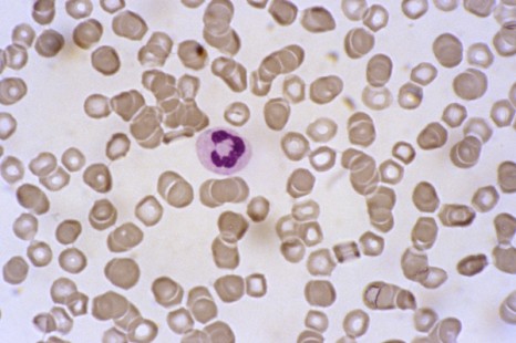
Neutrophile observed by optical microscopy ©Inserm/E.Cramer

The scientists have shown in an animal model that neutrophils alone are effective enough to induce the therapeutic effect of immunotherapy treatments often used to treat breast cancer. Neutrophils are drawn to the tumor after the injection of therapeutic antibodies and are activated after coming into contact with these antibodies. Once activated the neutrophils are capable of destroying the tumor cells. This discovery represents an important step towards the optimization and development of immunotherapy treatments used to treat these cancers.

The scientists made the following observations: the tumor mass did not regress in mice that were neutropenic (having a significantly low level or total lack of neutrophils) or in mice with neutrophils that could no longer be activated through contact with therapeutic antibodies. Then, scientists showed that the anti-tumor effect achieved via immunotherapy could be reproduced in deficient mice by administering them with neutrophils taken from healthy mice. These observations were made using models for both skin and breast cancer leaving us to assume that neutrophils play an important role in immunotherapy treatments used to fight various other cancers.

In their conclusion, the scientists highlighted current immunotherapy treatments used to fight cancer. These treatments are often coupled with other treatments. Because of this, a good strategy might be to favor treatment pairings that increase neutrophil count and activity.

A new therapeutic strategy to combat prion and Alzheimer’s diseases

A work performed by the teams headed by Benoit Schneider and Odile Kellermann (INSERM Unit 747, team “Stem cells, Signalling and Prions”, Université Paris Descartes) as well as Jean-Marie Launay’s team (INSERM Unit 942 Hôpital Lariboisière and the FondaMental Foundation) was published this week in the magazine Nature Medicine. The article revealed that in neurons, an enzyme, the kinase PDK1, is involved in the accumulation of the pathological proteins involved in prion and Alzheimer’s diseases. The researchers show that the pharmacological inhibition of this enzyme exerts a beneficial effect towards both pathologies.

Details of this research were published in the magazine Nature Medicine

Différenciation cellulaire

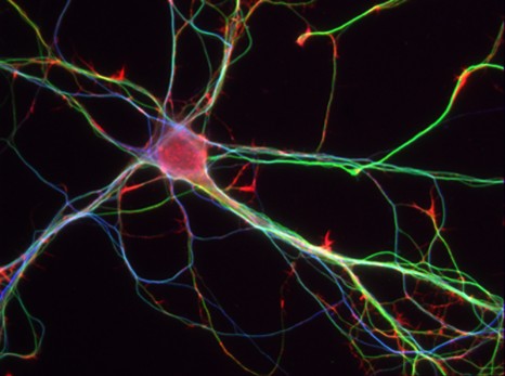
Mouses Neurones – 7 days – © Inserm/L.Peris

Prion diseases (Creutzfeld-Jakob disease in humans) and Alzheimer’s disease are associated with an accumulation of abnormal proteins in the brain. These are the scrapie prion protein (PrPSc) in the case of prion diseases and Aß amyloid peptides in the case of Alzheimer’s. In the brain, PrPSc and Aß peptides exert their toxic effect by causing the death of neurons, at the root of the clinical symptoms associated with these diseases.

It is acknowledged that the production of the pathological proteins PrPSc and Aß40/42 originate from a defect in the physiological cleavage of the entire, non-pathological prion protein (PrPC) or of the amyloid peptides precursor (APP). However, it remained unsolved why this cleavage, which normally protects neurons, is altered in prion and Alzheimer’s diseases.

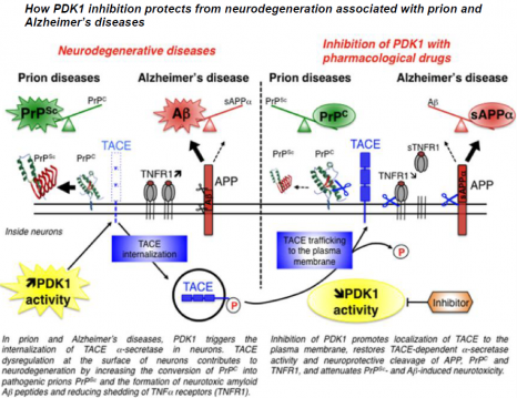
The work performed by Benoit Schneider (CNRS researcher in INSERM Unit 747, “Stem cells, Signalling and Prions”, Université Paris Descartes) and Jean-Marie Launay (INSERM Unit 942 Hôpital Lariboisière) in collaboration with other French teams working in the prion field has just identified a cascade of reactions that blocks the beneficial cleavage of PrPC and APP by the alpha-secretase TACE (an acronym for the TNFα Converting Enzyme). The researchers demonstrate how TACE dysregulation contributes to neurodegeneration by causing the accumulation of the PrPSc and Aß40/42 pathological proteins and exacerbating neuron sensitivity to inflammation.

Under normal physiological conditions, TACE is present on the surface of neurons, where it cleaves PrPC, APP and the receptors to TNFα inflammatory factor (TNFR), thus restricting the production of the pathological proteins PrPSc and Aß and protecting neurons from the toxic effects of TNFα.

schéma Prions Alzheimer en

© Benoit Schneider & Mathéa Pietri, August 2013

In neurons infected by pathogenic prions as in the “Alzheimer’s” neurons, the TACE protease is no longer present on the cell surface but is found inside neurons. This internalization diverts TACE away from its substrates, that is PrPC, APP and TNFR, and thereby cancels its neuroprotective activity. The researchers reveal for the first time that the kinase PDK1 plays a key role in controlling the localization of TACE in neurons. The overactivation of PDK1 is responsible for the internalization of TACE in diseased neurons (those infected by prions and Alzheimer’s neurons) as in the brains of patients suffering from Alzheimer’s disease.

The pharmacological blockade of PDK1 relocates TACE to the surface of neurons and restores its neuroprotective function. The inhibition of PDK1 protects neurons from neurodegeneration by rescuing the physiological cleavage of PrPC, APP and TNFR by TACE.

“Based on our work on prion infection, we succeded in identifying PDK1 as a new therapeutic target not only for Creutzfeld-Jakob disease but also for Alzheimer’s disease” explained the researchers.

The action of PDK1 on TACE was demonstrated in vitro using a neuronal cell line and cultures of neurons isolated from the brains of mice that had been infected by pathogenic prions, and in vivo using animal models. Treatment with PDK1 pharmacological inhibitor attenuates the motor deficits and extends the life span of mice infected with prions. Using three mouse models of Alzheimer’s pathology, the researchers showed that the treatment also counteracted memory and cognitive deficits associated with Alzheimer pathogenesis.“Because treatments available to combat prion and Alzheimer’s diseases are few and their efficacy limited, these results could open up new avenues for the treatment of these neurodegenerative diseases”, conclude the researchers.

By demonstrating that the inhibition of PDK1 alleviates both prion and Alzheimer’s diseases, these data argue that at a mechanistic level AD links to prion diseases. Dysregulation of PDK1-dependent TACE cleavage activity emerges as a central event in neurodegenerative pathways involved in both diseases.

The challenge is now to understand how pathogenic prions or amyloid Ab peptides trigger PDK1 activation.

Testosterone responsible for worsening iron overload in chronic liver diseases

A research team from Toulouse has just elucidated the mechanisms behind the differences in iron absorption between men and women. The team used mice to demonstrate how the action of testosterone can be “countered” with a drug already used in the treatment of some bronchial cancers.  The results are published this month in the Hepatology review.

artere

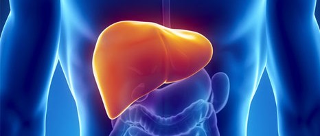
The regulation of the iron metabolism differs between men and women. As a consequence, chronic liver diseases associated with a reduced hepcidin production capacity – a hormone that inhibits iron absorption by the duodenum – are often more severe in men than in women.

This is the case for hepatitis C, alcoholic liver disease and hereditary hemochromatosis.

With a view to better understanding the underlying mechanisms of this sexual dimorphism, Marie-Paule Roth and her co-workers at the Centre for Physiopathology in Purpan (Mixed Inserm/CNRS/Université Paul Sabatier Research Unit, Toulouse) have taken advantage of the major differences in hepcidin expression and iron overload observed in male and female mice with iron metabolism disorders.

The researchers showed that testosterone robustly represses hepcidin transcription in these mice since it activates the growth factor receivers in the liver (EGF). Yet, when these EGF receivers are inhibited in male mice by a drug used in the treatment of some bronchial cancers, testosterone-induced repression ceases.   Hepcidin production is then significantly increased. Castrating male mice leads to an increase in hepcidin production, as well as a very strong reduction in iron overload in the heart and pancreas of these mice.

To conclude, Marie-Paule Rothe and her team emphasise: “These results indicate that the testosterone-induced repression of hepcidin may have clinical repercussions for patients who, for different reasons, are only capable of producing hepcidin in limited quantities. Inhibiting the activation of EGF receivers in these patients could help them to produce more hepcidin, thus limiting iron overload that worsens the prognosis of chronic liver disease” they explain.

A gene has been identified that is at cause in several forms of epilepsy with language dysfunction.

A team of researchers from Inserm led by Pierre Szepetowski (INMED: “Institut de Neurobiologie de la Méditerranée” combined inserm/ University of Aix-Marseille unit) has just succeeded in identifying a gene whose mutations are responsible for a wide spectrum of epilepsies and epileptic encephalopathies with language dysfunction in children.
This work has been published in the journal “Nature Genetics”.

Épilepsie

An epilepsy crisis is caused by sudden, short-lived, excessive activity of a group of neurons. It causes paroxysmal clinical symptoms, such as convulsions. Normally, epilepsy does not alter the cognitive capacities. However, in certain forms known as epileptic encephalopathies, the epileptic component can cause or worsen serious cognitive and behavioural problems (mental handicaps, language dysfunctions, autistic regression, etc.). This is the difference between these disorders and “conventional” epilepsy.

The team and the network of researchers led by Pierre Szepetowski tried to get a better understanding of the relationships between epilepsy and the numerous other problems related to the illness: autistic problems, cognitive problems, language dysfunction, speech impairment, dyslexia, voluntary movement disorders, migraines, etc.

Up until now, the cause of three rare forms of epilepsy and epileptic encephalopathies (acquired epileptic aphasia, continuous wave spike in slow sleep syndrome, and Rolandic epilepsy with speech disorders), had been under debate for over fifty years in the medical and scientific world and had remained unknown.

Thanks to a wide-ranging genetic analysis, the researchers, working as part of an extended network of epileptologists and scientists associating different hospitals and research centres , have just demonstrated that 20% of these cases of epilepsy often associated with language dysfunction have a common genetic cause. In all these forms of the disorder, there is mutation of the gene GRIN2A that codes for a glumatate receptor, a crucial neurotransmitter in the brain.

According to Pierre Szepetowski, this new light shed on the problem shows that “these three symptoms can be viewed as different clinical expressions of one and the same pathology at the crossroads between epilepsy, language dysfunction and cognitive and behavioural disorders”.

Identifying the gene GRIN2A as a major gene responsible for these epileptic encephalopathies provides the first crucial indications towards future understanding of the underlying mechanisms.

“These encephalopathies normally start around 4-5 years of age, after a period of normal development. Thereafter, development is variable and highly unpredictable. The identification of a first major cause will help us to better explain to parents how the disorder occurs, in particular for genetic counselling. We can also hope to see early therapeutic strategies set up in the future once we have a better understanding of the mechanisms. These will be crucial to improving the prognosis in cases of associated neuropsychological deficiencies”‘ adds Pierre Szepetowski.

Will in utero prevention of later epilepsy be possible one day?

During pregnancy, defects in the development of the cerebral cortex of the future baby can tigger subsequent epilepsy crises. Pierre Szepetowski’s team has just demonstrated that the absence of a protein known as Srpx2 disturbs the migration of neurons in the brains of developing rats. Even better, they succeeded in counter-acting these defects and their post-natal epileptic consequences in rats by administering Tubacin to the mother. Tubacin is capable of modifying the functioning of tubulins – molecules that are vital to building the architecture of neurons and to neuron migration.
These works, published in early July in the journal “Brain”, look like being a first step in the direction of theoretically preventing in utero the occurrence of certain types of epilepsy.

Caffeine consumption during pregnancy and its effects on the brain during development

Caffeine is the most consumed psychoactive substance in the world, including during pregnancy. Christophe Bernard, Inserm research director, and his team within the “1106 Institut de Neurosciences des Systèmes” unit (Inserm/Aix-Marseille University), have recently described certain harmful effects after caffeine consumption by female mice during pregnancy on the brains of their offspring. This work, despite performed in rodents, suggests that careful studies should be performed to assess the consequences of caffeine consumption by women during pregnancy.

These results are being published in the Science Translational Medicine review of 7th of August 2013.

Many substances have a direct effect on brain function, by modifying the activity of neurons. This applies to antidepressants, anti-anxiety drugs, nicotine, alcohol and recreational drugs such as cannabis, heroin, cocaine, etc. These substances, known as psychoactive substances, bind to proteins present in brain cells and modify their activity. When consumed during pregnancy some of these psychoactive substances can affect the construction of the fetal brain, as the proteins to which they bind play key roles in brain development. The consumption of some of these substances is thus strongly discouraged during pregnancy.

Researchers from the “1106 Institut de Neurosciences des Systèmes” unit (Inserm/Aix-Marseille University) reproduced regular coffee consumption in female mice (equivalent to 3 cups of coffee a day for a human), throughout pregnancy until the weaning of the offspring, by adding caffeine to the drinking water.

“The baby mice showed enhanced susceptibility to epileptogenic conditions and, when reaching adulthood, we detected significant spatial memory problems, i.e. difficulty in identifying their position in their environment” explained Christophe Bernard, Inserm research director.

Migration Neurones

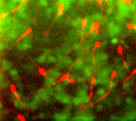
Migration of neurons©Inserm / Christine Métin – Christophe Bernard

The research team managed to identify the mechanism responsible for the deleterious effects of caffeine on the developing brain. During development, some cells are generated in specific cerebral regions, and later migrate to the regions where they will function. This is the case for neurons releasing GABA – a principal chemical mediator in the brain – which later migrate to, among other locations, the hippocampus, a brain region that plays a key role in memory formation.

Caffeine directly influences the migration of these neurons, which contain a particular receptor called A2A. When caffeine binds to these A2A receptors, the migration speed of these neurons is decreased. The cells therefore reach their intended destination later than planned. This delayed migration affects the construction of the brain with effects seen at birth (cellular excitability and susceptibility to seizures) and during adulthood (loss of neurons and memory deficits on certain tests).

©Inserm / Christine Métin – Christophe Bernard

Given their observations in mice, the authors suggest developing longitudinal studies—both short-term and, above all, long-term—to assess the consequences for newborns. Newborns may be exposed to caffeine, either during pregnancy and/or during breastfeeding, or if the child is treated for sleep apnea using a caffeine citrate based treatment.

“This study is the first demonstration of the harmful effects of exposure to caffeine on the developing brain. Although this study raises the question of caffeine consumption by pregnant women, it is necessary to reiterate the difficulty of extrapolating these results obtained in rodents to the human population without taking into consideration the differences in brain development and maturation between the species”

, highlighted the main author of the study.

fermer