Menu

New immunotherapy-based approach for post-transplant leukaemia relapse

An Inserm team from the Mondor Institute for Biomedical Research (IMRB) has just identified a key switch in the immune response, and proposes a new immunotherapy-based approach for combating leukaemia. And maybe other cancers in time. This work is published in the journal Blood.


Towards a new immunotherapy for cancer? That may well emerge from the work carried out at Inserm Unit 955, “Mondor Institute for Biomedical Research” (IMRB) by a team led by Prof. José Cohen, Coordinator of the Clinical Investigation Centre – Biotherapy at Henri Mondor Hospital AP-HP, results of which have just been published in Blood. Although it works on the treatment of leukaemias, this team has discovered a key to the regulation of the immune system that makes it possible to stimulate T lymphocyte action, and probably increase the elimination of malignant cells.

It was their work on graft versus host disease, a serious complication in leukaemia patients who receive blood cell transplants, that set them on the track. This complication is due to the recipient’s cells being attacked by overactive T cells present in the transplant. Moreover the researchers found that the presence in the transplant of T regulatory cells (T regs), a specific subpopulation of T lymphocytes, the role of which is to down-regulate the immune response, limited this phenomenon. In a mouse model, when they injected additional T regs during transplantation, they prevented the transplant from attacking the host. They therefore decided to go further, and find out how these T reg cells were controlled.


TNFR2, an immune response switch

To do this, they performed different experiments, and finally demonstrated the existence of a closed circuit between conventional T lymphocytes and T reg lymphocytes, involving a key receptor called TNFR2. When conventional T lymphocytes are active, they secrete tumour necrosis factor (TNF), which binds to the TNFR2 receptors present on the surface of T regs. This signal stimulates the latter, which then inhibit the conventional T lymphocyte response. Conversely, when the TNFR2 receptor is blocked, T regs become inactive and T lymphocytes are activated.

“This TNFR2 receptor acts on the immune response just like a real switch. When it is in the ‘on’ position, it inhibits it. When it is in the ‘off’ position, it stimulates it,” explains José Cohen, who supervised this work. Having made this discovery, the researchers now intend to block this TNFR2 receptor in an attempt to improve the immune response against cancer, which will have direct application in immunotherapies. “The role of immunotherapy is to target ‘check-points’ in the immune response in order lift inhibition and allow it to more effectively eliminate malignant cells. The treatments currently available are specific for a population of T lymphocytes known as effectors, while we propose a new target, the regulators. These treatments could therefore be complementary,” adds the researcher.


The team has already filed a patent to protect the exploitation of this receptor in the context of post-transplant leukaemia relapse. The idea is now to develop a human anti-TNFR2 antibody to test this therapeutic strategy in a so-called “humanised” mouse model. If the results are conclusive, clinical trials will be conducted. At the same time, this approach is being evaluated for other types of cancers, including solid tumours, by the team led by Dr Benoit Salomon (CIMI-Paris), a co-author of the study.

The gut microbiota to the rescue of chemotherapies

inserm_18659

© Inserm/U510

Two species of bacteria present in the gut boost the efficacy of cyclophosphamide-based chemotherapies by optimising the antitumour immunity induced by this drug. This is reported by researchers from Inserm, Gustave Roussy, CNRS, Institut Pasteur Lille, and the Universities of Paris Sud and of Lille in an article published on 4 October in the journal Immunity.


Recent studies have shown that certain gut microbes encourage tumours to grow, whereas others contribute to making cancer treatments more effective. It remained necessary to identify the nature and mode of action of the bacterial species capable of optimising the antitumour response induced by chemotherapy.

In this new study, Mathias Chamaillard[1], Laurence Zitvogel[2] and their collaborators showed that two gut bacteria, Enterococcus hirae and Barnesiella intestinihominis, together potentiate the therapeutic effects of cyclophosphamide, a chemotherapeutic agent used to treat many cancers.

How? Chemotherapy has secondary effects that include increased permeability of the intestinal barrier and, consequently, the entry of the bacteria constituting the microbiota into the bloodstream. To combat this abnormal entry of bacteria into the bloodstream, an immune response is initiated. Against all expectations, this response is beneficial for patients, since it can also lead to the destruction of the tumour cells. The tumour is therefore attacked directly by cyclophosphamide and indirectly by this “booster” effect of the bacteria.

Several preclinical models enabled the researchers to demonstrate that the antitumour immune response induced by cyclophosphamide is optimised after oral administration of E. hirae. Treatment by oral administration of B. intestinihominis enabled a similar effect to be obtained.

The researchers then analysed the immune profile of the blood lymphocytes from 38 patients with advanced stage cancer of the lung or ovary, and treated by chemoimmunotherapy. They discovered that the presence of T memory lymphocytes specific for E. hirae and B. intestinihominis makes it possible to predict the period for which a patient lives with a cancer without it getting worse, during and after a treatment.

“The efficacy of a cancer drug relies on a complex interaction between the patient’s microbiome and his/her ability to mount an effective immune response against certain bacteria of the gut microbiota,” explains one of the main authors of the study, Mathias Chamaillard, Inserm Research Director.

“These results allow us to consider increasing the efficacy of these treatments by optimising the use of antibiotics, but also by supplementing the numbers of certain bacteria, known as oncobiotics (or their active substances), which are able to enhance the effect of cancer drugs.”

The researchers have planned to identify, in future studies, the specific parts of the bacteria responsible for enhancing the effects of cyclophosphamide. “If we succeed in answering this question, we can perhaps find a way of improving the survival of the patients treated using this chemotherapy by giving them drugs derived from these bacteria,” concludes Mathias Chamaillard.

[1] Unit 1019, “Center for Infection and Immunity of Lille” (Inserm/CNRS/University of Lille/Institut Pasteur Lille)

[2] Unit 1015, “Immunology of Tumours and Immunotherapy” (Inserm/Gustave Roussy Institute/Paris-Sud University)

Eating well to grow well: discovery of a missing link

Rénald Delanoue, Inserm Researcher, and his colleagues at the Institute of Biology Valrose in Nice (Inserm-CNRS-Université Côte d’Azur) have identified the missing links in the process that regulates the size of an organism based on the richness of its diet. Their research was conducted on Drosophila, an insect that seems very distant from humans, but the study of which has nonetheless enabled many advances in biomedical research. This work is published in the 30 September 2016 issue of the journal Science.


The size of an organism depends on its nutrient intake during development. In the event of a nutrient deficiency during this period, animals modify their growth and become adults of small size, while retaining the correct proportions. This coupling between nutrition and growth involves hormones from the insulin family and IGFs (insulin growth factors); however, the molecular mechanisms that govern this regulation are still not well understood.

Work done by Inserm researchers at the Institute of Biology Valrose (Inserm-CNRS-UCA) has enabled the identification of the substances on which this coupling is based at molecular level in the fruit fly (Drosophila melanogaster). Despite 700 million years of evolutionary divergence, this insect is a relevant model for biomedical research, because it possesses the same physiological processes as mammals.

For example, it is interesting to know that the fat body in Drosophila fulfils the same roles as the liver and adipose tissues in humans. The neurosecretory insulin producing cells (IPCs) are located in the larval brain of the insect, and correspond functionally to the pancreatic β-cells in humans.

Using the Drosophila model, these researchers have already shown that the coupling of nutrition and growth requires communication between these two organs (ndlr fat body and IPCs). Depending on the quantity of amino acids available in the diet, the fat body sends different signals to the brain. The IPCs are able to interpret these, and to secrete the appropriate amount of insulin. A low level of amino acids induces a reduction in insulin secretion and a slowing in growth, and vice versa.

It remained necessary to identify the nature of these remotely acting signals, and the IPC molecule capable of interpreting them to determine the quantity of insulin to be secreted. To do this, the researchers inhibited, one by one, the known receptors on IPCs, and identified the Methuselah receptor, inhibition of which blocks insulin secretion.

The Stunted protein, which binds to this receptor, was known, but had not been linked to the regulation of insulin secretion. And for a reason! This protein is usually found mainly inside the cells, and plays a role in ATP synthesis. It was therefore surprising to find it freely circulating in the haemolymph of the insect (the equivalent of the bloodstream). The researchers showed that the level of circulating Stunted varies with the quantity of amino acids present in the diet. Its suppression disrupts insulin secretion, and gives rise to adults of small size. Finally, they also demonstrated that this “signalling” function of Stunted in communication between two organs is a novel one, and independent of its function in ATP synthesis.


These results, although very fundamental, could nonetheless guide the study of the molecular circuits of certain diseases such as diabetes, obesity or some forms of cancer that depend on hormones and receptors from the same family as those described in Drosophila. Stunted proteins, which have been found on the surface of many cell types, could also play a signalling role in humans.

Drosophile2

The fluorescent protein GFP (green) is expressed in insulin producing neurons, IPCs (dotted white lines). Secretion of Drosophila insulins (Dilp2, in red) is regulated in these specialised neurons by the presence of the Methuselah receptor. The absence of this membrane protein in Mth- IPCs leads to blocking of insulin (Dilp2) secretion and its accumulation in IPCs (bottom row), compared with the control (top row).

(c) Rénald Delanoue/Inserm

1 in 2 French people suffers from joint pain

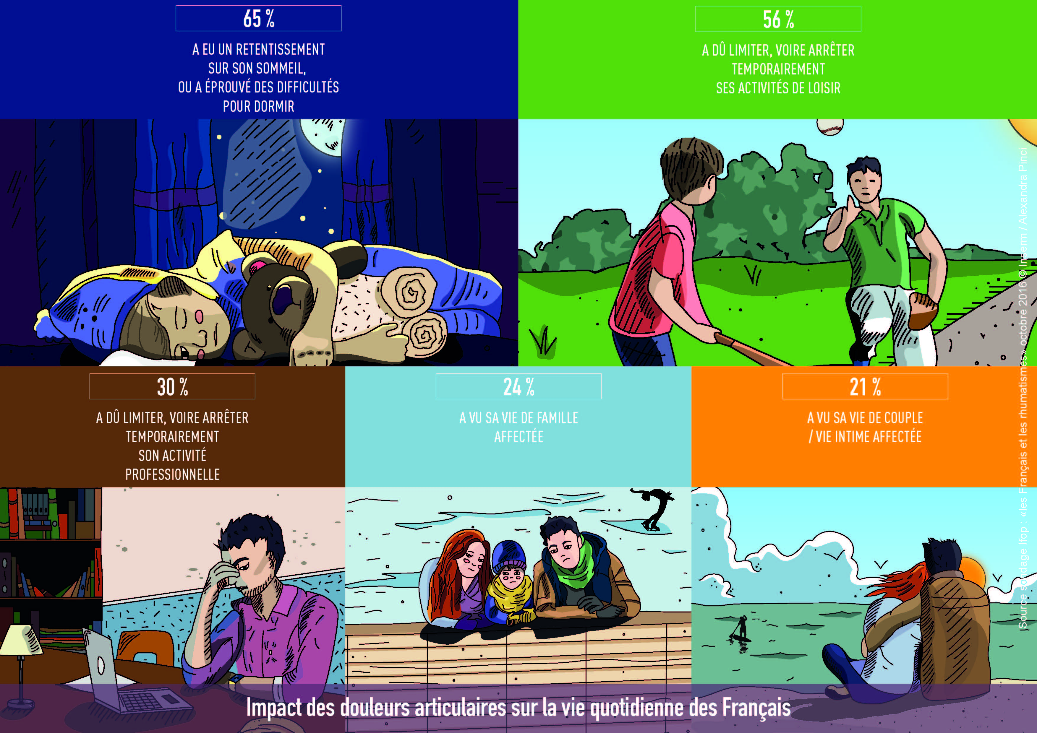
According to the results of an IFOP survey, “Les Français et les Rhumatismes” (Rheumatic Diseases and the French), conducted by Inserm and “Ensemble Contre les Rhumatismes” (Together Against Rheumatic Diseases), 93% of French people report having already suffered from joint pain[1] and one in two were suffering from it at the time of responding to the survey. This joint pain strongly affects their daily life. Quality of sleep and the ability to perform leisure activities are the two aspects of daily life that are most affected, ahead of working life and family life.

The results of this survey emphasise the need to continue to conduct ambitious research programmes to find new treatments, and encourage the French to act from the earliest age possible to retain their “joint capital.”


This press file has been prepared to mark World Arthritis Day and the French initiative “Ensemble Contre les Rhumatismes” (ECR, Together Against Rheumatic Diseases) which will take place on 12 October next.

ECR brings together the 3 most important contributors to research on joint diseases in France. Created at the initiative of the National Alliance for Life Sciences and Health (Inserm-AVIESAN), Fondation Arthritis and the French Society for Rheumatology (SFR), Ensemble Contre les Rhumatismes (ECR, Together Against Rheumatic Diseases) has the mission of promoting research in Rheumatology in order to prevent, relieve and cure diseases of the bones and joints, which affect one quarter of the European population.

Rheumatic diseases

Rheumatic diseases comprise all diseases of the musculoskeletal system (bones / cartilages / muscles tendons / ligaments).

Osteoarthritis and Arthritis are multigenerational diseases that affect the young and less young.

Rheumatoid Arthritis and Ankylosing Spondylitis are the most common inflammatory rheumatic diseases.

12-13 million French people suffer from rheumatic diseases including:

9-10 million from osteoarthritis

600,000 from chronic inflammatory rheumatic diseases (rheumatoid arthritis, spondylitis)

4,000 children suffer from idiopathic juvenile arthritis, a serious form of juvenile arthritis


True / False

Joint pain affects only a small part of the population / FALSE

More than 1 in 2 French people presently suffer from joint or bone pain
1/3 of 18-24 year olds presently suffer from joint pain*

WHO has declared lumbago to be the leading cause of disability worldwide? In France nearly 4,000 children suffer from inflammatory rheumatic diseases, nearly 600,000 adults suffer from the same diseases, and nearly 10 million French people suffer from osteoarthritis. These diseases are therefore clearly not only due to the ageing population.

Joint diseases are not serious / FALSE

65% of French people have had their sleep affected by joint pain.
Of those under 25 years of age, more than 1 in 2 young people consider rheumatism to have had an impact on their social and family life*

Joint diseases have a major impact on quality of life and sleep, but also on mortality. They are the main reason for sick leave. Rheumatic diseases are the leading cause of chronic pain. They have a direct impact on mobility and an indirect impact by increasing the risk of cardiovascular disease and cancer because of the inactivity they generate.

One should remain as still as possible / FALSE

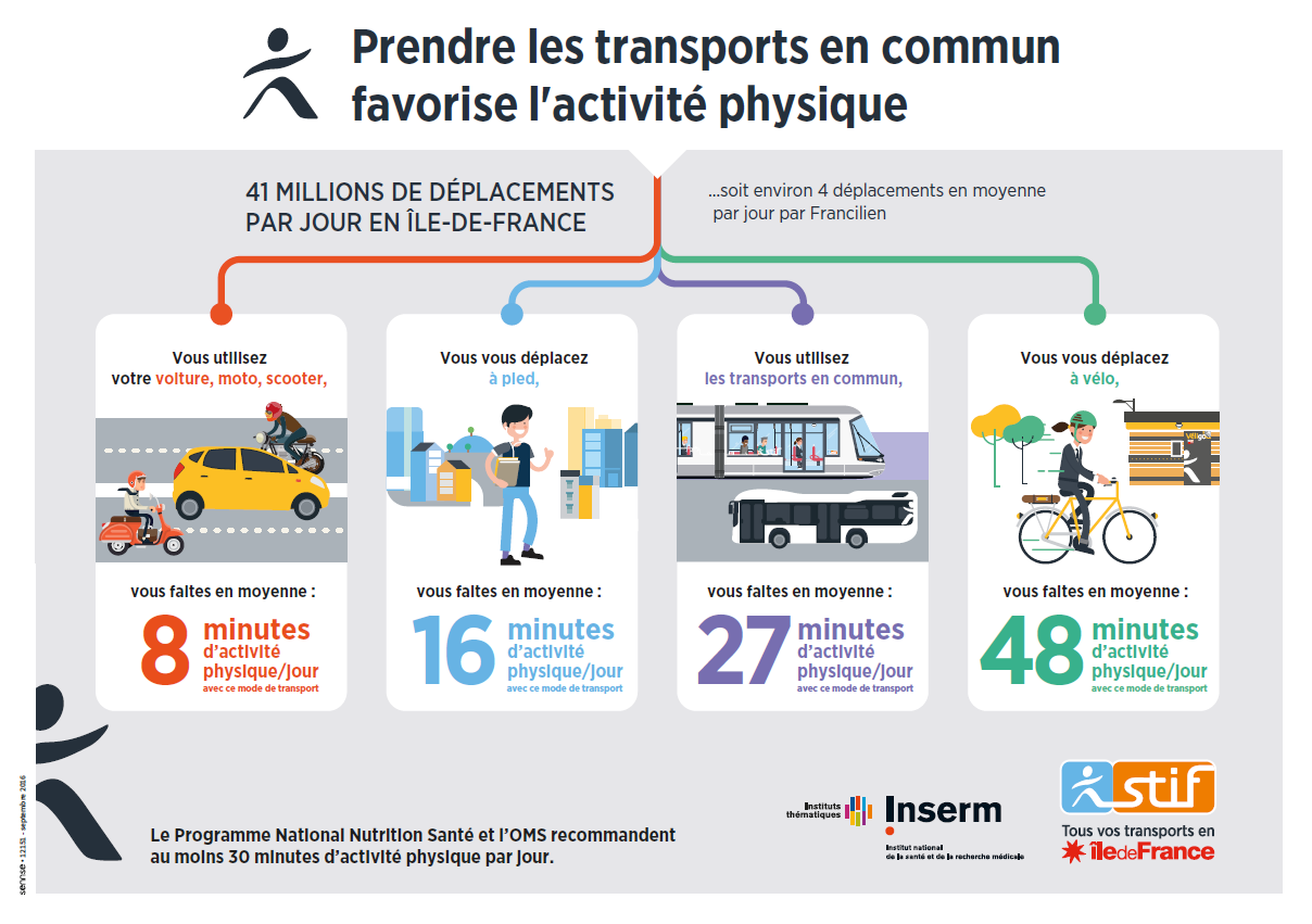
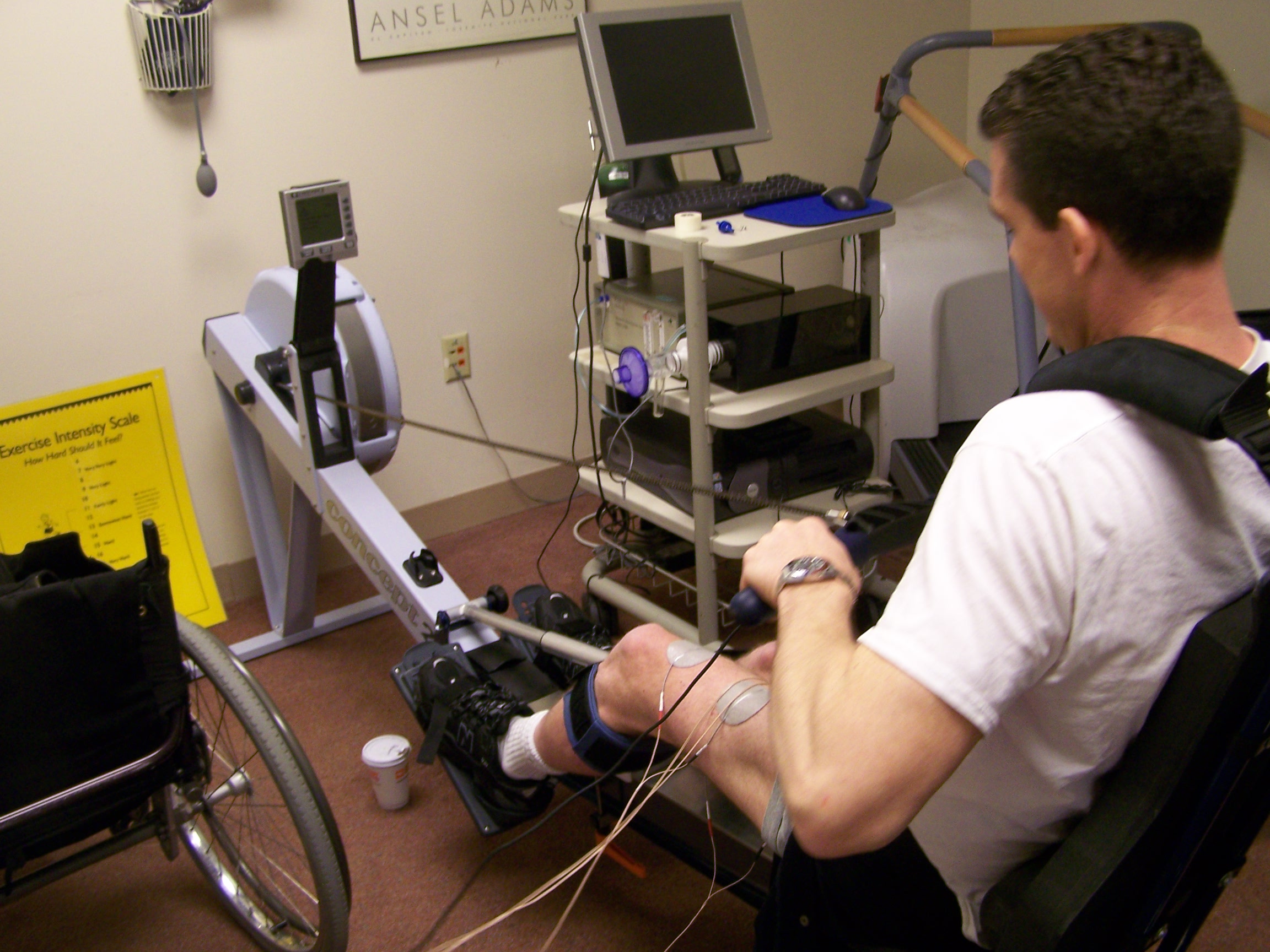
On the contrary, physical activity, which is impeded by mobility problems in people affected by these diseases, is also a remedy for maintaining one’s bone and joint capital.


What should be remembered from the IFOP survey:

How common rheumatic diseases are

A large majority of respondents (93%) have already had joint pain at least once in their lifetime. 8 out of 10 French people have even experienced it several times.

1 in 2 French people and 1 in 3 young people (18-24 years) are presently suffering (at the time of responding to the survey) from joint pain.

These figures, when compared to those for the prevalence of rheumatic diseases in Europe (one in four people has rheumatic disease), may suggest that the French are “patients” without knowing it. Most of them do not associate their bone and joint pain with the possibility of being treated for rheumatic disease.

The researchers therefore recommend speaking more and more to French people about “osteoarticular/bone and joint diseases” rather than “rheumatism.” First, because the word “diseases” gets away from the concept of being destined to have this pain. Second, because the words “osteoarticular” or “bone and joint” specifically describe the location of the problems (bones and joints), in the same manner as “cardiovascular disease” or “lung disease.”

Received ideas

8 out of 10 French people wrongly believe that rheumatic diseases are never fatal. In reality, due to the resulting inactivity, mortality is increased by 50% when walking is limited by these diseases.

The majority of respondents believe that rheumatic diseases are age-related, and cannot affect children. In reality, 4,000 children in France suffer from idiopathic juvenile arthritis.

Impact on daily life

Joint pain strongly affects the daily life of people who suffer from it. Three quarters of them mention at least one impact on the following activities: sleep, leisure activities, working life, family life, and life with spouse/partner.

These diseases have a particular impact on quality of sleep and the inability to take part in a leisure activity.


What about research?

Medical research is a major asset in combating rheumatic diseases, the leading cause of disability worldwide, and which affect more than one quarter of the European population.

Some twenty years ago, for example, collaboration between basic and clinical research enabled the discovery of innovative drugs. These are biotherapies such as anti-TNF agents, which specifically target inflammatory molecules. These discoveries revolutionised the treatment of patients with rheumatoid arthritis or spondylitis.

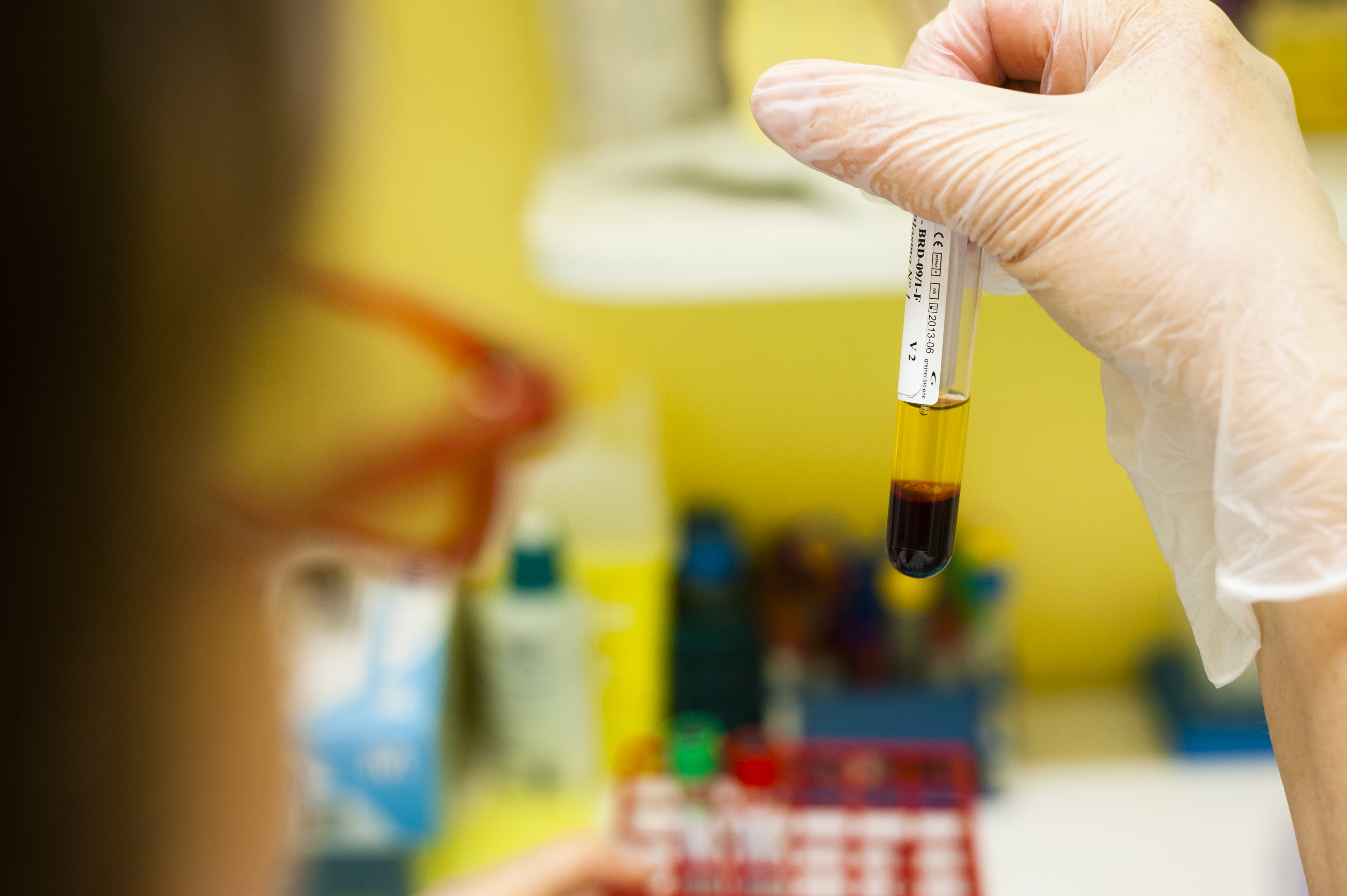
Also through research, some years ago a blood test to diagnose rheumatoid arthritis, anti-CCP, enabled physicians to diagnose the condition early, thereby allowing them to initiate treatment before the joints become damaged.

Many other bone and joint diseases have not yet benefited from advances in research. Inserm researchers are working to try to find new treatments. In France, highly ambitious projects are supported by the 3 bodies involved in the ECR initiative.

MIRIAD PROJECT

“MIcrobiome Research Initiative on spondyloArthritis and Dysbiosis”

THE MIRIAD project, led by Prof. Maxime Bréban, a researcher at Inserm Unit 1173, “Infection and Inflammation,” and a physician at Ambroise-Paré Hospital, Paris (AP-HP), in collaboration with INRA and the Flanders Institute of Biotechnology, Ghent (Belgium), will study the influence of environment on inflammatory rheumatic diseases and has the following specific objectives: 1) To identify a diagnostic marker, 2) To test a new therapeutic strategy that involves restoring the equilibrium of the microbiota in patients with spondylitis by administering a healthy microbiota.

ROAD PROJECT

“Research on Osteoarthritis Diseases”

The ROAD project, led by Jérôme Guicheux, Inserm Research Director in Nantes (Inserm Unit 791, “Laboratory of Osteoarticular and Dental Tissue Engineering:” LIOAD), is the first networked research project on osteoarthritis in France, bringing together the 7 best academic and clinical laboratories. It has the following objectives: 1) To classify the different types of osteoarthritis in mice, 2) To classify the types of osteoarthritis of the knee in humans, and to create the first National Bank of human osteoarthritic tissues, 3) To identify new biomarkers and specific targets, in order to develop innovative therapeutic strategies.

These two projects constitute pioneering initiatives in Europe. Microbiota and ageing are, moreover, two of the three priority cross-disciplinary research programmes in Inserm’s 2016-2020 strategic plan.

ESPOIR AND DESIR (Hope and Desire) COHORTS

Two national multicentre cohorts for rheumatoid arthritis (ESPOIR cohort) and spondylitis (DESIR cohort), in which several Inserm teams are participating, have already given rise to many discoveries regarding the risk factors for these diseases, such as smoking or poor oral hygiene. Prof. Xavier Mariette (Inserm Unit 1084) is coordinating a clinical trial in the ESPOIR cohort aimed at improving the oral status of patients suffering from rheumatoid arthritis.


Can rheumatic diseases be prevented?

In the absence of treatment, it is possible to conduct preventive action on rheumatic diseases. Developing the notion of “Joint Capital,” the capital that must be maintained for life at any cost, and particularly from childhood, is vital. This capital is essential, since once it is affected by one or other of these diseases, loss of this capital ends in inactivity, with known harmful consequences for the heart and blood vessels, among other things.

This “Bone and Joint Capital” relies on:

  • Specific physical activity, that needs to be tailored for patients with a bone or joint disease.
  • A balanced diet, to avoid both overweight, a major risk factor for rheumatic disease, and deficiency in calcium and vitamin D, which increases the risk of osteoporosis and hence of fracture.
  • Smoking cessation and good oral hygiene, to eliminate 2 demonstrated risk factors for the initiation and aggravation of rheumatoid arthritis.
  • Trauma prevention programmes, from childhood/adolescence:
    • Writing textbooks (junior and senior secondary school level, in Life and Earth Sciences) explaining the physiological functioning of the musculoskeletal system, and the environmental risk factors for the main bone and joint diseases, to help incorporate the concept of “Bone and Joint Capital”
    • Training sports coaches in the consequences of trauma for the bones and joints (targeted warm-ups, strengthening the muscles to stabilise the joints, appropriate interruption of sports in the event of trauma, etc.)
  • In older people, prevention of falls, which cause fractures in patients with osteoporosis.


A day to talk about it

World Arthritis Day (WAD) takes place on 12 October every year. On this day Inserm, Aviesan (National Alliance for Life Sciences and Health), Fondation Arthritis and the French Society for Rheumatology (SFR), 3 major bodies that fund rheumatology research in France, have joined forces to organise the
ECR 2016 (Ensemble Contre les Rhumatismes) day. Its objective will be to speak with one voice to the public and relevant politicians about the importance of supporting research on rheumatic diseases.

It will be held at the Daunou building from 10 am to 4 pm, in the form of round table discussions broadcast live on the Web and in 2 French cities.

Each theme of the “ECR 2016” day will be successively presented by a researcher and by a representative of a patient association, and followed by a discussion to enable the exchange of points of view between researchers, physicians and patients.

The programme has been prepared in very close collaboration with the patient associations, which were invited to a first meeting at Inserm to propose and select themes and speakers to meet their needs.

Further information: www.ensemblecontrelesrhumatismes.org

Email: vasb@rafrzoyrpbageryrfeuhzngvfzrf.bet

senapvforeraonhz@rafrzoyrpbageryrfeuhzngvfzrf.bet

[1] Rheumatic diseases comprise all diseases of the musculoskeletal system (bones / cartilages / muscles / tendons / ligaments)

Nobel Prize in medicine 2016

The Nobel Prize in Physiology or Medicine 2016 is awarded to Yoshinori Ohsumi for his discoveries of mechanisms for autophagy.

jap

Yoshinori Ohsumi. Ill. Niklas Elmehed.

© Nobel Media 2016.

Read the press release

Zika virus found inside spermatozoa

Zika spermaozoides (c) Suberbielle

Spermatozoa infected by Zika virus (green; arrowhead) 

© Elsa Suberbielle, CPTP / Inserm

Recent work has shown that Zika virus persists in semen for up to 6 months after infection[1]. In a correspondence published in The Lancet Infectious Diseases, the researchers, in addition to confirming its long persistence in semen (in this case for more than 130 days, i.e., over 4 months), reveal the presence of the virus even within spermatozoa. This work results from collaboration between researchers from Inserm, CNRS, and academic practitioners from University Toulouse III – Paul Sabatier and Toulouse University Hospital.


In this letter, the scientists report the case of a 32-year-old man returning from French Guyana with symptoms suggestive of Zika virus infection, namely, moderate fever, rash, and muscle and joint pain. Zika virus was detected in the patient’s plasma and urine 2 days after the onset of these symptoms. Samples of semen (11 samples), blood (10) and urine (5) were taken and analysed over a total period of 141 days.

Upon analysis, Zika virus was found to be present in all samples up to the 37th day. Beyond that point, the virus was found only in the semen, where it persisted for over 130 days, while the patient was in good health. This result was confirmed in two other patients, in whom the virus persisted for 69 and 115 days in the semen. For the moment, the factors responsible for this variation in persistence amongst individuals are unknown. From the time of diagnosis, however, these patients were advised to use protection during sexual relations.


The research team subsequently analysed the semen from the patient and examined the spermatozoa it contains using various microscopy techniques.

“We detected the presence of Zika virus inside approximately 3.5% of this patient’s spermatozoa,explains Guillaume Martin-Blondel, Inserm researcher at the Toulouse Purpan Pathophysiology Centre (Inserm/CNRS/University of Toulouse III – Paul Sabatier) and physician at the Infectious and Tropical Diseases Department of Toulouse University Hospital.

The researchers explain that for other sexually-transmitted viruses, such as HIV, the virus remains “stuck” at the surface of spermatozoa. For purposes of in vitro fertilization, it is therefore possible to “wash” the spermatozoa from HIV-infected patients, whereas this seems to be excluded for patients positive for Zika virus. However, the “active” nature of the Zika virus present in spermatozoa remains to be determined, as well as the ability of these spermatozoa to transmit infection (since the virus is also present outside the spermatozoa, in the seminal fluid).


In conclusion, analysis of this case has important implications for the prevention of sexual transmission of this virus, by means that remain unknown at present. These observations also raise many questions on the need to include screening for Zika virus in the testing of sperm donations in fertility centres.

[1] https://www.eurosurveillance.org/ViewArticle.aspx?ArticleId=22556

Towards an improved diagnosis of certain cancers

Asthme à l'étude

(c) Inserm/Latron, Patrice

Paris Descartes University, AP-HP, CNRS and Inserm have unveiled a royalty-free method that enables wide-scale use of circulating DNA in patients with lung and pancreatic cancer. This study is published in the journal Clinical Chemistry.


Detection of genetic alterations in tumour tissues helps to guide patient management for certain cancers, such as lung cancer. At present, these genetic studies are performed on tumour samples taken during biopsies and surgical procedures. Since tumour cells release very small quantities of their DNA into the plasma (circulating tumour DNA) a blood sample may be sufficient for the study of genetic alterations, allowing a “liquid biopsy” to be performed. The objective is not to replace the biopsy indispensable for diagnosing the cancer, but to identify the mutations and guide the prescription of cancer treatment by analysing the circulating tumour DNA. Finally, this analysis makes it possible to monitor the progression of the disease with time, and in some cases to avoid the need for further biopsies.

Before now, the molecular biology techniques needed to perform liquid biopsies were expensive, because their implementation was complex. Indeed, they require great sensitivity because they have to detect very small quantities of DNA, and great specificity to identify them as being of tumour origin. These techniques are developing at a rapid rate, and a team from Paris Descartes University UMR_S1147 has recently developed ultrasensitive techniques, based on droplet digital PCR, which study the most common genetic alterations and analyse epigenetic modifications.

Currently, this same team, working with physicians and biologists from Georges Pompidou European Hospital (HEGP) and Pitié Salpêtrière Hospital, of the Paris Public Hospitals (AP-HP), propose a method for detecting circulating tumour DNA based on next generation sequencing (NGS). It uses data from any type of NGS sequencer. It does not require the installation of expensive new machines, nor does it generate any additional costs compared with detection of mutations within tissues. It performs optimal processing of NGS data using a statistical method based on comparison of the patient plasma sample with samples from a cohort of control subjects.

Compared with digital PCR, this method obtains a comparable mutation rate, while being applicable to a greater number of genes and a greater number of patients.

This method could ultimately be used widely in the different molecular biology platforms that cover the whole territory, and facilitate the analysis of circulating tumour DNA as part of the care and follow-up of cancer patients. Additional clinical studies are needed in order to validate the ability of this new technique to guide the management of patients.


This study involved researchers from the laboratories “Personalized Medicine, Pharmacogenomics, Therapeutic Optimization” (UMR-S1147, Paris Descartes University/Inserm, CNRS SNC 5014), and “Mother and Child Facing Tropical Infections” (MERIT; UMR 216, French Institute for Development Research [IRD] and Paris Descartes University) and physicians from the Georges Pompidou European Hospital and Pitié-Salpêtrière Hospital AP-HP.

A new way to combat viruses

The team “Physiopathology and therapeutics of chronic viral hepatitis and related cancers” of the Mondor Institute of Biomedical Research (Inserm/UPEC), located on the premises of Henri Mondor Hospital AP-HP, in collaboration with the researchers of the Centre for Structural Biochemistry (CNRS/Inserm/Montpellier University), with the support of ANRS, has created a whole new family of molecules that inhibit cyclophilins – proteins indispensable to cell metabolism – and have strong therapeutic potential as broad-spectrum antiviral drugs. This discovery, published in Nature Communications on 22 September 2016, also opens the possibility of using these new inhibitors to protect cells in the context of ischaemia-reperfusion (organ transplants, recovery after ischaemic accidents, or neurodegenerative diseases).


Cyclophilins are cellular proteins that play complex and indispensable roles in cell metabolism. They are very often used by viruses to help complete their life cycle. Cyclophilins therefore represent a potential target for broad-spectrum antiviral drugs, i.e. drugs that are effective against many virus families. Cyclophilin inhibitors also have cell protection properties associated with inhibiting the opening of the mitochondrial pore. Unfortunately, the existing inhibitors, all derived from cyclosporin A, present considerable problems that impede their clinical development.


The teams led by Professor Jean-Michel Pawlotsky (Mondor Institute of Biomedical Research [IMR], Henri Mondor Hospital AP-HP, Inserm/University Paris-Est Créteil [UPEC]) and Dr Jean-François Guichou (Centre for Structural Biochemistry [CBS], Montpellier University/CNRS/Inserm), have joined forces and used innovative techniques to create and optimise a whole new family of chemicals that directly target cyclophilins. This work was supported by the French National Agency for Research on AIDS and Viral Hepatitis (ANRS). The new inhibitors thus specifically block the action of cyclophilins by inhibiting the multiplication of viruses from different viral families such as hepatitis C virus, HIV, hepatitis B virus and several coronavirus strains.

This work therefore made it possible to create a whole new family of cyclophilin inhibitors with strong therapeutic potential as broad-spectrum antiviral agents, i.e. active against many viral families for which there is currently no treatment. This discovery also provides an opportunity to use new inhibitors to protect cells in the context of ischaemia-reperfusion (organ transplants, recovery after ischaemic disease, or neurodegenerative diseases).


This work was carried out as part of a close collaboration between Dr Abdelhakim AHMED-BELKACEM and Prof. Jean-Michel PAWLOTSKY for the team “Physiopathology and therapeutics of chronic viral hepatitis and related cancers” (Mondor Institute of Biomedical Research, Inserm/UPEC). It also involves Dr Lionel COLLIANDRE and Dr Jean-François GUICHOU from the Centre for Structural Biochemistry (CBS; Montpellier University/Inserm/CNRS) and researchers from the Institute of Analytical Sciences (ISA; CNRS/ENS Lyon/Claude Bernard Lyon 1 University).

The team “Physiopathology and therapeutics of chronic viral hepatitis and related cancers” at the Mondor Institute of Biomedical Research is developing research on two main themes: the development of broad-spectrum antiviral approaches (building on its experience in the development of new treatments for hepatitis C), and the role of inflammation and the liver microenvironment in the occurrence and progression of primary liver cancers.

Meanwhile, the team “Structures and screening of therapeutic and environmental targets” at the Centre for Structural Biochemistry is developing an integrated approach to spur and rationalise the development of new molecules with potential therapeutic applications, using X-ray crystallography and structural bioinformatics.

Too much fat rapidly unbalances the intestinal flora

Visuel sansonneti microbiote

Localisation of the bacteria the ileum of a mouse fed a standard diet (left) or a high-fat diet (right).

© Institut Pasteur

A disruptive element such as a change in diet, and the whole intestinal flora becomes disrupted, with possible repercussions for health. An international research study conducted by the Molecular Microbial Pathogenesis Unit (Institut Pasteur/Inserm), directed by Philippe Sansonetti, has just demonstrated, in mice, the direct influence of a diet too high in fat on the intestinal flora and its environment. In response to this new diet, the bacterial communities reorganise themselves and the small intestine itself is transformed. And this happens in the first month. These results were published in the journal PNAS on 16 September.


The billions of bacteria that populate our intestine – known as the microbiota – play a central role in digestion, but also have a role in some diseases such as type 2 diabetes or obesity. These diseases have often been associated with an imbalance of the intestinal flora, with some bacteria becoming clearly predominant, and a permeable intestine, likely to release inflammatory substances into the bloodstream. But although many studies have been conducted on the state of the microbiota once the disease has become established, few have focused on the development of this intestinal imbalance, e.g. when a high-fat diet is introduced. For this reason, an international research team has addressed the question. “We wanted to see, from an early stage, how the intestinal bacteria behave in the presence of a high-fat diet,” emphasises Thierry Pédron, a research engineer in the Molecular Microbial Pathogenesis Unit (Institut Pasteur/Inserm). “And we soon focused our research on the small intestine, because that was where we saw the most obvious variations!”


Some mice in the study were therefore fed an ordinary diet, whereas others were fed a diet containing 70% lipid. Using genomic techniques, the researchers were able to identify the different bacterial species contained in faecal samples, and monitor the changes in composition of the microbiota in time. They also localised and accurately identified the bacteria within the small intestine.

“Only one month after starting this new high-fat diet, we observed changes in the composition of the microbiota,” says Thierry Pédron. “Some bacterial species proliferated, whereas others declined, and the species Candidatus arthromitus actually disappeared completely. Furthermore, and as never seen before, we observed a massive concentration of bacteria between the intestinal villi.”


Ordinarily, bacteria cannot come close to or even cross the intestinal wall because the epithelium releases antimicrobial peptides and is lined with a protective mucus. The researchers then focused on these intestinal wall defences: they found that production of antimicrobial peptides fell following massive ingestion of fat, and that the mucus layer became thinner. In other words, not only does the microbiota become reorganised under the influence of lipids, but the intestine itself undergoes transformation. And the modifications do not end there. Additional measurements made it possible to demonstrate an increase in the permeability of the small intestine, and a reduction in PPAR-γ activity. “PPAR-γ is a molecule with many functions. It plays an important role in fatty acid metabolism, as well as in inflammation and embryonic development,” explains Thierry Pédron. “This drop seems closely related to the drop in antimicrobial peptide level.” And although the connections between all these results and their potential involvement in some dietary imbalances have not yet been established, it is reassuring to note that when the mice are put back on a balanced diet, everything returns to normal within a month!

Ile-de-France residents + public transport = Champions of daily exercise

tramway à strasbourg

(c) Fotolia

Ile-de-France Transport Union (STIF), the public transport authority for the Ile-de-France (Greater Paris) region, and Inserm, the French National Institute of Health and Medical Research, have revealed the results of a study* measuring the physical activity associated with the daily journeys of Ile-de-France residents. This study shows that the use of public transport contributes significantly to the physical activity of Ile-de-France residents, an important behaviour for staying healthy.


In Ile-de-France, taking public transport generates 27 minutes of physical activity for users!

The study reveals that although cycling represents the greatest source of moderate or vigorous activity involved in daily journeys, only 2% of the Ile-de-France population are bike users. In second place, public transport generates the most physical activity for users: the 29% of Ile-de-France residents who use public transport daily actually get an average of 27 minutes’ physical activity per day using this mode of transport, i.e. an expenditure of 180 kcal.

We tend to forget that travellers using bus, métro (underground), tramway, RER (suburban rail), or a combination of these modes during a single journey, generally make part of the trip on foot. Public transport offers travellers many opportunities to walk, either between the departure or arrival points and stations, or when making connections.

On average, journeys involving public transport generate even more daily physical activity for users than journeys made entirely on foot, since people who travel on foot get an average of 16 minutes of physical activity per day during these walks.


Parisians are the most active in their daily journeys

The average duration of physical activity performed by Ile-de-France residents during their daily commute depends on where they live. Because they have the longest journeys, and are the most likely to use modes of transport other than cars, Parisians are the most active, getting 29 minutes of physical activity per day while commuting. Indeed, 30% of their journeys are done on public transport, and over half on foot. Ile-de-France residents from the inner and outer suburbs respectively get 22 minutes and 18 minutes of physical activity during their daily commutes.


Encouraging use of public transport to combat inactivity

“Our study shows that Ile-de-France commuters who choose public transport get an average of nearly 30 minutes of exercise per day on weekdays, i.e. the average duration recommended by WHO and the national health authorities in order to stay healthy and fit. Apart from the strictly active modes of transport such as walking and cycling, promotion of public transport use seems to constitute a particularly effective lever for increasing the level of physical activity in the population,” adds Basile Chaix, scientific manager of the study and Inserm Research Director, and Ruben Brondeel, a PhD student working on the project.

Combating inactivity is currently a real challenge for society and for public health. WHO also considers inactivity to be the 4th leading risk factor for mortality worldwide. Insufficient physical activity is a cause of overweight and obesity, but is also the main cause of many cancers and cardiovascular diseases, which represent over 55% of the 550,000 annual deaths in France. Walking in all its forms is the simplest form of exercise, and is accessible to all.


STIF is working to improve the integration of transport systems in Ile-de-France

The study conducted with Inserm demonstrates that the use of public transport has advantages in combating inactivity among Ile-de-France residents. The encouragement of public transport use, and hence of daily physical activity, among Ile-de-France residents is undergoing further integration. To accomplish this, no type of mobility will be neglected in increasing and facilitating the links between the different types of public transport, the future Grand Paris Express network and the car, bike and motorised two-wheel vehicles. To enhance the provision of services that facilitate and promote active modes of transport and public transport, STIF is committed to:
• developing and encouraging cycling, with the development of parking spaces for Véligo bikes near stations, offering a secure collective storage facility accessible by Navigo card, and a self-service covered shelter,
• increasing park and ride options to facilitate access for travellers to the rail network by creating new car-parks close to stations,
• and establishing more balanced road-sharing, especially for buses, in order to provide a safer and more efficient service.


KEY FIGURES from the study:

• 41 million journeys per day in Ile-de-France, i.e. an average of 3.87 journeys per person per day

Duration of physical activity as a function of the transport modes used by Ile-de-France residents
• 22 minutes  mean duration of daily physical activity by Ile-de-France residents during journeys, for all types of transport combined
• 8 minutes  mean duration of daily physical activity by users of individual motorised modes of transport during journeys by these modes (car, motorised two-wheel vehicles, taxi, etc.), i.e. 58 kcal
• 16 minutes  mean duration of daily physical activity by Ile-de-France residents who travel entirely on foot during these journeys, i.e. 103 kcal
• 27 minutes  mean duration of daily physical activity by users of public transport during journeys by this mode, i.e. 180 kcal
• 48 minutes  mean duration of daily physical activity by bike users during journeys by this mode

Duration of physical activity of Ile-de-France residents as a function of place of residence
• Residents living in Paris do 29 min. of physical activity during their daily journeys
• Residents living in the inner suburbs do 22 min. of physical activity during their daily journeys
• Residents living in the outer suburbs do 18 min. of physical activity during their daily journeys


*Methodology of the study:
The study is based on the intersecting results of the Record GPS Study conducted by Inserm in 2012-2013, which is based on an objective and precise measurement of physical activity performed during movement using accelerometry, and the 2010 General Transport Survey (EGT) conducted by STIF and DRIEA (Regional and Interdepartmental Directorate of Equipment and Development) under Omnil (Ile-de France Observatory for Mobility). Results relate to Ile-de-France residents aged 35-83 years.

About STIF (Ile-de France Transport Union)
STIF designs, organises and funds public transport for all Ile-de-France residents. At the heart of the Ile-de-France transport network, STIF brings together all the stakeholders (travellers, elected representatives, builders, carriers, infrastructure managers etc.), and invests and innovates to improve the service provided to travellers. It selects and manages projects for the development of the networks and modernisation of all forms of transport that it entrusts to carriers.
Chaired by Valérie Pécresse, President of Ile-de-France Regional Council, STIF is made up of the Ile-de-France Region and eight other Ile-de-France departments, and is thus responsible for the overall vision of Ile-de-France’s transport systems (train, RER, métro, tramway, T Zen [high-quality bus service] and bus). www.stif.info and www.stif.org

About Inserm (French National Health and Medical Research Institute)
Created in 1964, Inserm is a public scientific and technical research institution, under the joint supervision of the French Ministry of Education, Higher Education and Research and the French Ministry of Social Affairs, Health and Women’s Rights. Inserm is the only French public body dedicated to biological and medical research and human health, and occupies a position along the entire pathway from the laboratory to the patient’s bedside. Its researchers study all diseases from the most common to the most rare.
Inserm supports nearly 300 laboratories distributed throughout the French territory. Together the teams comprise nearly 15,000 researchers, engineers, technicians, managers, clinician-researchers, post-doctoral fellows, etc.


The Nemesis team, led by Basile Chaix, Inserm Research Director, focuses on the relationships between environment, mobility and health, primarily studying the effects of living quarters on health, and secondly the impact of transport habits on health. The Nemesis team is part of the Pierre Louis Institute of Epidemiology and Public Health (IPLESP), which reports to both Inserm (French National Institute of Health and Medical Research, www.inserm.fr) and Pierre and Marie Curie University (UPMC, www.upmc.fr).


Other partners in the study: the Directorate-General for Infrastructure, Transport and the Sea (DGITM) of the Ministry of Ecology, the Centre for Studies and Expertise on Risks, Environment, Mobility, and Urban and Country Planning (CEREMA), RATP (Autonomous Operator of Parisian Transports), Ile-de-France Regional Council, and the Regional and Interdepartmental Directorate of Equipment and Development (DRIEA).

A tailored sports training programme for people with paraplegia

handisport

(c) Gaëlle Deley


Now that the 2016 Summer Olympic Games are over, Rio de Janeiro will be hosting the Paralympic Games from 7 to 18 September. The participating athletes will be attempting to win Olympic medals, thus testing their bodies to the limit.

What are the risks for the top athletes? What lines of prevention can be developed? What techniques could optimise their physical abilities ?


Gaëlle Deley, who is involved year-round in research on the interactions between exercise, sport and health, has, with her colleagues at Inserm Unit 1093 and in collaboration with the Cardiovascular Research Center (CVRC) of Harvard University in Boston, established a training programme called “Stimule ton Handicap”, to enable people with paraplegia to exercise using all their muscles. Every week, participants perform an exercise using a rowing machine connected to an electrical stimulation system, in an individually tailored session. Electrodes taped to the participants’ thighs transmit a low-intensity current. Their muscles contract and propel the seat backwards. Arm movements and stimulation of the hamstrings then make it possible to return the seat to the front.

After 6 months of training, the muscle strength of people with paraplegia increased by over 170%, their muscle mass by over 102% and their aerobic capacity by over 77%, and a real impact on their quality of life was observed.

Following these encouraging results, the researchers adapted the programme to people with quadriplegia (cared for in Boston), hemiplegia or cerebral palsy.

If added to their training, this method would enable top level athletes, such as the Paralympic athletes, to develop their physical performances.


To obtain further information about this programme, you can contact Gaëlle Deley.

fermer